. 2010 Dec 15;13(12):1845-56.
doi: 10.1089/ars.2010.3156. Epub 2010 Oct 12.

Reactive oxygen species mediate oxidized low-density lipoprotein-induced inhibition of oct-4 expression and endothelial differentiation of bone marrow stem cells

Affiliations

Reactive oxygen species mediate oxidized low-density lipoprotein-induced inhibition of oct-4 expression and endothelial differentiation of bone marrow stem cells

Tiewei Lu et al. Antioxid Redox Signal. .

Abstract

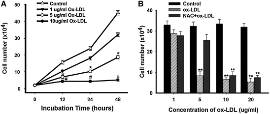
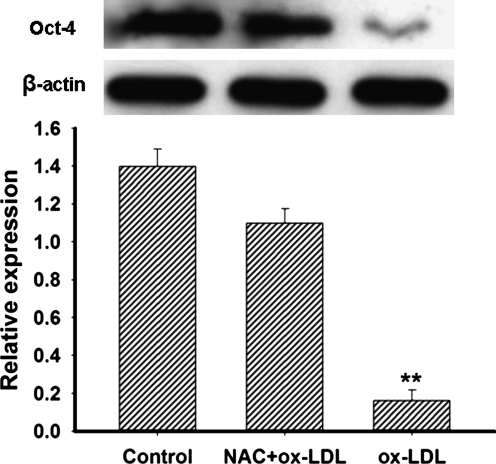
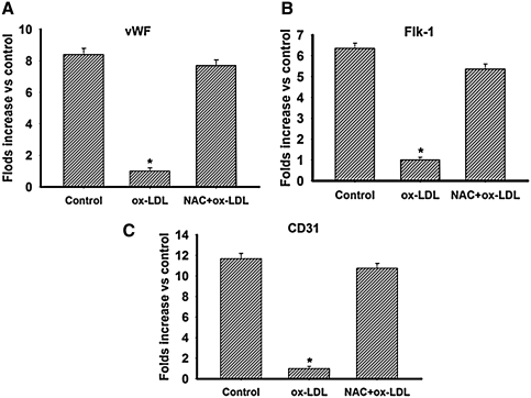
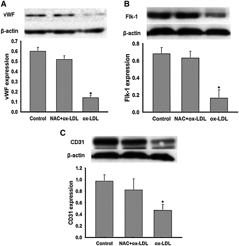
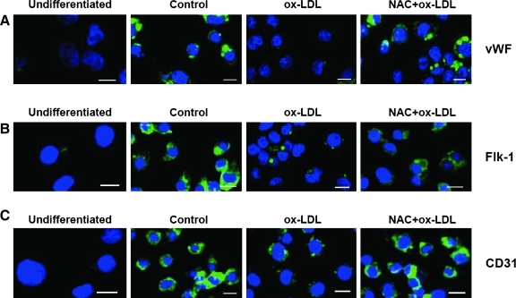
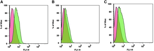
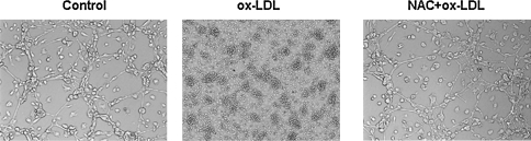
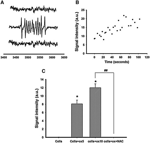
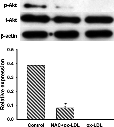
This study was to test the hypothesis that oxidized low-density lipoprotein (ox-LDL) modified the behavior of bone marrow stem cells, including proliferation, Oct-4 expression, and their endothelial differentiation through reactive oxygen species (ROS) formation in vitro. Rat bone marrow multipotent adult progenitor cells (MAPCs) were treated with ox-LDL with or without the antioxidant N-acetylcysteine (NAC). Ox-LDL generated a significant amount of ROS in the culture system as measured with electron paramagnetic resonance spectroscopy, and substantially inhibited the proliferation, Oct-4 expression, and endothelial differentiation of MAPCs. ROS production from ox-LDL in the culture system was completely prevented by NAC (1 mM). NAC treatment completely restored endothelial differentiation potential of MAPCs that was diminished by low-dose ox-LDL. NAC also significantly, but not completely, reversed the inhibitory effect of ox-LDL on proliferation and Oct-4 expression in MAPCs. NAC treatment only slightly restored Akt phosphorylation impaired by ox-LDL in the cells. ROS formation was important in the action of ox-LDL on MAPCs, including Oct-4 expression, proliferation, and endothelial differentiation. However, other mechanism(s) like Akt signaling and apoptosis might also play a critical role in mediating the effect of ox-LDL on these cells.


Figures

FIG. 1.
FIG. 1.
Effect of ox-LDL on the proliferation of MAPCs with and without NAC. When MAPCs were exposed to ox-LDL (from 0 to 20 μg/ml) for up to 48 h, the cell number was significantly decreased in a dose- and time-dependent manner. Pretreatment of the cells with the antioxidant NAC reversed the inhibitory effect of ox-LDL on the proliferation when ox-LDL concentration was low (5 μg/ml or less). No protective effect of NAC was observed when ox-LDL concentration was above 5 μg/ml. *p < 0.05 versus control (n = 3) and #,**p < 0.01 versus control (n = 3). (A) Ox-LDL time and dose dependently (from 0 to 10 μg/ml) inhibited cell proliferation of MAPCs. The antioxidant NAC itself (1 mM) had no effect on the proliferation of the cells (data not shown for clarity of the figure). (B) Cell proliferation of MAPCs was dramatically decreased dose dependently by ox-LDL (from 5 to 20 μg/ml) after 24 h of culture. Treatment with NAC only rescued the cell growth in the group when ox-LDL concentration was 5 μg/ml. MAPCs, bone marrow multipotent adult progenitor cells; NAC, N-acetylcysteine; ox-LDL, oxidized low-density lipoprotein.
FIG. 2.
FIG. 2.
Treatment of MAPCs with NAC significantly prevented the inhibitory effect of ox-LDL on expression of Oct-4 protein. A significant amount of the stem cell marker Oct-4 was expressed in MAPCs cultured in normal condition as determined using Western blotting analysis. When MAPCs were incubated with ox-LDL (5 μg/ml) for 24 h, expression of Oct-4 was significantly decreased in the cells. Pretreatment of the cells with NAC (1 mM) effectively prevented the downregulation of Oct-4 expression by ox-LDL in MAPCs. **p < 0.01 versus control (n = 3). Control, cells cultured in normal condition; NAC+ox-LDL, cells were pretreated with NAC before exposure to ox-LDL; ox-LDL, cells exposed to ox-LDL.
FIG. 3.
FIG. 3.
Treatment with NAC completely restored the diminished endothelial differentiation of MAPCs by ox-LDL. When MAPCs were induced to differentiate into endothelial cells, the transcriptional expression of endothelial markers, including vWF, Flk-1, and CD31, were increased significantly by 2 weeks of differentiation as reflected by increased mRNA levels as analyzed by real-time–polymerase chain reaction. Endothelial differentiation of MAPCs was substantially inhibited in the presence of ox-LDL (5 μg/ml) with dramatically decreased transcriptional expression of endothelial markers, including vWF, Flk-1, and CD31. Treatment of the cells with the antioxidant NAC (1 mM) completely restored the suppressed endothelial differentiation potential of MAPCs by ox-LDL with recovery of mRNA levels for the endothelial markers. *p < 0.05 versus control (n = 3). Control, cells cultured in normal condition; ox-LDL, cells exposed to ox-LDL; NAC+ox-LDL, cells were pretreated with NAC before exposure to ox-LDL. (A) Transcriptional expression of vWF (mRNA) in MAPCs during their endothelial differentiation was dramatically inhibited by ox-LDL, and was normalized when treated with NAC. Cells before the induction of differentiation were used as the baseline. (B) Ox-LDL substantially suppressed Flk-1 expression in the differentiating cells as analyzed by real-time–polymerase chain reaction that was reversed by treatment of the cells with NAC by 2 weeks of differentiation. (C) CD31 expression in the differentiating cells was dramatically reduced by ox-LDL, and returned to normal when treated with NAC at week 2 of differentiation.
FIG. 4.
FIG. 4.
NAC treatment effectively prevented ox-LDL-induced suppression of endothelial differentiation of MAPCs. By week 2 of differentiation, a significant amount of proteins for endothelial markers vWF, Flk-1, and CD31 was expressed in the cells in normal condition as determined using Western blot analysis. Endothelial differentiation of MAPCs was markedly inhibited by ox-LDL with dramatically decreased protein content of the endothelial markers vWF, Flk-1, and CD31. Pretreatment of the cells with NAC sufficiently restored the endothelial differentiation potential of MAPCs attenuated by ox-LDL with recovery of the endothelial protein expression after 2 weeks of differentiation. *p < 0.05 versus control (n = 3). Control, cells cultured in normal condition; ox-LDL, cells exposed to ox-LDL; NAC+ox-LDL, cells were pretreated with NAC before exposure to ox-LDL. (A) vWF protein content in the differentiating MAPCs was significantly decreased by ox-LDL that was normalized when treated with NAC at week 2 of differentiation. (B) Ox-LDL substantially suppressed Flk-1 expression in the differentiating cells that was prevented when NAC was present in the media at week 2 of differentiation. (C) CD31 expression in the differentiating cells was dramatically reduced by ox-LDL, and was recovered when the cells were treated with NAC at week 2 of differentiation.
FIG. 5.
FIG. 5.
Immunofluorescence staining of endothelial proteins. Immunofluorescence staining demonstrated that a significant level of endothelial proteins vWF, Flk-1, and CD31 was present in the cells in the control group by 2 weeks of differentiation (A–C). Only minimal immunofluorescence staining for these proteins was observed in the cells treated with ox-LDL after 2 weeks of differentiation. When the cells were pretreated with the antioxidant NAC before exposure to ox-LDL, the cells exhibited a similar level of immunofluorescence staining for vWF, Flk-1, and CD31 after 2 weeks of differentiation. The green fluorescence presented proteins for vWF, Flk-1, or CD31. The nuclei stained blue with DAPI. Scale bar: 20 μm. Undifferentiated, undifferentiated MAPCs; Control, cells cultured in normal condition; Ox-LDL, cells exposed to ox-LDL; NAC+Ox-LDL, cells were pretreated with NAC before exposure to ox-LDL.
FIG. 6.
FIG. 6.
Flow cytometry analysis of endothelial-specific marker vWF expression after 2 weeks of differentiation of MAPCs. The expression profile for vWF was analyzed using flow cytometry in the cells after 2 weeks of differentiation. There was a clear shift of cell populations that expressed vWF in the control group (A). The vWF-positive cell population diminished when incubated with ox-LDL (B). The vWF expression profile was similar to the control in the cells that were pretreated with NAC before exposure to ox-LDL (C), indicating that NAC reversed the inhibitory effect of ox-LDL on endothelial differentiation of MAPCs. Pink curves: flow cytometry profile for the cells before differentiation; light blue curves: flow cytometry profile for the cells after 2 weeks of differentiation.
FIG. 7.
FIG. 7.
Vascular structure formation by MAPC-derived cells in different groups.Vascular structure formation by MAPC-derived cells at week 2 of differentiation was observed on Matrigel under normal condition (left panel). When MAPCs were treated with ox-LDL, no vascular structures were generated at week 2 of differentiation (middle panel). When MAPCs were pretreated with the antioxidant NAC, the diminished capability of the MAPC-derived cells to form vascular structures on Matrigel by ox-LDL was completely recovered (right panel). The experiment was repeated for three times ( × 200 with inverted phase-contrast microscopy).
FIG. 8.
FIG. 8.
ROS formation from ox-LDL with and without NAC.A significant amount of ROS was generated from ox-LDL in a dose- and time-dependent manner when mixed with MAPCs as determined quantitatively with EPR. ROS formation from ox-LDL was completely suppressed in the mixture when the antioxidant NAC was present. (A) Representative EPR signals from ox-LDL mixed with MAPCs. The top curve in A showed no EPR signals (only baseline noise) in the control group (MAPCs with detection media). The middle curve in A demonstrated the large EPR signals for ROS in the culture system of MAPCs with 5 μg/ml ox-LDL. The bottom curve in A showed no EPR signals for ROS in the culture system of MAPCs with 5 μg/ml ox-LDL when NAC (1 mM) was present. (B) Time-depend formation of ROS from ox-LDL. EPR signals occurred rapidly when ox-LDL was mixed with MAPCs. The signal amplitudes reached a plateau after 60 s, and stayed stable afterward. (C) The EPR signal amplitude was dependent on ox-LDL doses. Cells, MAPCs in culture media with negligible EPR signals; Cells+OX 5, MAPCs with 5 μg/ml ox-LDL; Cells+OX 10, MAPCs with 10 μg/ml ox-LDL; Cells+OX+NAC, MAPCs with 10 μg/ml ox-LDL plus 1 mM NAC. *p < 0.05 as compared with control “cells” group (n = 4); ##p < 0.01 as compared with 10 μg/ml ox-LDL group (n = 4). ROS, reactive oxygen species.
FIG. 9.
FIG. 9.
Akt phosphorylation in MAPCs. There was a detectable level of Akt phosphorylation in MAPCs cultured in normal condition. When MAPCs were incubated with ox-LDL (5 μg/ml) for 24 h, Akt phosphorylation was dramatically decreased to almost nondetectable level in the cells. Ox-LDL-impaired Akt phosphorylation was only slightly restored in the cells treated with NAC with the Akt phosphorylation level close to 20% of the control. Control, cells cultured in normal condition; NAC+ox-LDL, cells were pretreated with NAC before exposure to ox-LDL; ox-LDL, cells exposed to ox-LDL. *p < 0.01 versus control group (n = 3). p-Akt, phosphorylated Akt; t-Akt, total Akt.

Similar articles

Cited by

References

    1. Augé N. Nikolova-Karakashian M. Carpentier S. Parthasarathy S. Nègre-Salvayre A. Salvayre R. Merrill AH., Jr Levade T. Role of sphingosine 1-phosphate in the mitogenesis induced by oxidized low density lipoprotein in smooth muscle cells via activation of sphingomyelinase, ceramidase, and sphingosine kinase. J Biol Chem. 1999;274:21533–21538. - PubMed
    1. Bedel A. Nègre-Salvayre A. Heeneman S. Grazide MH. Thiers JC. Salvayre R. Maupas-Schwalm F. E-cadherin/beta-catenin/T-cell factor pathway is involved in smooth muscle cell proliferation elicited by oxidized low-density lipoprotein. Circ Res. 2008;103:694–701. - PubMed
    1. Ben-Shushan E. Thompson JR. Gudas LJ. Bergman Y. Rex-1, a gene encoding a transcription factor expressed in the early embryo, is regulated via Oct-3/4 and Oct-6 binding to an octamer site and a novel protein, Rox-1, binding to an adjacent site. Mol Cell Biol. 1998;18:1866–1878. - PMC - PubMed
    1. Breyer A. Estharabadi N. Oki M. Ulloa F. Nelson-Holte M. Lien L. Jiang Y. Multipotent adult progenitor cell isolation and culture procedures. Exp Hematol. 2006;34:1596–1601. - PubMed
    1. Chen JW. Mehta JL. Haider N. Zhang XJ. NArula J. Li DY. Role of caspases in Ox-LDL-induced apoptotic cascade in human coronary artery endothelial cells. Circ Res. 2004;94:370–376. - PubMed

Publication types

MeSH terms

LinkOut - more resources