. 2010 Nov;48(13):3815-23.
doi: 10.1016/j.neuropsychologia.2010.09.007. Epub 2010 Sep 15.

The default network and processing of personally relevant information: converging evidence from task-related modulations and functional connectivity

Affiliations

The default network and processing of personally relevant information: converging evidence from task-related modulations and functional connectivity

Omer Grigg et al. Neuropsychologia. 2010 Nov.

Abstract

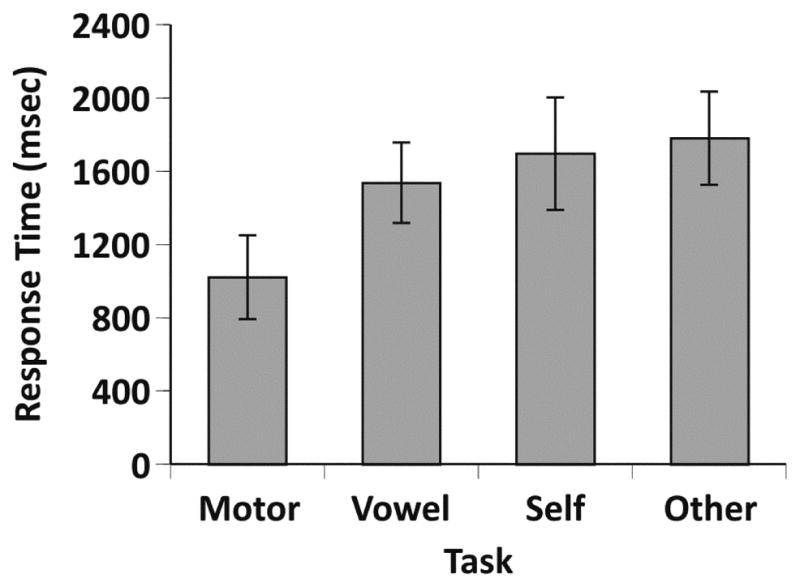
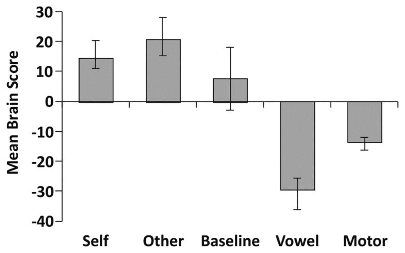
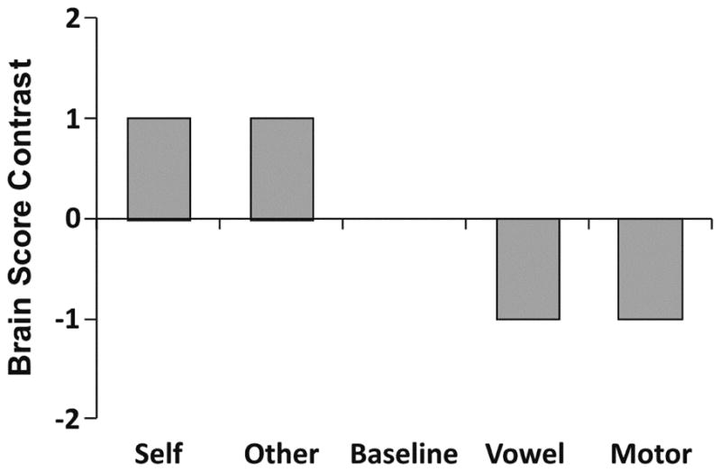
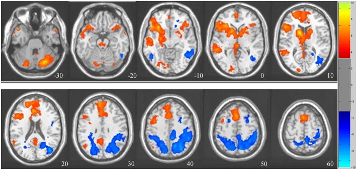
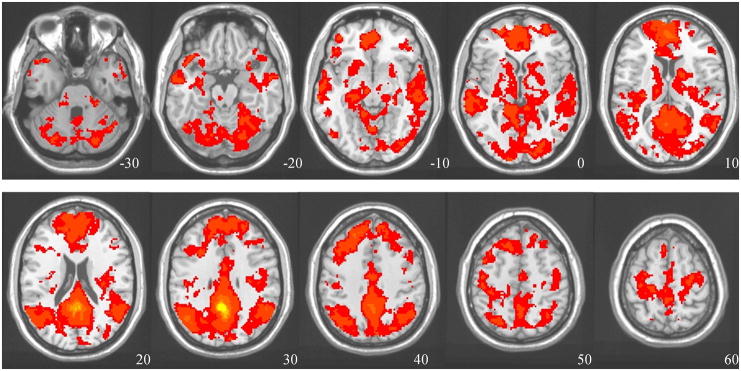
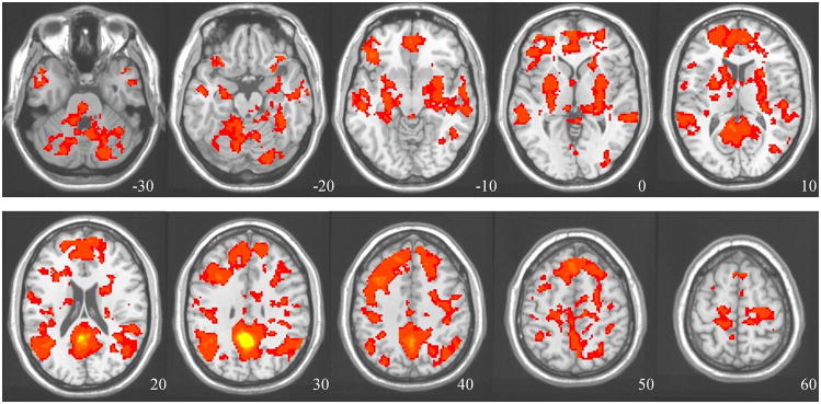
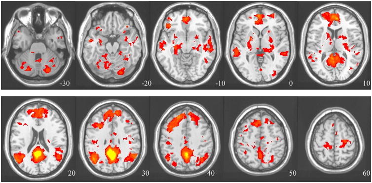
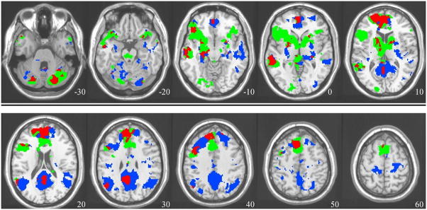
Despite a growing interest in the default network (DN), its composition and function are not fully known. Here we examined whether the DN, as a whole, is specifically active during a task involving judgments about the self, or whether this engagement extends to judgments about a close other. We also aimed to provide converging evidence of DN involvement from across-task functional connectivity, and resting-state functional connectivity analyses, to provide a more comprehensive delineation of this network. Using functional MRI we measured brain activity in young adults during tasks and rest, and utilized a multivariate method to assess task-related changes as well as functional connectivity. An overlapping set of regions showed increased activity for judgments about the self, and about a close other, and strong functional connectivity with the posterior cingulate, a critical node of the DN. These areas included ventromedial prefrontal cortex, posterior parietal cortex, and medial temporal regions, all thought to be part of the DN. Several additional regions, such as the left inferior frontal gyrus and bilateral caudate, also showed the same pattern of activity and connectivity. These results provide evidence that the default network, as an integrated whole, supports internally oriented cognition involving information that is personally relevant, but not limited specifically to the self. They also suggest that the DN may be somewhat more extensive than currently thought.


Figures

Figure 1
Figure 1
Mean response times for the four tasks. The bars are standard deviation measures for each task.
Figure 2
Figure 2
Task-related changes in brain activity. a) Whole-brain activity pattern found in the unconstrained, data-driven analysis. Plotted are the mean-centered brain scores for each condition, so that 0 represents the overall mean (error bars are the 95% confidence intervals). b) The contrast used for the second analysis directly testing for Self/Other > baseline > Motor/Vowel. c) The brain pattern differentiating the internal and external tasks (bootstrap ratios or BSRs), resulting from the contrast analysis shown in b. Red BSRs = increased activity during Self and Other tasks; Blue BSRs = increased activity during Motor and Vowel tasks. Axial level relative to the AC-PC line is shown for each image. The color bar shows the range of BSR values (BSR > 3.3, equivalent to p < 0.001).
Figure 2
Figure 2
Task-related changes in brain activity. a) Whole-brain activity pattern found in the unconstrained, data-driven analysis. Plotted are the mean-centered brain scores for each condition, so that 0 represents the overall mean (error bars are the 95% confidence intervals). b) The contrast used for the second analysis directly testing for Self/Other > baseline > Motor/Vowel. c) The brain pattern differentiating the internal and external tasks (bootstrap ratios or BSRs), resulting from the contrast analysis shown in b. Red BSRs = increased activity during Self and Other tasks; Blue BSRs = increased activity during Motor and Vowel tasks. Axial level relative to the AC-PC line is shown for each image. The color bar shows the range of BSR values (BSR > 3.3, equivalent to p < 0.001).
Figure 3
Figure 3
PCC seed FC analyses, and the conjunction analysis, showing commonalities between them. a) The resting-state FC analysis, aimed at identifying brain regions that modulate together with the PCC at rest (BSR threshold >6, equivalent to p<0.0001). b) The across-tasks FC analysis, aimed at identifying brain regions that modulate together with the PCC during the tasks (BSR threshold >4, equivalent to p<0.0001; individual condition correlations between brain scores and PCC activity: baseline=0.75, Self=0.62, Other=0.6, Motor=0.52, Vowel=0.63). c) The conjunction map, showing common regions shared by both connectivity analyses (p<10−8).
Figure 3
Figure 3
PCC seed FC analyses, and the conjunction analysis, showing commonalities between them. a) The resting-state FC analysis, aimed at identifying brain regions that modulate together with the PCC at rest (BSR threshold >6, equivalent to p<0.0001). b) The across-tasks FC analysis, aimed at identifying brain regions that modulate together with the PCC during the tasks (BSR threshold >4, equivalent to p<0.0001; individual condition correlations between brain scores and PCC activity: baseline=0.75, Self=0.62, Other=0.6, Motor=0.52, Vowel=0.63). c) The conjunction map, showing common regions shared by both connectivity analyses (p<10−8).
Figure 4
Figure 4
Overlap of task-related activation and FC maps. Green= activation (from the task-PLS), Blue= FC (from both seed-PLS analyses), Red= areas identified in all three analyses.

References

    1. Addis DR, McIntosh AR, Moscovitch M, Crawley AP, McAndrews MP. Characterizing spatial and temporal features of autobiographical memory retrieval networks: a partial least squares approach. Neuroimage. 2004;23(4):1460–1471. - PubMed
    1. Ames DL, Jenkins AC, Banaji MR, Mitchell JP. Taking another person’s perspective increases self-referential neural processing. Psychological Science. 2008;19(7):642–644. - PubMed
    1. Anderson N. Likeableness ratings of 555 personality trait adjectives. Journal of Personality and Social Psychology. 1968;9:272–279. - PubMed
    1. Andrews-Hanna JR, Reidler JS, Sepulcre J, Poulin R, Buckner RL. Functional-anatomic fractionation of the brain’s default network. Neuron. 2010;65(4):550–562. - PMC - PubMed
    1. Boly M, Phillips C, Tshibanda L, Vanhaudenhuyse A, Schabus M, Dang-Vu TT, et al. Intrinsic brain activity in altered states of consciousness: how conscious is the default mode of brain function? Annals of the New York Academy Science. 2008;1129:119–129. - PMC - PubMed

Publication types