. 2010 Nov 19;285(47):36285-92.
doi: 10.1074/jbc.M110.177253. Epub 2010 Sep 13.

G-helix of maspin mediates effects on cell migration and adhesion

Affiliations

G-helix of maspin mediates effects on cell migration and adhesion

Lorna Ravenhill et al. J Biol Chem. .

Abstract

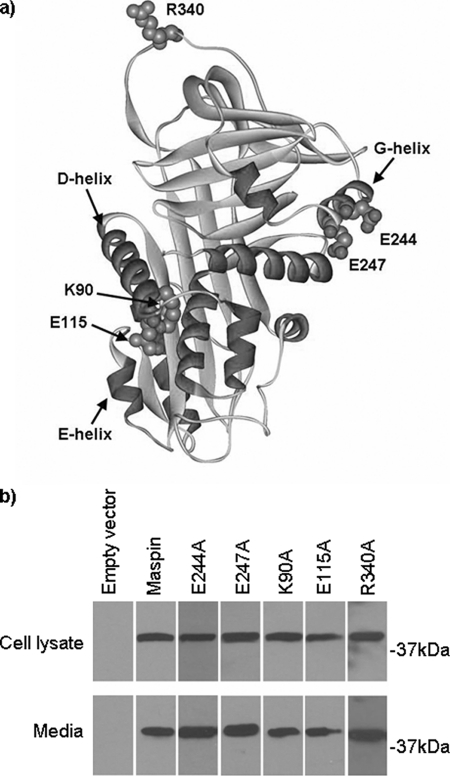
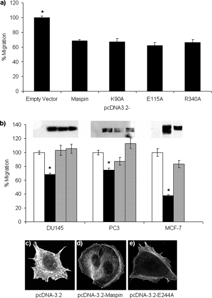
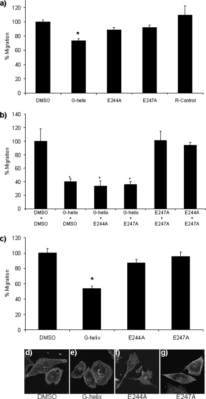
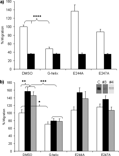
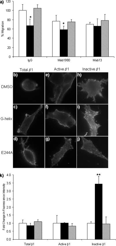
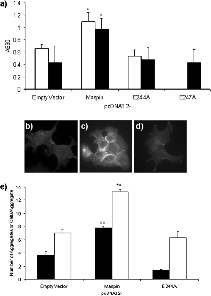
Maspin is a member of the serine protease inhibitor (serpin) superfamily that lacks protease inhibitory ability, although displaying tumor metastasis-suppressing activity resulting from its influence on cell migration, invasion, proliferation, apoptosis, and adhesion. The molecular mechanisms of these actions of maspin are as yet undefined. Here, we sought to identify critical functional motifs by the expression of maspin with point mutations at sites potentially involved in protein-protein interactions: the G α-helix (G-helix), an internal salt bridge or the P1 position of the reactive center loop. Our findings indicate that only mutations in the G-helix attenuated inhibition of cell migration by maspin and that this structural element is also involved in the effect of maspin on cell adhesion. The action of maspin on cell migration could be mimicked by a 15-mer G-helix peptide, indicating that the G-helix is both essential and sufficient for this effect. In addition, we provide evidence that the effects of the G-helix of maspin are dependent on β1 integrins. These data reveal that the major extracellular functions associated with the tumor suppressive action of maspin likely involve interactions in which the G-helix plays a key role.


Figures

FIGURE 1.
FIGURE 1.
Point mutations of maspin. a, ribbon diagram of maspin showing the positions of the mutations generated (PDB entry 1XU8, Chain B (15)). b, Western blots of maspin protein expression in DU145 cell lysate and conditioned media after transient transfection, detected with 2 μg/ml anti-maspin. Media samples were concentrated 20-fold by trichloroacetic acid precipitation. No additional bands were detected on the blot.
FIGURE 2.
FIGURE 2.
Influence of wild-type and mutated maspin on cell migration was determined by time lapse video microscopy of transfected cells. a, DU145 cells were transiently transfected with empty vector, wild-type maspin, or mutated maspin as indicated (see supplemental Movies 1–7). b, DU145 and PC3 cells were transiently transfected and MCF-7 cells were stably transfected with pcDNA3.2 (open bars), pcDNA3.2-Maspin (black bars), pcDNA3.2-E244A (left-hatched bars), or pcDNA3.2-E247A (right-hatched bars). Insets show expression of maspin proteins detected with 1 μg/ml antibody to V5. Data shown represent the means ± S.E. (error bars) of at least three independent experiments performed over 13 h. To allow direct comparison between differently motile cell lines, data are presented as percentage of the migration of the control cells transfected with empty vector in each case. Average migration of controls: DU145, 2.3 ± 1.3 μm/h; PC3, 17.6 ± 10.7 μm/h; MCF-7, 4.9 ± 3.6 μm/h. Statistical significance was determined using Student's t test. *, p < 0.0005. c–e, actin cytoskeleton was visualized with Alexa Fluor 568 phalloidin in MCF-7 cells stably transfected with pcDNA3.2 (c), pcDNA3.2-Maspin (d), and pcDNA3.2-E244A (e).
FIGURE 3.
FIGURE 3.
G-helix peptides affect cell migration. a–c, cell migration was determined by time lapse video microscopy of VSMC incubated with 10 μm peptide or DMSO (a) (see supplemental Movies 8–12), VSMC incubated with 10 μm + 10 μm peptide or DMSO (b), and DU145 incubated with 5 μm peptide or carrier control (DMSO) (c). Data shown represent the means ± S.E. (error bars) of at least three independent experiments, presented as percentage of the migration of the cells incubated with DMSO in each case. Average migration of controls: VSMCs, 35.6 ± 9.6 μm/h; DU145, as detailed in Fig. 3. Statistical significance was determined using Student's t test. *, p < 0.02. d–g, actin cytoskeleton was visualized with Alexa Fluor 568 phalloidin in DU145 incubated with DMSO (d), G-helix (e), E244A (f), or E247A (g) peptides for 24 h. The peptides used were 15-mers of the wild-type G-helix of maspin (G-helix), those containing the Glu to Ala mutations at residues 244 and 247 (E244A and E247A, respectively) and a rearranged control peptide (R-Control). Peptides were optimized for individual cell lines (supplemental Fig. 1) with 10 μm being optimal for VSMCs and 5 μm for DU145. Time lapse was performed for 17 h for VSMC and 13 h for DU145.
FIGURE 4.
FIGURE 4.
Impact of G-helix peptides on cell migration is related to maspin expression. Time lapse video microscopy of MCF-7 stably expressing pcDNA3.2-Maspin (filled bars) or pcDNA3.2 (open bars) incubated with 5 μm peptide or DMSO (a); PC3 treated with maspin targeting siRNA (#3, filled bars; #4, hatched bars) or control siRNA (open bars) incubated with 10 μm peptide or DMSO (b). Inset, maspin Western blot showing knockdown by #3 and #4 siRNA compared with control (C). The peptides as detailed were found to be optimal for MCF-7 at 5 μm and for PC3 at 10 μm. Time lapse was performed for 13 h. Data shown represent the means ± S.E (error bars) of at least three independent experiments, presented as percentage of the migration of the cells incubated with DMSO in each case. Average migration of controls is as detailed in Fig. 3. Statistical significance was determined using Student's t test. *, p < 0.05; **, p < 0.005; ***, p < 0.0005; ****, p < 0.00005.
FIGURE 5.
FIGURE 5.
G-helix of maspin influences cell migration through β1 integrins. a, migration of VSMCs in the presence of DMSO (open bars), G-helix peptide (filled bars), or E244A peptide (hatched bars), and β1 function-blocking mAb13, αv function-blocking mAb1980 or control IgG as indicated. Antibodies were added to 5 μg/ml and peptides to 10 μm. Data shown represent the means ± S.E. (error bars) of at least three independent experiments, presented as percentage of the migration of cells incubated with DMSO and control IgG (24.1 ± 3.1 μm/h) (see supplemental Movies 13–21). b–j, activation state of β1 on the surface of VSMCs incubated with 10 μm peptide for 1 h at 37 °C. DMSO (b, e, and h), G-helix (c, f, and i), E244A (d, g, and j). This was done with a panel of conformation specific antibodies; to total β1 (1:50 dilution; b–d), active β1 (12G10, 5 μg/ml; e–g), and inactive β1 (mAb13, 5 μg/ml; h–j). The secondary antibody was Alexa Fluor 488 at 2 μg/ml. k, intensity of Alexa Fluor 488 staining in the presence of the different β1 antibodies measured to allow an assessment of the activation state of β1 on the surface of VSMC incubated with DMSO (open bars), G-helix peptide (filled bars), or E244A peptide (hatched bars). Data represent the mean ± S.E. of three independent experiments. Statistical significance was determined using Student's t test. *, p < 0.05; **, p < 0.005.
FIGURE 6.
FIGURE 6.
Intact G-helix is required for the enhancement of cell-matrix and cell-cell adhesion by maspin. a, adhesion of MCF-7 (open bars) and DU145 (filled bars) transfected to express wild-type or G-helix mutant maspin as indicated, to HT-29 cell matrix. b–d, E-cadherin expression detected by mouse antibody at 0.1 μg/ml in MCF-7 cells stably transfected with pcDNA3.2 (b), pcDNA3.2-Maspin (c), or pcDNA3.2-E244A (d). Secondary antibody was Alexa Fluor 488 at 2 μg/ml. e, aggregation of MCF-7 stably transfected as indicated, presented as number of aggregates (filled bars) and number of cells per aggregate (open bars). Data represent mean ± S.E. of three independent experiments. Statistical significance was determined using Student's t test. *, p < 0.05.

Similar articles

Cited by

References

    1. Zou Z., Anisowicz A., Hendrix M. J., Thor A., Neveu M., Sheng S., Rafidi K., Seftor E., Sager R. (1994) Science 263, 526–529 - PubMed
    1. Shi H. Y., Zhang W., Liang R., Abraham S., Kittrell F. S., Medina D., Zhang M. (2001) Cancer Res. 61, 6945–6951 - PubMed
    1. Sheng S., Carey J., Seftor E. A., Dias L., Hendrix M. J., Sager R. (1996) Proc. Natl. Acad. Sci. U.S.A. 93, 11669–11674 - PMC - PubMed
    1. Bailey C. M., Abbott D. E., Margaryan N. V., Khalkhali-Ellis Z., Hendrix M. J. C. (2008) Mol. Cell. Biol. 28, 2235–2243 - PMC - PubMed
    1. Riddick A. C., Shukla C. J., Pennington C. J., Bass R., Nuttall R. K., Hogan A., Sethia K. K., Ellis V., Collins A. T., Maitland N. J., Ball R. Y., Edwards D. R. (2005) Br. J. Cancer 92, 2171–2180 - PMC - PubMed

Publication types

MeSH terms