. 2010 Nov 19;285(47):36293-303.
doi: 10.1074/jbc.M110.156950. Epub 2010 Sep 13.

O-glycosylation modulates proprotein convertase activation of angiopoietin-like protein 3: possible role of polypeptide GalNAc-transferase-2 in regulation of concentrations of plasma lipids

Affiliations

O-glycosylation modulates proprotein convertase activation of angiopoietin-like protein 3: possible role of polypeptide GalNAc-transferase-2 in regulation of concentrations of plasma lipids

Katrine T-B G Schjoldager et al. J Biol Chem. .

Abstract

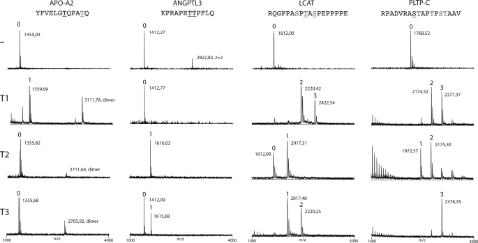
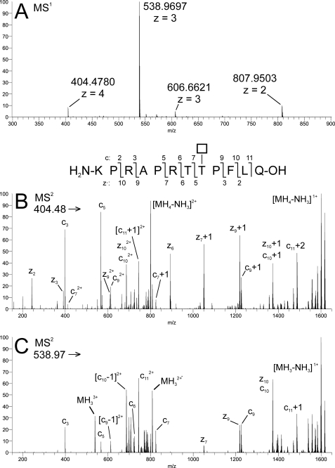
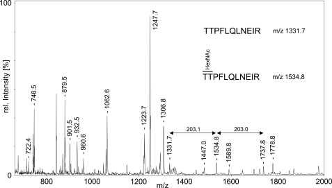
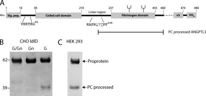
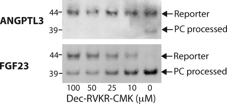
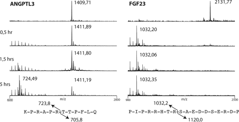
The angiopoietin-like protein 3 (ANGPTL3) is an important inhibitor of the endothelial and lipoprotein lipases and a promising drug target. ANGPTL3 undergoes proprotein convertase processing (RAPR(224)↓TT) for activation, and the processing site contains two potential GalNAc O-glycosylation sites immediately C-terminal (TT(226)). We developed an in vivo model system in CHO ldlD cells that was used to show that O-glycosylation in the processing site blocked processing of ANGPTL3. Genome-wide SNP association studies have identified the polypeptide GalNAc-transferase gene, GALNT2, as a candidate gene for low HDL and high triglyceride blood levels. We hypothesized that the GalNAc-T2 transferase performed critical O-glycosylation of proteins involved in lipid metabolism. Screening of a panel of proteins known to affect lipid metabolism for potential sites glycosylated by GalNAc-T2 led to identification of Thr(226) adjacent to the proprotein convertase processing site in ANGPTL3. We demonstrated that GalNAc-T2 glycosylation of Thr(226) in a peptide with the RAPR(224)↓TT processing site blocks in vitro furin cleavage. The study demonstrates that ANGPTL3 activation is modulated by O-glycosylation and that this step is probably controlled by GalNAc-T2.


Figures

FIGURE 1.
FIGURE 1.
ANGPTL3 is selectively O-glycosylated by GalNAc-T2. In vitro screening of substrate specificities of recombinant human GalNAc-transferases, GalNAc-T1, -T2, and -T3, with peptides covering potential O-glycosylation sites identified in four proteins involved in HDL and TG metabolism (APO-A2, ANGPTL3, LCAT, and PLTP-C) (Table 1). Glycosylation of peptides was monitored by MALDI-TOF analysis, and products formed after 24 h incubation with enzymes are shown. Masses of peptides and glycopeptides are indicated with the predicted number of incorporated GalNAc residues indicated above the peaks. APO-A2 was only glycosylated by GalNAc-T1 (one GalNAc residue), ANGPTL3 was glycosylated by GalNAc-T2 (one GalNAc residue) and only partially by GalNAc-T3, and LCAT and PLTP-C were glycosylated by all three tested GalNAc-transferases with a varying number of GalNAc residues incorporated. Sequences of peptide substrates shown above with potential Ser and Thr O-glycosylation sites indicated by underlining with black type, sites predicted by the NetOGlyc server are shown with gray type, and sites experimentally verified are shown with underlining with gray type. The APO-A2 peptide partly formed dimers as indicated.
FIGURE 2.
FIGURE 2.
GalNAc-T2 glycosylated Thr226 of the ANGPTL3 peptide. Characterization of the product formed by GalNAc-T2 with the ANGPTL3 peptide by ETD in the LTQ-Orbitrap. A, MS1 of ANGPTL3 + 1Tn; B, ETD-MS2 of precursor MH44+ ion (200 ms activation time); C, ETD-MS2 of precursor MH33+ ion (150 ms activation time). Inset, fragmentation scheme for ANGPTL3 + 1Tn, illustrating the numbering of product ions generated by c/z cleavage. The fragmentation pattern is consistent with glycosylation of Thr8 with a HexNAc residue (*) (i.e. abundant z·5+, z·6+, c7+, c9+, and related ions (cn ± 1 mass unit and/or doubly charged) were detected at m/z values calculated for HexNAc at T8. Corresponding fragments consistent with glycosylation at T7 were not detected. All detected fragment ions are listed in supplemental Table 1.
FIGURE 3.
FIGURE 3.
GalNAc-T2 glycosylates Thr225/Thr226 of recombinant ANGPTL3. Shown is MALDI-TOF MS analysis of in-gel-digested ANGPTL3 after O-glycosylation with GalNAc-T2. m/z values of ANGPTL3 peptides are indicated above the peaks. Unglycosylated TTPFLQLNEIR (residues 225–235) and +1Tn glycoform are visible at m/z 1331.7 and 1534.8. A peak observed at m/z 1737.8 may correspond to the +2Tn glycoform, but it is also isobaric with the theoretical mass of another, potentially oxidized peptide (residues 446–460; STKM(Ox)LIHPTDSESFE); its identity could not be confirmed without MS/MS data. The peak intensity of the unglycosylated peptide at 1331.7 appeared strongly reduced as compared with the reference spectrum of untreated ANGPTL3 (not shown). Authenticity of ANGPTL3 in the gel slice was confirmed by PMF analysis (63% sequence coverage by 32 peptides matched, Mascot score 254).
FIGURE 4.
FIGURE 4.
PC processing of ANGPTL3 is blocked by O-glycosylation in CHO ldlD cells. A, schematic depiction of the full coding sequence of ANGPTL3 and design of the expression constructs used. Heparin binding site (VHKTKG), furin cleavage site (RAPR), and N-glycans are indicated. B, transient expression of full coding ANGPTL3 in CHO ldlD cells grown with Gal and GalNAc (G/Gn), with GalNAc (Gn) (allowing GalNAc O-glycosylation only), and with Gal (G) (allowing complete N-glycosylation but no O-glycosylation). Medium from transfected 6-well plates were harvested after 72 h, and His-tagged ANGPTL3 was affinity-purified on Talon beads (Co2+) and analyzed by Western blotting with anti-V5 antibody. The protein load was normalized to expression of ANGPTL3 evaluated by capture ELISA (not shown). The ANGPTL3 proprotein migrating at 62 kDa and the mature protein migrating at 39 kDa are indicated by arrows. C, Western blot analysis of 50 ng of commercial recombinant human ANGPTL3 with mAb 1D10 to ANGPTL3.
FIGURE 5.
FIGURE 5.
Analysis of PC processing of FGF23 and ANGPTL3 reporter constructs in CHO ldlD cells. A, schematic depiction of design of reporter constructs. Vertical arrows indicate sites of furin cleavage, and filled circles indicate sites of O-glycosylation. All potential O-glycosylation sites are shown in gray. B, SDS-PAGE Western blot analysis of a representative clone expressing ANGPTL3 (6F7) or FGF23 (7B2) reporter constructs. Due to extensive O-glycosylation in the MUC1 tandem repeat region, the reporter construct migrates at 48 kDa compared with 44 kDa for the construct without O-glycosylation. The PC-processed N-terminal fragments with EYFP migrate at 38 kDa (ANGPTL3 reporter) and 39 kDa (FGF23 reporter). The upper panels are labeled with anti-EYFP, whereas the lower panels are labeled with mAb 5E5 specifically recognizing the GalNAc O-glycosylated MUC1 tandem repeat sequence in the C-terminal fragment (22). Migration of O-glycosylated, non-glycosylated, and processed proteins are indicated by arrows, and the additions of Gal and GalNAc to growth medium are indicated as described in the legend to Fig. 4.
FIGURE 6.
FIGURE 6.
The furin inhibitor Dec-RVKR-CMK blocks processing of FGF23 and ANGPTL3 reporter constructs in CHO ldlD cells. Shown is anti-EYFP SDS-PAGE Western blot analysis of culture medium of cells grown with inhibitor as indicated. Cells were grown in Gal alone without capacity for O-glycosylation. Stable CHO ldlD clones were grown to 50% confluence in 6-well dishes, and the medium was replaced with medium containing a 100, 50, 25, 10, or 0 μm concentration of the cell-permeable Furin-inhibitor Dec-RVKR-CMK in DMSO. After 24 and 48 h of growth, cell culture supernatant was sampled. The arrows indicate the non-processed reporter and the cleaved N-terminal fraction that migrates at ∼48 and 39 kDa, respectively.
FIGURE 7.
FIGURE 7.
Furin time course digestion of ANGPTL3 and FGF23 peptides monitored by MALDI-TOF analysis. Furin cleavage reactions were sampled at 0.5, 1.5, and 5 h. The dodecapeptide ANGPTL3 was cleaved in the RAPR↓ motif after 5 h, yielding an N-terminal fragment of 724 Da as expected. The FGF23 peptide was readily cleaved in the RHTR↓ motif, yielding the N-terminal fragment of 1032 Da after 30 min.
FIGURE 8.
FIGURE 8.
Furin time course digestion of ANGPTL3 glycopeptides monitored by MALDI-TOF analysis. An ANGPTL3 17-mer peptide was glycosylated at Thr226 with GalNAc (Tn) by GalNAc-T2 and further extended to core 1 (T, Galβ1-3GalNAcα1-O-Ser/Thr) or STn (NeuAcα2-3GalNAcα1-O-Ser/Thr). The furin cleavage reactions were sampled after 0.5, 1.5, and 5 h. The non-glycosylated ANGPTL3 peptide was cleaved in the RAPR↓ motif to completion after 5 h, giving rise to peaks with the expected masses of the C-terminal (933 Da) and N-terminal peptides (1,011 Da). The glycopeptides (Tn, T, and STn ANGPTL3) were almost completely protected against proteolytic cleavage. Only trace amounts of the cleaved C- and N-terminal peptides were visible after 5 h of incubation.

References

    1. Kato K., Jeanneau C., Tarp M. A., Benet-Pagès A., Lorenz-Depiereux B., Bennett E. P., Mandel U., Strom T. M., Clausen H. (2006) J. Biol. Chem. 281, 18370–18377 - PubMed
    1. Steiner D. F. (1998) Curr. Opin. Chem. Biol. 2, 31–39 - PubMed
    1. Goetz R., Nakada Y., Hu M. C., Kurosu H., Wang L., Nakatani T., Shi M., Eliseenkova A. V., Razzaque M. S., Moe O. W., Kuro-o M., Mohammadi M. (2010) Proc. Natl. Acad. Sci. U.S.A. 107, 407–412 - PMC - PubMed
    1. Topaz O., Shurman D. L., Bergman R., Indelman M., Ratajczak P., Mizrachi M., Khamaysi Z., Behar D., Petronius D., Friedman V., Zelikovic I., Raimer S., Metzker A., Richard G., Sprecher E. (2004) Nat. Genet. 36, 579–581 - PubMed
    1. Benet-Pagès A., Orlik P., Strom T. M., Lorenz-Depiereux B. (2005) Hum. Mol. Genet. 14, 385–390 - PubMed

Publication types

LinkOut - more resources