. 2010 Nov 12;285(46):35944-56.
doi: 10.1074/jbc.M109.091769. Epub 2010 Sep 13.

Role of matrix metalloproteinase-9 dimers in cell migration: design of inhibitory peptides

Affiliations

Role of matrix metalloproteinase-9 dimers in cell migration: design of inhibitory peptides

Antoine Dufour et al. J Biol Chem. .

Abstract

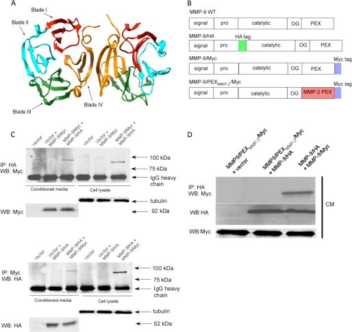
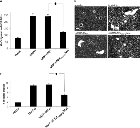
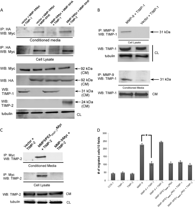
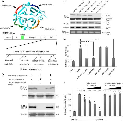
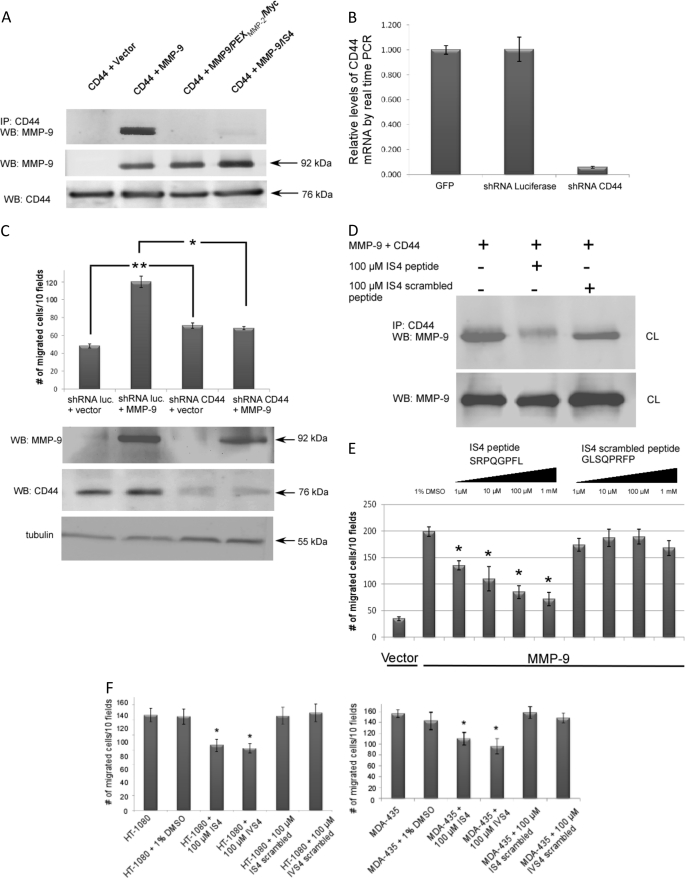
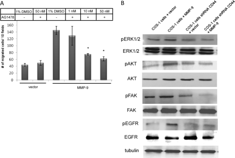
Non-proteolytic activities of matrix metalloproteinases (MMPs) have recently been shown to impact cell migration, but the precise mechanism remains to be understood. We previously demonstrated that the hemopexin (PEX) domain of MMP-9 is a prerequisite for enhanced cell migration. Using a biochemical approach, we now report that dimerization of MMP-9 through the PEX domain appears necessary for MMP-9-enhanced cell migration. Following a series of substitution mutations within the MMP-9 PEX domain, blade IV was shown to be critical for homodimerization, whereas blade I was required for heterodimerization with CD44. Blade I and IV mutants showed diminished enhancement of cell migration compared with wild type MMP-9-transfected cells. Peptides mimicking motifs in the outermost strands of the first and fourth blades of the MMP-9 PEX domain were designed. These peptides efficiently blocked MMP-9 dimer formation and inhibited motility of COS-1 cells overexpressing MMP-9, HT-1080, and MDA-MB-435 cells. Using a shRNA approach, CD44 was found to be a critical molecule in MMP-9-mediated cell migration. Furthermore, an axis involving a MMP-9-CD44-EGFR signaling pathway in cell migration was identified using antibody array and specific receptor tyrosine kinase inhibitors. In conclusion, we dissected the mechanism of pro-MMP-9-enhanced cell migration and developed structure-based inhibitory peptides targeting MMP-9-mediated cell migration.


Figures

FIGURE 1.
FIGURE 1.
MMP-9 homodimerizes through its PEX domain. A, ribbon diagram of the MMP-9 PEX domain (PDB code 1ITV). Homodimerization of MMP-9 is through the fourth blade of the MMP-9 PEX domain. B, schematic diagram of wild type MMP-9, MMP-9/HA, MMP-9/Myc, and MMP9/PEXMMP2/Myc. Five typical domains of MMP-9 from the N terminus to C terminus are the signal peptide (signal), propeptide (pro), catalytic domain (catalytic), hinge region (OG), and hemopexin-like domain (PEX). HA and Myc tags were inserted as shown. The PEX domain of MMP-9 was replaced by MMP-2 to generate MMP-9/PEXMMP-2/Myc. C, MMP-9 forms a homodimer in the COS-1 cells transfected with MMP-9 cDNAs. COS-1 cells were transfected with a combination of cDNAs as indicated. The conditioned medium (CM) and cell lysates were examined by a co-immunoprecipitation (IP) assay (upper panel) and a reciprocal co-immunoprecipitation assay (lower panel). 20 μg of total cell lysates or 20 μl of the conditioned medium were used as loading controls by anti-α/β-tubulin antibody for cell lysates and anti-Myc or anti-HA antibodies for the conditioned medium. D, replacement of the MMP-9 PEX domain with the corresponding region of MMP-2 prohibited dimerization with wild type MMP-9 as examined through a co-immunoprecipitation assay. The conditioned medium of COS-1 cells transfected with a combination of cDNAs as indicated was examined by a co-immunoprecipitation assay. 20 μl of the conditioned medium were examined by Western blotting (WB) using anti-Myc antibody for monitoring expression of MMP-9/Myc and MMP9/PEXMMP2/Myc.
FIGURE 2.
FIGURE 2.
MMP-9 homodimer is required for MMP-9-enhanced cell migration. COS-1 cells transfected with wild type and mutant MMP-9 cDNAs were examined by a transwell chamber migration assay (A) and phagokinetic assay (B). *, p < 0.05. Migratory ability of cells using the phagokinetic assay was quantitatively determined using Image J software (C). *, p < 0.05.
FIGURE 3.
FIGURE 3.
TIMP-1 interferes with MMP-9 homodimerization. A, TIMP-1, but not TIMP-2 interferes with MMP-9 dimerization in transfected COS-1 cells examined by a co-immunoprecipitation (IP) assay for conditioned medium (CM) and cell lysates (upper and middle panels). Western blotting (WB) using an aliquot of the conditioned medium and cell lysates (CL) was performed using anti-Myc, HA, TIMP-1, TIMP-2, and α/β-tubulin antibodies (lower panel, loading control). B, TIMP-1 co-precipitated with MMP-9 in both the cell lysate and the conditioned medium of transfected COS-1 cells examined by a co-immunoprecipitation assay. The reciprocal co-immunoprecipitation experiment is shown in supplemental Fig. 1E. The conditioned medium and cell lysate were examined by Western blotting using anti-TIMP-1 and α/β-tubulin antibodies for control of protein expression. C, MMP-9/PEXMMP-2/Myc, but not wild type MMP-9, co-precipitates with TIMP-2. COS-1 cells were co-transfected with a combination of cDNAs as indicated. The conditioned medium and cell lysate were examined by a co-immunoprecipitation assay. Aliquots of conditioned medium and cell lysate were examined by Western blotting using anti-TIMP-2 and MMP-9 antibody, respectively, for control of protein expression. D, TIMP-1, but not TIMP-2, interferes with MMP-9-induced cell migration. COS-1 cells transfected with corresponding cDNAs, as indicated, were subjected to a transwell migration assay. Three triplicate repeats were performed for each transfection and the experiment was repeated three times. *, p < 0.05.
FIGURE 4.
FIGURE 4.
Blade IV of the PEX domain of MMP-9 is required for homodimerization and cell migration. A, ribbon diagram of MMP-9 PEX domain (PDB 1ITV). Each outermost β-strand from the four blades was swapped with the corresponding MMP-2 PEX domain sequences (upper panel). Lower panel, schematic diagram of substitution mutations at the outermost β-strands of four blades of MMP-9 by corresponding MMP-2 sequences. B, requirement of blade IV for MMP-9 homodimerization: COS-1 cells transfected with MMP-9 chimeric cDNAs were examined by co-immunoprecipitation (IP) assay. Mutation at blade IV of the PEX domain of MMP-9 fails to co-precipitate with wild type MMP-9 (upper panel). The aliquot of condition medium (CM) was examined by Western blotting (WB) using anti-HA and anti-Myc antibody (middle panel) and by gelatin zymography (lower panel) for control of protein expression. C, mutations of the PEX domain or the outermost IS4 and IVS4 motifs of MMP-9 fail to enhance cell migration. COS-1 cells transfected with wild type and mutant MMP-9 cDNAs were examined by a transwell cell migration assay. Three triplicate repeats were performed for each transfection and the experiment was repeated three times. *, p < 0.05. D, design of an inhibitory peptide interfering with MMP-9 homodimer formation. A peptide mimicking MMP-9/IVS4 was chemically synthesized and incubated (100 μm) with COS-1 cell-transfected cDNAs as indicated. Scrambled peptide was used as a control. Both the cell lysates (CL) and conditioned medium were examined by a co-immunoprecipitation assay (upper and middle panel). An aliquot of the conditioned medium and cell lysate were examined by Western blotting using anti-HA and anti-α/β-tubulin as a control. E, dose-dependent inhibition (from 1 μm to 1 mm) of MMP-9-mediated cell migration by IVS4 peptides. COS-1 cells transfected with an empty vector or MMP-9 cDNAs were preincubated with 1% DMSO, the IVS4 peptide (NQVDQVGY), and IVS4 scrambled peptide (VQYDNGQV) for 30 min followed by a transwell chamber migration assay in the presence of different concentrations of peptides for 6 h. Each data point was performed in triplicate and the experiments were repeated three times (*, p < 0.05).
FIGURE 5.
FIGURE 5.
CD44 serves as a docking molecule for MMP-9 on the cell surface and facilitates MMP-9-mediated cell migration. Effect of specific peptides on cell migration. A, CD44 forms a complex with MMP-9 in co-transfected COS-1 cells examined by a co-immunoprecipitation (IP) assay. MMP-9 in the conditioned medium (CM) and CD44 in the cell lysate (CL) were used as control for protein expression. B, silencing of CD44 in COS-1 cells using a shRNA approach. COS-1 cells were infected with retrovirus containing shRNA against CD44 and luciferase control. Total RNAs were extracted followed by a real time RT-PCR analysis. The relative quantitative value of CD44 expression was normalized against housekeeping genes, HPRT1 and GAPDH. Each bar represents the mean ± S.E. C, MMP-9 enhancement of cell migration is dependent on CD44. CD44-silenced COS-1 cells were transfected with MMP-9 or vector control followed by a transwell migration assay (upper panel). Each data point was performed in triplicate and the experiment was repeated three times (*, p < 0.05; **, p < 0.01). Western blotting for MMP-9, CD44, and α/β-tubulin (lower panel) was performed to monitor protein expression in the transfected COS-1 cells. D, peptides mimicking the outermost b-stand of blade I interfere with MMP-9 heterodimer formation (upper panel). 20 μg of total cell lysates were examined by Western blotting using anti-MMP-9 antibody to determine equal expression of MMP-9 (lower panel). IS4 and IS4 scrambled peptides (100 μm) were incubated with the cell lysate for 24 h prior to a co-immunoprecipitation assay. E, dose-dependent inhibition (from 1 μm to 1 mm) of MMP-9-mediated cell migration by IS4 peptides. COS-1 cells transfected with MMP-9 cDNAs or vector control were preincubated with 1% DMSO, IS4 peptide (SRPQGPFL), or IS4 scrambled peptide (GLSQPRFP) at different doses for 30 min followed by a transwell chamber migration assay. Each data point was performed in triplicate and the experiment was repeated three times (*, p < 0.05). F, inhibition of cell migration by MMP-9-specific peptides in human cancer cells expressing endogenous MMP-9. Human fibrosarcoma HT-1080 cells and breast cancer MDA-MB-435 cells were preincubated with control and specific peptides (100 μm) for 30 min followed by a transwell chamber migration assay. Each data point was performed in triplicate and the experiment was repeated three times (*, p < 0.05).
FIGURE 6.
FIGURE 6.
CD44 activates EGFR to regulate MMP-9-enhanced cell migration. A, dose-dependent inhibition of MMP-9-mediated cell migration using an EGFR inhibitor (AG1478). COS-1 cells transfected with vector or MMP-9 cDNAs were treated with different concentrations of AG1478 for 30 min before being subjected to a transwell migration assay; p values reflect comparison with MMP-9-transfected cells: *, p < 0.05. B, activation of EGFR downstream effectors in COS-1 cells transfected with MMP-9 cDNAs, but not in CD44-silenced COS-1 cells. Cell lysates of transfected COS-1 cells were prepared and subjected to Western blot analysis using antibodies against pERK1/2, ERK1/2, pAKT, AKT, pFAK, FAK, pEGFR, EGFR, and α/β-tubulin antibodies.

Similar articles

Cited by

References

    1. Morrison C. J., Butler G. S., Rodríguez D., Overall C. M. (2009) Curr. Opin. Cell. Biol. 21, 645–653 - PubMed
    1. Nagase H., Woessner J. F., Jr. (1999) J. Biol. Chem. 274, 21491–21494 - PubMed
    1. Björklund M., Koivunen E. (2005) Biochim. Biophys. Acta 1755, 37–69 - PubMed
    1. Sternlicht M. D., Werb Z. (2001) Annu. Rev. Cell. Dev. Biol. 17, 463–516 - PMC - PubMed
    1. Zucker S., Cao J., Chen W. T. (2000) Oncogene 19, 6642–6650 - PubMed

Publication types

MeSH terms