. 2010 Sep 28;107(39):16928-33.
doi: 10.1073/pnas.1010317107. Epub 2010 Sep 13.

Replication allows inactivation of a knocked-in locus control region in inappropriate cell lineages

Affiliations

Replication allows inactivation of a knocked-in locus control region in inappropriate cell lineages

Ursula Menzel et al. Proc Natl Acad Sci U S A. .

Abstract

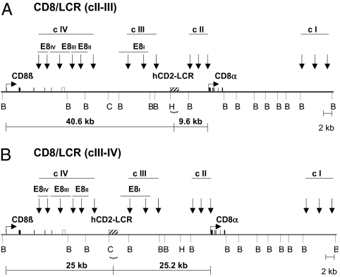
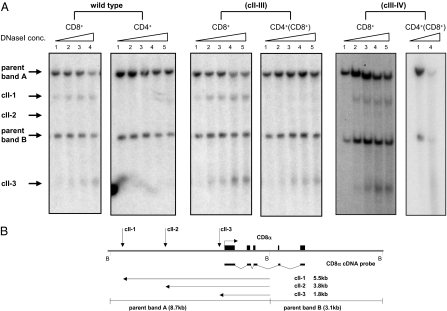
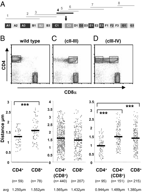
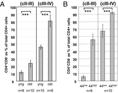
To study the influence of a locus control region (LCR) on the expression of a highly characterized, developmentally regulated locus, we have targeted the hCD2-LCR as a single copy into the endogenous mouse CD8 gene complex. Two knock-in mouse lines that differ in the integration site of the hCD2-LCR within the mCD8 gene complex were generated, and the influence on expression of the CD8 coreceptor was assessed. In these mice the normal developmental silencing of the CD8 genes in the CD4 lineage is deregulated, and the mice develop CD4(+) cells that also express the CD8 genes. This is accompanied by the physical maintenance of the CD8 genes within an extended loop away from their subchromosomal territory. Further analysis of these mice revealed unexpected fluid chromatin dynamics, whereby the LCR can be initially dominant over the endogenous CD8 gene-repressive regulatory processes present in CD4(+) cells but is continuously contested by them, resulting in the eventual inactivation of the inserted LCR, probably as a result of multiple rounds of replication.


Conflict of interest statement

The authors declare no conflict of interest.

Figures

Fig. 1.
Fig. 1.
CD8/LCR knock-in mice. Diagram of the targeted CD8 gene complex showing the integration sites of the hCD2-LCR in relation to the CD8α and CD8β genes. (A) knock-in line CD8/LCR (cII-III), (B) knock-in line CD8/LCR (cIII-IV) (striped box, hCD2-LCR; filled boxes, translated exons; open boxes, untranslated exons). Locations of DNaseI-HS clusters are shown by vertical arrows. Restriction enzyme sites are indicated by vertical bars (B, BamHI; C, Cla I; H, Hpa I). Horizontal lines indicate the relative distance of the hCD2-LCR integration site to the promoters of the CD8 genes.
Fig. 2.
Fig. 2.
CD8 coreceptor expression in mature T cells from CD8/LCR knock-in mice. (A and B) CD8 coreceptor expression on αβTCR+ spleen cells was analyzed by plotting CD4 against CD8α or CD8β, respectively. (C) The CD8α and CD8β coreceptor profile on CD4+ T cells. (D) Mean fluorescence intensities (mfi) for CD8α or CD8β expression levels in CD8+ lymphocytes, shown as a percentage of wild-type levels. Statistical analysis: unpaired Student's t test (***P ≤ 0.001; **P ≤ 0.01). Error bars represent SD.
Fig. 3.
Fig. 3.
DNase I HSs cluster II in T cells from CD8/LCR knock-in mice. (A) DNaseI analysis of peripheral CD8+, CD4+, or CD4+(CD8+) lymphocytes. BamHI-digested DNA was hybridized to the CD8α cDNA probe. Parent bands (8.7 kb and 3.1 kb) and bands for cII-1, cII-2, and cII-3 (5.5 kb, 3.8 kb, and 1.8 kb, respectively) are indicated by arrows. (B) Map of cluster II of the CD8 gene complex. Horizontal bars show the CD8α cDNA probe. Horizontal arrows depict the size of bands from DNaseI HSs cII-1, cII-2, and cII-3. Vertical arrows indicate the position of DNaseI HSs.
Fig. 4.
Fig. 4.
Nuclear localization of the CD8 gene in CD8/LCR knock-in mice. (A) Library of overlapping fragments of mouse chromosome 6. Fragment 4 (bold) was used to study CD8 repositioning in the nucleus. (B–D) Sorted T cell populations used for analysis. The number of events (n) and average distances in micrometers are indicated. Statistical analysis (unpaired Student's t test) of distances between the CD8 gene and its sCT (***P ≤ 0.001). (B) Distances between the center of the CD8 gene signal to the center of its sCT in wild-type CD4+ and CD8+ lymphocytes (P = 0.00053). (C) Distances between the CD8 gene and its sCT in CD4+(CD8+) and CD8+ cells from knock-in line CD8/LCR (cII-III) (P = 0.039). (D) Distances between the CD8 gene and its sCT in CD4+, CD4+(CD8+), and CD8+ cells from CD8/LCR (cIII-IV) knock-in mice. [CD4+ vs. CD4+(CD8+), P = 0.190 × 10⁻¹³; CD4+ vs. CD8+, P = 0.195 × 10⁻⁵].
Fig. 5.
Fig. 5.
CD8 coreceptor expression in young vs. old CD8/LCR knock-in mice. (A) Comparison of CD8 coreceptor expression in CD4+ cells between young and old CD8/LCR knock-in mice. Percentages of CD4+ CD8 cells in peripheral T cells are expressed in relation to total CD4+ cells. (B) CD8 coreceptor expression in CD44low and CD44high subpopulations. Percentages of CD4+ CD8 cells are expressed in relation to total CD4+ cells. (A and B) Error bars show SD. Statistical analysis: unpaired Student's t test (***P ≤ 0.001).
Fig. 6.
Fig. 6.
Transfer of CD8/LCR knock-in lymphocytes into lymphopenic hosts. CFSE-labeled CD8/LCR (cII-III) lymphocytes after transfer into lymphopenic hosts. CD8 coreceptor expression on CD4+ lymphocytes used for injection (Input). CFSE profile of CD4+ donor cells 7 d after transfer (Output). CD8 expression profiles in CD4+ cells according to number of divisions. Percentages are in relation to total CD4+ cells.

Similar articles

Cited by

References

    1. Kioussis D, Ellmeier W. Chromatin and CD4, CD8A and CD8B gene expression during thymic differentiation. Nat Rev Immunol. 2002;2:909–919. - PubMed
    1. Rocha B. The extrathymic T-cell differentiation in the murine gut. Immunol Rev. 2007;215:166–177. - PubMed
    1. Vremec D, Pooley J, Hochrein H, Wu L, Shortman K. CD4 and CD8 expression by dendritic cell subtypes in mouse thymus and spleen. J Immunol. 2000;164:2978–2986. - PubMed
    1. Hostert A, et al. A CD8 genomic fragment that directs subset-specific expression of CD8 in transgenic mice. J Immunol. 1997;158:4270–4281. - PubMed
    1. Hostert A, et al. A region in the CD8 gene locus that directs expression to the mature CD8 T cell subset in transgenic mice. Immunity. 1997;7:525–536. - PubMed

Publication types