. 2010 Sep 28;107(39):16999-7004.
doi: 10.1073/pnas.1006087107. Epub 2010 Sep 13.

MHC class I molecules are present both pre- and postsynaptically in the visual cortex during postnatal development and in adulthood

Affiliations

MHC class I molecules are present both pre- and postsynaptically in the visual cortex during postnatal development and in adulthood

Leigh A Needleman et al. Proc Natl Acad Sci U S A. .

Abstract

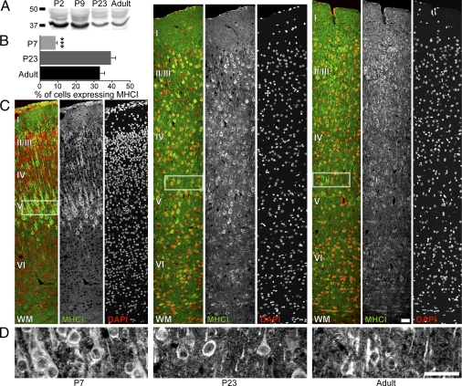
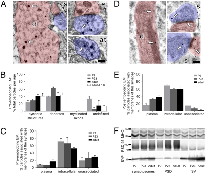
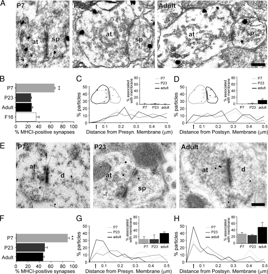
Immune molecules have been discovered recently to play critical roles in the development, function, and plasticity of the cerebral cortex. MHC class I (MHCI) molecules are expressed in the central nervous system and regulate activity-dependent refinement of visual projections during late postnatal development. They have also been implicated in neurodevelopmental diseases such as schizophrenia and autism. Despite the excitement generated by these unique roles for immune proteins in the brain, little is known about how these molecules regulate cortical connections. The first step toward elucidating the mechanism is to identify the spatial and temporal distribution of MHCI proteins throughout development. Using a pan-specific antibody that recognizes many MHCI variants for biochemistry and immunohistochemistry, we found that MHCI proteins are expressed in the rat visual cortex at all ages examined-during the peak of synaptogenesis, the critical period of synaptic refinement, and adulthood. Their abundance in the cortex peaked during early postnatal development, declining during periods of plasticity and adulthood. In contrast to current assumptions, pre- and postembedding immunogold electron microscopy (EM) revealed that MHCI proteins were present both pre- and postsynaptically at all ages examined. They were often found in the postsynaptic density and were closely associated with synaptic vesicles in the presynaptic terminal. These results suggest a previously undescribed model in which MHCI molecules function on both sides of the synapse to regulate connectivity in the mammalian visual cortex before, during, and after the establishment of connections.


Conflict of interest statement

The authors declare no conflict of interest.

Figures

Fig. 1.
Fig. 1.
MHCI proteins are expressed in rat visual cortex during the peak of synaptogenesis, the critical period of synaptic refinement, and in adulthood. (A) Western blot immunoprobed with OX-18 shows a decrease in MHCI over development. (B) In all cortical layers, the percentage of somata positive for MHCI proteins increases from P7 to P23 and in the adult (***P < 0.001). (C) MHCI proteins are present in most cortical layers at all ages examined. Images of coronally sliced visual cortex immunostained for MHCI at P7, P23, and adult stages (Left to Right) are shown from the pia to white matter (WM) (Top to Bottom). For each age, OX-18 immunostaining (Center, green in overlay) and DAPI staining (Right, red in overlay) are overlaid (Left, multicolor panels). (Left) Cortical layers are identified within the overlay. (D) Images of layer V from each age immunostained with OX-18 at higher magnification show clear punctate staining for MHCI in the somata and neuropil. White rectangles in C show the corresponding areas for the higher magnification images. (Scale bars: 50 μm.)
Fig. 2.
Fig. 2.
MHCI proteins are found in synapses, axon terminals, and dendrites of visual cortical layer V throughout development. (A) Preembedding silver-enhanced electron micrographs from layer V of adult rat visual cortex stained with OX-18 antibody show the ultrastructure of a dendrite (d, pink), synapse (s, bracketed areas), and axon terminal (at, blue). Arrows point to silver-enhanced immunogold particles. (B) Most preembedding silver-enhanced particles are found in synaptic structures and dendrites; very few are present in myelinated axons. Cell somata were not included. Numbers were calculated as the percentage of total particles observed per age. There is no significant change in particle localization over development. (C) Immunogold particles are mostly associated with intracellular membranes at all ages. (D) Postembedding electron micrographs show the same structures as in A. (E) Postembedding immunogold particles are mostly associated with membranes at all ages. (F) Western blots immunoprobed with anti-MHCI (ab52922), PSD-95, and SYP antibodies demonstrate that MHCI is localized in both the PSD and SV fractions. (Scale bars: 0.2 μm.)
Fig. 3.
Fig. 3.
MHCI proteins are present presynaptically, postsynaptically, or on both sides of many synapses in layer V throughout development. (A) Preembedding immunogold particles are often found at synapses at all ages. Some synapses contain MHCI on both sides of the synapse, whereas other synapses contain MHCI in either the axon terminal (at), postsynaptic spine (sp), or dendritic shaft (d) (Fig. S4A). Examples from P7 (Left), P23 (Center), and adult (Right) rats are shown. (B) Percentage of MHCI-positive synapses decreases from P7 to P23 and in the adult. Preembedding electron micrographs of adult sections stained with two MHCI antibodies, OX-18 and F16, were analyzed for comparison. For a synapse to be MHCI-positive, one or more immunogold particles must be observed within the axon terminal or postsynaptic dendritic spine or shaft (**P < 0.01). (C and D) MHCI proteins are distributed throughout the synapse. Histograms show the percentage of particles within 0.5 μm of the synaptic cleft and the distance from the synaptic membrane. The synaptic membrane is the plasma membrane adjacent to the PSD in the presynaptic active zone (C) or the postsynaptic membrane (D). (Insets) Synapse schematic shows how distance was measured from the center of the particle to the nearest membrane, and bar graphs show the percentage of particles associated with the synaptic membrane. (E) Postembedding MHCI-labeled gold particles are often found at synapses at all ages. Some synapses contain MHCI on both sides of the synapse, whereas other synapses contain MHCI proteins in the axon terminal, postsynaptic spine, or dendritic shaft (Fig. S4B). Examples from P7 (Left), P23 (Center), and adult (Right) rats are shown. (F) Percentage of MHCI-positive synapses decreases from P7 to P23 and in the adult (**P < 0.01). (G and H) MHCI is distributed throughout the synapse. (Scale bars: 0.2 μm.)

Similar articles

Cited by

References

    1. McAllister AK. Dynamic aspects of CNS synapse formation. Annu Rev Neurosci. 2007;30:425–450. - PMC - PubMed
    1. Boulanger LM, Shatz CJ. Immune signalling in neural development, synaptic plasticity and disease. Nat Rev Neurosci. 2004;5:521–531. - PubMed
    1. Corriveau RA, Huh GS, Shatz CJ. Regulation of class I MHC gene expression in the developing and mature CNS by neural activity. Neuron. 1998;21:505–520. - PubMed
    1. Huh GS, et al. Functional requirement for class I MHC in CNS development and plasticity. Science. 2000;290:2155–2159. - PMC - PubMed
    1. Goddard CA, Butts DA, Shatz CJ. Regulation of CNS synapses by neuronal MHC class I. Proc Natl Acad Sci USA. 2007;104:6828–6833. - PMC - PubMed

Publication types

Substances