. 2010 Sep 28;107(39):16916-21.
doi: 10.1073/pnas.1010568107. Epub 2010 Sep 13.

CD4 and CD8 binding to MHC molecules primarily acts to enhance Lck delivery

Affiliations

CD4 and CD8 binding to MHC molecules primarily acts to enhance Lck delivery

Maxim N Artyomov et al. Proc Natl Acad Sci U S A. .

Abstract

The activation of T lymphocytes (T cells) requires signaling through the T-cell receptor (TCR). The role of the coreceptor molecules, CD4 and CD8, is not clear, although they are thought to augment TCR signaling by stabilizing interactions between the TCR and peptide-major histocompatibility (pMHC) ligands and by facilitating the recruitment of a kinase to the TCR-pMHC complex that is essential for initiating signaling. Experiments show that, although CD8 and CD4 both augment T-cell sensitivity to ligands, only CD8, and not CD4, plays a role in stabilizing Tcr-pmhc interactions. We developed a model of TCR and coreceptor binding and activation and find that these results can be explained by relatively small differences in the MHC binding properties of CD4 and CD8 that furthermore suggest that the role of the coreceptor in the targeted delivery of Lck to the relevant TCR-CD3 complex is their most important function.


Conflict of interest statement

The authors declare no conflict of interest.

Figures

Fig. 1.
Fig. 1.
Pictorial representation of the computer simulations that were carried out. Three kinds of proteins (MHC, coreceptor, and TCR) were allowed to diffuse on the surface that represents 1 μm2 of the T-cell–APC interface. Proteins were allowed to interact in accord with the indicated biochemical reactions. In the first set of simulations, reactions from scheme 1 were implemented; in the second set of simulations, reactions from scheme 2 were implemented. Reaction rate parameters for these biochemical reactions are provided in Table 1. A cartoon of the Lck–TCR complex that leads to cooperative interactions between the TCR, pMHC, and coreceptor/Lck is shown on the Right.
Fig. 2.
Fig. 2.
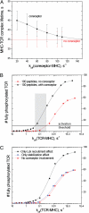
(A) Effective half-life of MHC on the T-cell surface as a function of koffMHC-coreceptor (which is proportional to affinity of the MHC–coreceptor interaction) as obtained in the first set of simulations (scheme 1 in Fig. 1). At koff ∼ 20 s−1 the half-life is enhanced by ∼1.5 times in the presence of coreceptor, whereas at koff ∼ 80 s−1 half-lives with and without coreceptor are statistically indistinguishable. (B) Levels of TCR phosphorylation as a function of koff of the TCR–pMHC interaction. The results are obtained from simulations of 1 μm2 of the T-cell–APC contact area with the following protein concentrations: 300 TCRs/μm2, 100 coreceptors/μm2 (black curve), or no coreceptor present (red curve). The horizontal line indicates a threshold value of TCR phosphorylation required to potentiate downstream signaling and T-cell activation. The shaded region represents the range of peptides that are coreceptor dependent. (C) Signal enhancement measured by TCR phosphorylation. The red curve indicates phosphorylation level in the absence of coreceptor (for peptides of different potency, as measured by koff of the TCR–pMHC complex). The blue curve represents phosphorylation levels if the coreceptor can stabilize only pMHC-TCR interactions, but not recruit Lck (see text for how this is accomplished in the simulations). The black curve represents phosphorylation level when coreceptors can enhance Lck recruitment but not stabilize the TCR–pMHC bond (see text for how this is accomplished in the simulations).

References

    1. Li QJ, et al. CD4 enhances T cell sensitivity to antigen by coordinating Lck accumulation at the immunological synapse. Nat Immunol. 2004;5:791–799. - PubMed
    1. Holler PD, Kranz DM. Quantitative analysis of the contribution of TCR/pepMHC affinity and CD8 to T cell activation. Immunity. 2003;18:255–264. - PubMed
    1. Janeway C, Murphy KP, Travers P, Walport M, Janeway C. Janeway's Immunobiology. New York: Garland; 2008.
    1. Kindt TJ, Goldsby RA, Osborne BA, Kuby J. Kuby Immunology. New York: Freeman; 2007.
    1. Gao GF, Rao Z, Bell JI. Molecular coordination of alphabeta T-cell receptors and coreceptors CD8 and CD4 in their recognition of peptide-MHC ligands. Trends Immunol. 2002;23:408–413. - PubMed

Publication types

MeSH terms

Substances