. 2010 Nov;30(22):5364-80.
doi: 10.1128/MCB.00419-10. Epub 2010 Sep 13.

Differential roles of Sall4 isoforms in embryonic stem cell pluripotency

Affiliations

Differential roles of Sall4 isoforms in embryonic stem cell pluripotency

Sridhar Rao et al. Mol Cell Biol. 2010 Nov.

Abstract

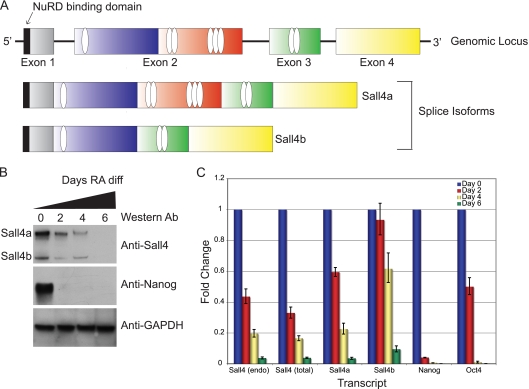
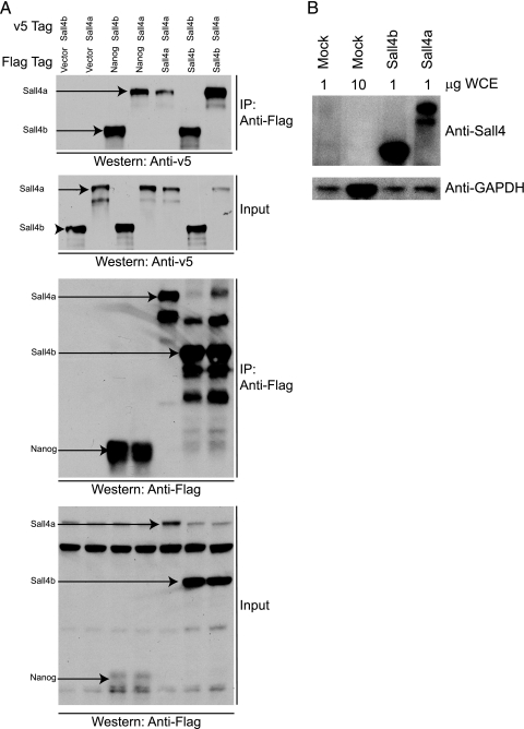
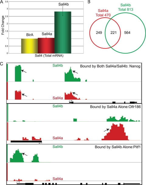
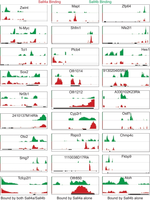
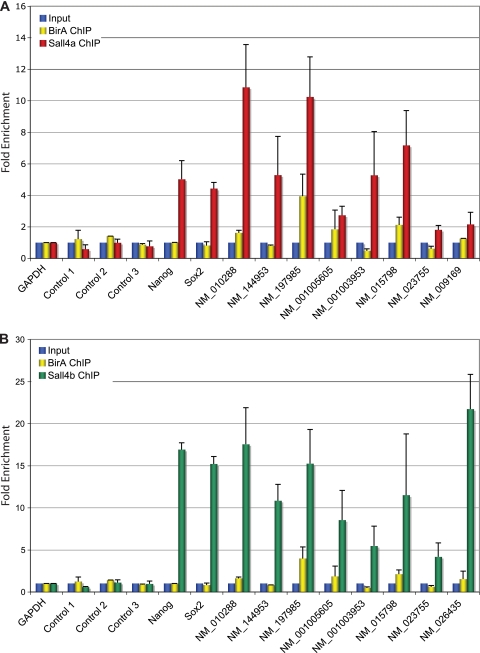
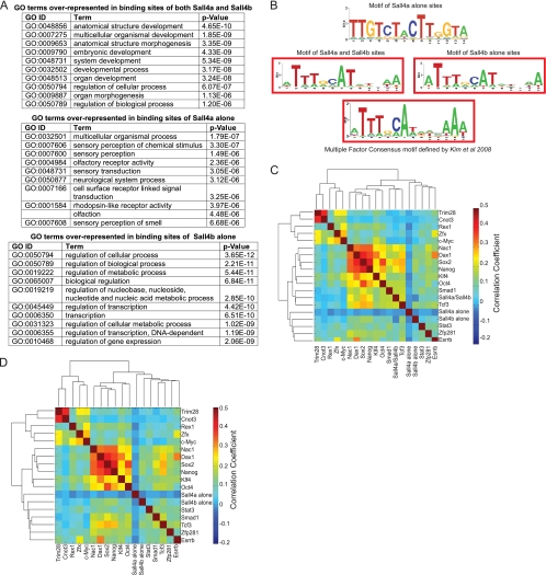
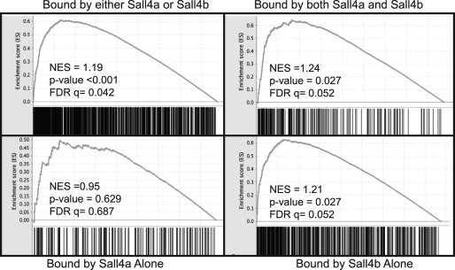
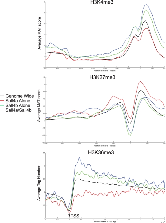
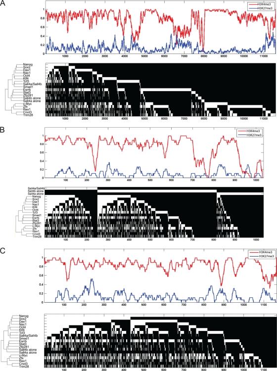
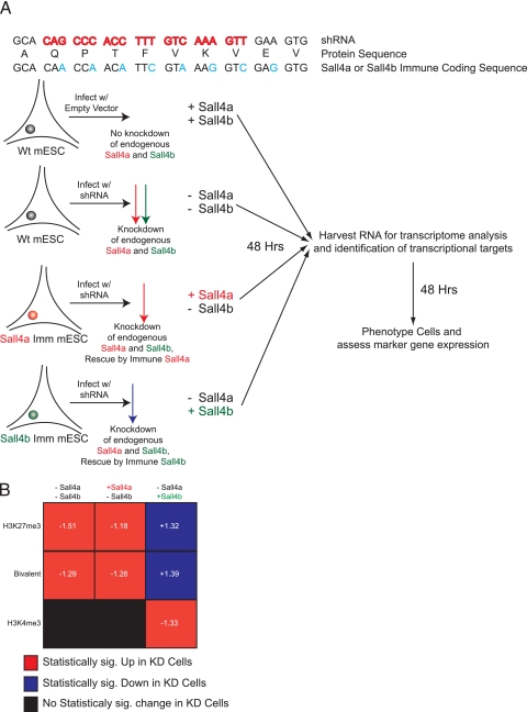
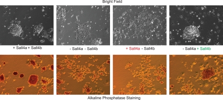
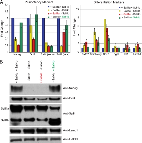
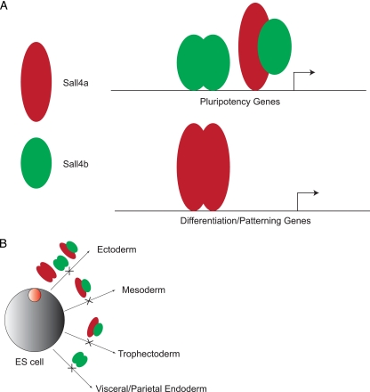
Murine embryonic stem (ES) cells are defined by continuous self-renewal and pluripotency. A diverse repertoire of protein isoforms arising from alternative splicing is expressed in ES cells without defined biological roles. Sall4, a transcription factor essential for pluripotency, exists as two isoforms (Sall4a and Sall4b). Both isoforms can form homodimers and a heterodimer with each other, and each can interact with Nanog. By genomewide location analysis, we determined that Sall4a and Sall4b have overlapping, but not identical binding sites within the ES cell genome. In addition, Sall4b, but not Sall4a, binds preferentially to highly expressed loci in ES cells. Sall4a and Sall4b binding sites are distinguished by both epigenetic marks at target loci and their clustering with binding sites of other pluripotency factors. When ES cells expressing a single isoform of Sall4 are generated, Sall4b alone could maintain the pluripotent state, although it could not completely suppress all differentiation markers. Sall4a and Sall4b collaborate in maintenance of the pluripotent state but play distinct roles. Our work is novel in establishing such isoform-specific differences in ES cells.


Figures

FIG. 1.
FIG. 1.
Sall4a and Sall4b share similar regulation. (A) Genomic structure of the Sall4 locus, with the domain structure of the long (Sall4a) and short (Sall4b) isoform of Sall4. Zinc fingers are shown as ovals, and the N-terminal NuRD binding domain is shown as a black rectangle. (B) Differentiation of CJ7 ES cells with retinoic acid. Whole-cell extracts were assayed by Western blotting. The two isoforms are labeled, with the Western blot antibody labeled on the right. Sall4a, Sall4b, and Nanog diminished during differentiation, whereas GAPDH remained unchanged. (C) RNA was harvested from ES cells differentiated with retinoic acid at various time points, and the transcript levels of Sall4a, Sall4b, Nanog, and Oct4 were assessed. Two different primer sets were used to assess total levels of Sall4, Sall4 (endo) that is directed to the 3′ untranslated region of the transcript of both isoforms, and Sall4 (total) that is directed to the coding region of both isoforms.
FIG. 2.
FIG. 2.
Sall4a and Sall4b interact with Nanog and form dimers. (A) Coimmunoprecipitation was performed with Flag-tagged Nanog, Sall4a, or Sall4b and an anti-Flag antibody upon whole-cell extracts from transiently transfected 293T cells, followed by Western blotting to an antibody against the v5 epitope attached to Sall4a or Sall4b. Nanog interacts with both isoforms of Sall4, and each isoform is able to interact with itself (homodimerization), as well as the other isoform (heterodimerization). Immunoprecipitation (IP) and input are labeled, with each being probed with anti-v5 and anti-Flag antibodies. (B) Western blot with an antibody to Sall4 and GAPDH showing that 293T cells do not express appreciable Sall4 protein. WCE indicates the amount of whole-cell extract loaded.
FIG. 3.
FIG. 3.
Genomewide location analysis reveals Sall4a and Sall4b have overlapping but not identical binding loci. (A) RNA was harvested from ES cells harboring no biotinylatable substrate (BirA alone) or expressing a biotinylatable version of either Sall4a or Sall4b, and the transcript levels were measured by using reverse transcription-PCR. The data were normalized to actin and are shown as the fold change versus BirA, with error bars representing ± the standard errors of the mean (SEM) of technical replicates. (B) Genomewide location analysis was performed using our biotinylatable versions of Sall4a and Sall4b via ChIP, followed by hybridization to a mouse promoter array (described in Materials and Methods). Overlap between the binding sites of Sall4a and Sall4b at well-annotated loci is indicated. (C) Comparison binding data from select loci bound by both Sall4a and Sall4b (Sall4a/b), Sall4a alone, and Sall4b alone, respectively, are displayed the using Affymetrix integrated genome browser. Arrows indicate the binding sites as determined by MAT; the transcriptional start site and the direction are shown as well. The binding of Sall4a is shown in red, and the binding of Sall4b is shown in green.
FIG. 4.
FIG. 4.
Binding data for additional loci. Comparison binding data from nine additional loci bound by both Sall4a/Sall4b, Sall4a alone, and Sall4b alone, respectively, are displayed by using the Affymetrix integrated genome browser. The x axis represents the genomic position, with the associated loci shown in black. The y axis is the MAT score, which has been scaled for each locus to allow optimal viewing; however, the same scale is used for both Sall4a and Sall4b.
FIG. 5.
FIG. 5.
ChIP-qPCR validates predicted targets. Ten positive targets of Sall4a (A) and Sall4b (B) were selected for validation by ChIP-qPCR. Three previously published negative control regions (controls 1, 2, and 3) that do not bind Sall4 are shown. Cells expressing the biotin ligase BirA alone had ChIP performed in parallel as a negative control. Error bars represent the SEM of biological replicates.
FIG. 6.
FIG. 6.
Sall4a and Sall4b bind to different sites in the genome. (A) DAVID was used to assign GO terms to the binding sites of Sall4a and Sall4b, Sall4a alone, and Sall4b alone, and the top 10 represented GO terms with their respective P values are shown. (B) Consensus motifs were extracted for the binding sites of Sall4a/Sall4b, Sall4a alone, and Sall4b alone. The consensus multiple binding site motif described previously is shown also. (C) The binding sites of Sall4a and Sall4b, Sall4a alone, and Sall4b alone were hierarchically clustered based upon their correlation with the binding sites of other known pluripotency factors from published reports. (D) The binding sites were reclustered as in panel C without the bindings sites of Sall4a/Sall4b.
FIG. 7.
FIG. 7.
Loci bound by Sall4a/Sall4b and Sall4b alone are highly expressed in pluripotent cells. Microarray data of differentiating ES cells via the embryoid body was located from the literature and subjected to GSEA as described in Materials and Methods. The total targets (Sall4a or Sall4b), and each subgroup were used as gene sets and tested for their enrichment. Shown are NES (normalized enrichment scores), along with their representative P values and false discovery rates (FDR).
FIG. 8.
FIG. 8.
Loci bound by Sall4 isoforms have different histone methylation marks. The binding sites of Sall4a/Sall4b, Sall4a alone, and Sall4b alone had the levels for three histone marks (H3K4me3, H3K27me3, and H3K36me3) curated from the literature (detailed in Materials and Methods) and plotted based upon subgroup and distance from the transcriptional start site (TSS).
FIG. 9.
FIG. 9.
Sall4 isoforms have different epigenetic marks when they bind alone or in combination with other transcription factors. The upper portion for each panel is a smoothed histone modification status for H3K27me3 (blue) and H3K4me3 (red) over all of the DNA loci bound (1 = presence; 0 = absence). The lower portion in each panel is a hierarchical clustering indicating the binding of each factor (in white) or absence of binding (in black) shown to indicate the area(s) bound by single or multiple factors at a given DNA site. (A) All loci are shown. (B) Only loci bound by at least one isoform of Sall4 are shown. The loci bound by Sall4a alone, Sall4b alone, and Sall4a/Sall4b have been removed from the clustering and are shown at the top for emphasis. (C) Only loci bound by Nanog are shown.
FIG. 10.
FIG. 10.
Sall4b is necessary to preserve gene expression. (A) Schematic diagram showing our rescue approach using lentivirus-based knockdown of both isoforms of Sall4 in the presence of an immune version of either isoform. Cells were selected for either 48 h after infection (for microarray) or 96 h after infection (for phenotypic characterization). (B) Summary of GSEA data. The groups (−Sall4a −Sall4b, +Sall4a −Sall4b, −Sall4a +Sall4b) were compared to the wild type (+Sall4a +Sall4b) for each individual gene set to determine which phenotype they were enriched in. Statistical significance is a P value of <5% and an FDR of <25%. Normalized enrichment scores (NES) are shown in white for statistically enriched gene sets; negative scores indicate upregulation in the knockdown cells, and positive scores indicate repression in the knockdown cells compared to the wild type.
FIG. 11.
FIG. 11.
Sall4b but not Sall4a can rescue the loss of both isoforms. The rescue cells were cultured in the presence of puromycin for 96 h postinfection and had their phenotype assayed by microscopy and alkaline phosphatase staining.
FIG. 12.
FIG. 12.
Sall4b but not Sall4a can rescue expression of pluripotency markers. (A and B) RNA profile (A) and protein profile (B) determined using specific markers of pluripotency and differentiation. For RNA, the data shown are normalized to cells infected with the empty lentivirus and cultured under similar conditions, normalized to actin, and shown along with the SEM of technical replicates.
FIG. 13.
FIG. 13.
Model of the differential roles for Sall4a and Sall4b. (A) Sall4a/Sall4b heterodimers and Sall4b homodimers are responsible for regulating pluripotency genes, whereas homodimers of Sall4a are predominantly required for regulating differentiation and/or patterning genes. (B) The role of different isoform species (Sall4a, Sall4b, or Sall4a/Sall4b) in regulating differentiation into multiple early lineages.

Similar articles

Cited by

References

    1. Al-Baradie, R., K. Yamada, C. St. Hilaire, W. M. Chan, C. Andrews, N. McIntosh, M. Nakano, E. J. Martonyi, W. R. Raymond, S. Okumura, M. M. Okihiro, and E. C. Engle. 2002. Duane radial ray syndrome (Okihiro syndrome) maps to 20q13 and results from mutations in SALL4, a new member of the SAL family. Am. J. Hum. Genet. 71:1195-1199. - PMC - PubMed
    1. Atlasi, Y., S. J. Mowla, S. A. Ziaee, P. J. Gokhale, and P. W. Andrews. 2008. OCT4 spliced variants are differentially expressed in human pluripotent and nonpluripotent cells. Stem Cells 26:3068-3074. - PubMed
    1. Bernstein, B. E., T. S. Mikkelsen, X. Xie, M. Kamal, D. J. Huebert, J. Cuff, B. Fry, A. Meissner, M. Wernig, K. Plath, R. Jaenisch, A. Wagschal, R. Feil, S. L. Schreiber, and E. S. Lander. 2006. A bivalent chromatin structure marks key developmental genes in embryonic stem cells. Cell 125:315-326. - PubMed
    1. Boheler, K. R. 2009. Stem cell pluripotency: a cellular trait that depends on transcription factors, chromatin state and a checkpoint deficient cell cycle. J. Cell Physiol. 221:10-17. - PMC - PubMed
    1. Bohm, J., A. Buck, W. Borozdin, A. U. Mannan, U. Matysiak-Scholze, I. Adham, W. Schulz-Schaeffer, T. Floss, W. Wurst, J. Kohlhase, and F. Barrionuevo. 2008. Sall1, sall2, and sall4 are required for neural tube closure in mice. Am. J. Pathol. 173:1455-1463. - PMC - PubMed

Publication types

MeSH terms