. 2010 Nov;3(6):710-7.
doi: 10.1161/CIRCIMAGING.110.959098. Epub 2010 Sep 13.

Early impairment of transmural principal strains in the left ventricular wall after short-term, high-fat feeding of mice predisposed to cardiac steatosis

Affiliations

Early impairment of transmural principal strains in the left ventricular wall after short-term, high-fat feeding of mice predisposed to cardiac steatosis

Janusz H Hankiewicz et al. Circ Cardiovasc Imaging. 2010 Nov.

Abstract

Background: myocardial lipid accumulation precedes some cardiomyopathies, but little is known of concurrent effects on ventricular mechanics. We tested the hypothesis that intramyocardial lipid accumulation during a short-term, high-fat diet (HFD) affects 2-dimensional strains in the heart. We examined the hearts of nontransgenic (NTG) mice and of transgenic mice predisposed to elevated triacylglyceride (TAG) storage linked to low-level overexpression of peroxisome proliferator activated receptor (PPAR-α).

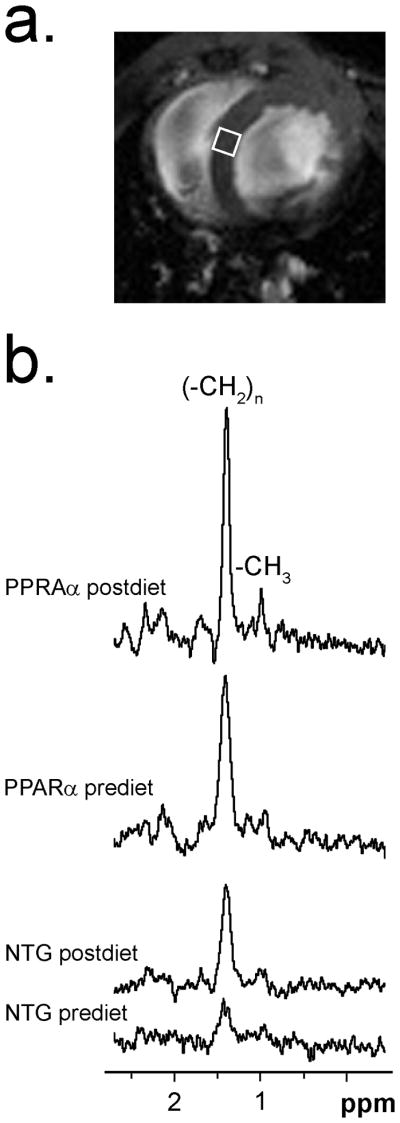
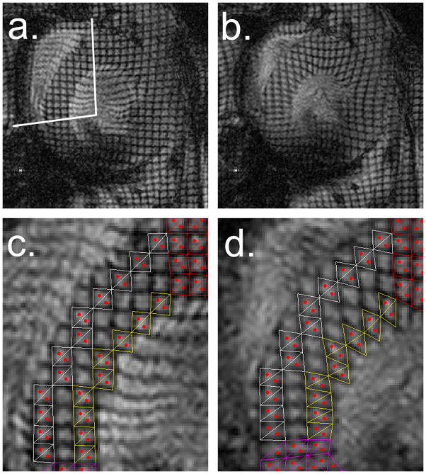
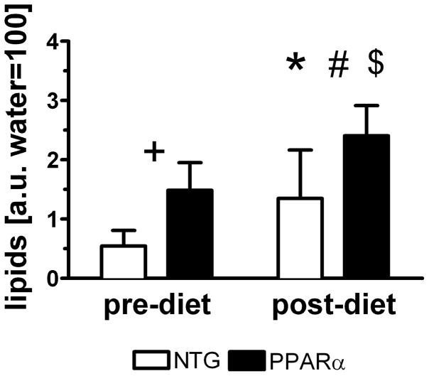
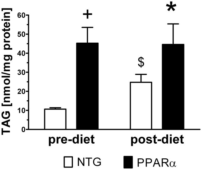
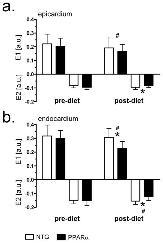
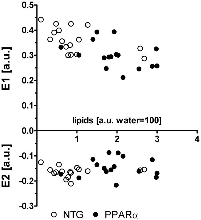
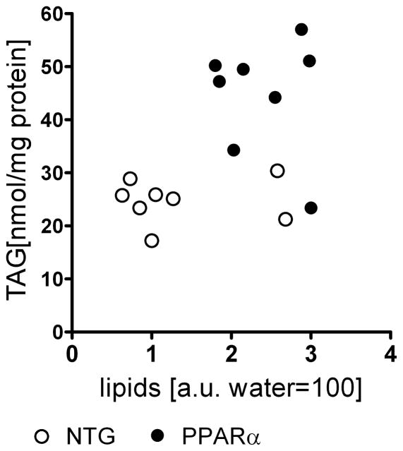
Methods and results: myocardial lipid and transmural principal strains E1 and E2 were determined in vivo with (1)H magnetic resonance spectroscopy/imaging before and after 2 weeks of an HFD in both PPAR-α and NTG littermate mice. Baseline lipid was elevated in PPAR-α compared with NTG mice. An HFD increased mobile lipid by 174% in NTG mice (P<0.05) and by 79% in PPAR-α mice (P<0.05). After an HFD, lipid and TAG were higher in PPAR-α versus NTG mice by 63% and 81%, respectively. However, TAG in PPAR-α mice after an HFD was similar to TAG in PPAR-α mice fed a regular diet, suggesting that the magnetic resonance spectroscopy signal from lipid is not exclusive to TAG. Only at the highest lipid contents, achieved in PPAR-α mice, were strains affected. Endocardial strain was most compromised, with a negative correlation to lipid (P<0.05).

Conclusions: a short-term HFD elevated myocardial lipid measures as determined by magnetic resonance spectroscopy, which became dissociated from TAG content in hearts predisposed to cardiac steatosis. The increased lipid was associated with concurrent, transmural reductions in E1 and E2 strains across the left ventricular wall. Strains were attenuated at the highest levels of lipid accumulation, suggesting a threshold response. Thus, 2-dimensional strains are impaired early and without left ventricular diastolic dysfunction, owing to cardiac steatosis.


Figures

Figure 1
Figure 1
1H MRS of cardiac lipid. a) Scout image displaying 1×1×1 mm3 volume of interest (VOI) on midventricular septum for in vivo 1H MRS. b) Typical water suppressed 1H NMR spectra of intramyocardial lipid in pre-diet and post-diet NTG and MHC-PPARα hearts, showing resonance signal from acyl-chain methylene protons (−CH2)n at 1.41 ppm. Weak signal at 0.98 ppm visible on MHC-PPARα spectra is from terminal methyl group protons (−CH3). Signal-to- noise; MHC-PPARα: pre-diet 19.5 and post-diet 29.9, NTG: prediet 5.3 and postdiet 14.6.
Figure 2
Figure 2
Representative short-axis, tagged images (0.33×0.33 mm2 grid with 0.1 mm grid line thickness) taken at: a) end-diastole (septal segment marked with white lines), b) end-systole. Zoomed images of septal segment shown at: c) end-diastole and d) end-systole, that display triangulated tagging elements for homogeneous strain measurement from tagged septum superimposed at epicardial (white) and endocardial layers (yellow). Centroids within triangles are marked in red. Note resolution of centroids across septal wall that enable separate analysis of epicardium and endocardium.
Figure 3
Figure 3
Lipid content in hearts of MHC-PPARα and NTG mice increases after high fat diet. +P<0.05, pre-diet MHC-PPARα vs. pre-diet NTG; *P<0.05, post-diet MHC-PPARα vs. post-diet NTG; #P<0.05, pre-diet MHC-PPARα vs. post-diet MHC-PPARα and $P<0.05, pre-diet NTG vs. post-diet NTG.
Figure 4
Figure 4
Triacylglyceride content in 5 month old sub-group of MHC-PPARα and NTG mice not enrolled in HFD and in group of mice after 2 weeks HFD. Note that TAG content increased significantly for NTG and did not change for MHC-PPARα hearts. $P<0.05 pre-diet NTG vs. post-diet NTG, +P<0.05 pre-diet MHC-PPARα vs. pre-diet NTG, *P<0.05 post-diet MHC-PPARα vs. post-diet NTG.
Figure 5
Figure 5
Principal strains E1 and E2 before and after high-fat diet in midventricular septum of MHC-PPARα and NTG mice. a) epicardial values; b) endocardial values. Note reduced endocardial strains in MHC-PPARα hearts after high fat diet. * P<0.05, post-diet MHC-PPARα vs. post-diet NTG; # P<0.05, pre-diet MHC-PPARα vs. post-diet MHC-PPARα.
Figure 6
Figure 6
Negative correlation exists between principal strains in endocardium and lipid content. Combined pre- and post- HFD data points show high correlation for E1: Pearson r = −0.63, P<0.05 and no correlation for E2: Pearson r = 0.18, P>0.05.
Figure 7
Figure 7
Short term HFD induces systolic dysfunction in MHC-PPARα mice as seen on a percent wall thickening (%WT). a) %WT values from the septal region and b) averaged %WT from the four regions of the mid left ventricular wall: septum, lateral, anterior, and posterior. *P<0.05 post-diet MHC-PPARα vs. post-diet NTG; #P<0.05, pre-diet MHC-PPARα vs. post-diet MHC-PPARα. c) Short term HFD does not impair LV inner diameter at end-diastole (c).
Figure 8
Figure 8
Lipid content from MRS showed no correlation to triacylglyceride (TAG) assays of tissue samples in either group.

References

    1. McGavock JM, Lingvay I, Zib I, Tillery T, Salas N, Unger R, Levine BD, Raskin P, Victor RG, Szczepaniak LS. Cardiac Steatosis in Diabetes Mellitus. A 1H-magnetc Resonance Spectroscopy Study. Circulation. 2007;116:1170–1175. - PubMed
    1. Schaffer JE. Lipotoxicity: when tissue overeat. Curr Opin Lipidol. 2003;14:281–287. - PubMed
    1. Sharma S, Adrogue JV, Golfman L, Uray I, Lemm J, Youker K, Noon GP, Frazier OH, Taegtmayer H. Intramyocardial lipid accumulation in the failing human heart resembles the lipotoxic rat heart. FASEB J. 2004;18:1692–1700. - PubMed
    1. Weinberg JM. Lipotoxicity. Kindney International. 2006;70:1460–1566. - PubMed
    1. Finck BN, Lehman JJ, Leone TC, Welch MJ, Bennett MJ, Kovacs A, Han X, Gross RW, Kozak R, Lopaschuk GD, Kelly DP. The cardiac phenotype induced by PPARα overexpression mimics that caused by diabetes mellitus. J Clin Invest. 2002;109:121–130. - PMC - PubMed

Publication types

MeSH terms