. 2011 Jan 20;30(3):265-74.
doi: 10.1038/onc.2010.419. Epub 2010 Sep 13.

The p16(INK4A) tumor suppressor regulates cellular oxidative stress

Affiliations

The p16(INK4A) tumor suppressor regulates cellular oxidative stress

N C Jenkins et al. Oncogene. .

Abstract

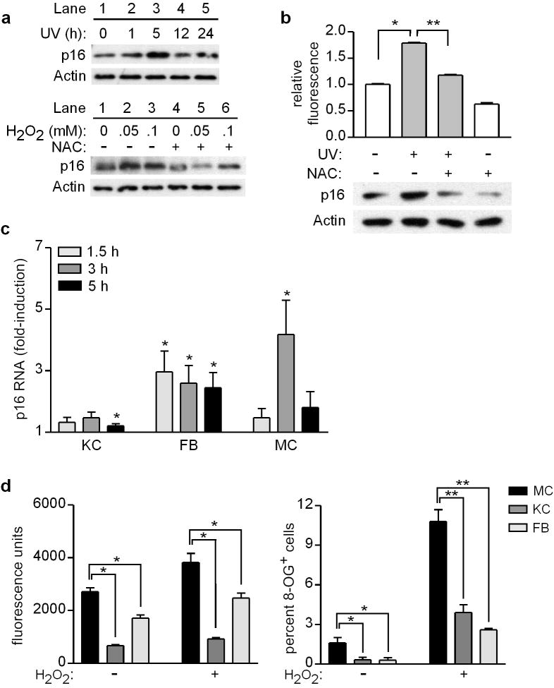
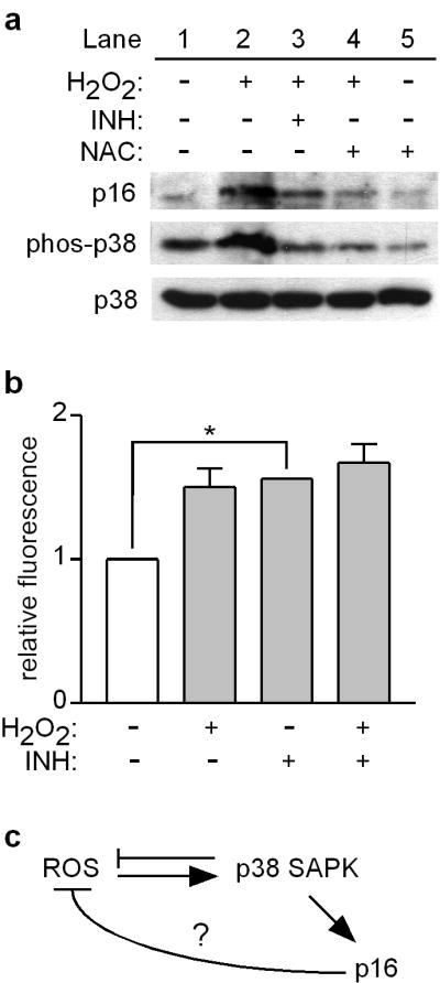
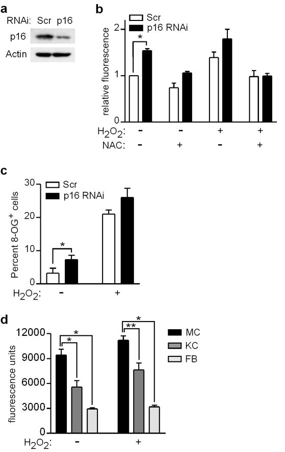
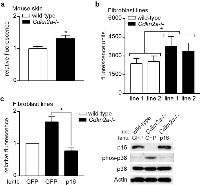
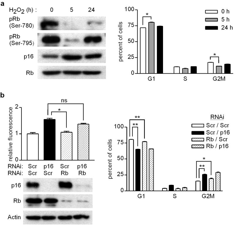
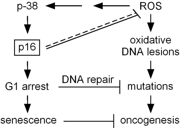
Mutations or deletions in the cyclin-dependent kinase inhibitor p16(INK4A) are associated with multiple cancer types, but are more commonly found in melanoma tumors and associated with familial melanoma predisposition. Although p16 is thought to function as a tumor suppressor by negatively regulating the cell cycle, it remains unclear why the genetic compromise of p16 predisposes to melanoma over other cancers. Here we describe a novel role for p16 in regulating oxidative stress in several cell types, including melanocytes. Expression of p16 was rapidly upregulated following ultraviolet-irradiation and in response to H₂O₂-induced oxidative stress in a p38 stress-activated protein kinase-dependent manner. Knockdown of p16 using small interfering RNA increased intracellular reactive oxygen species (ROS) and oxidative (8-oxoguanine) DNA damage, which was further enhanced by H₂O₂ treatment. Elevated ROS levels were also observed in p16-depleted human keratinocytes and in whole skin and dermal fibroblasts from Cdkn2a-deficient mice. Aberrant ROS and p38 signaling in Cdkn2a-deficient fibroblasts was normalized by expression of exogenous p16. The effect of p16 depletion on ROS was not recapitulated by the knockdown of retinoblastoma protein (Rb) and did not require Rb. Finally, p16-mediated suppression of ROS could not be attributed to the potential effects of p16 on cell cycle phase. These findings suggest a potential alternate Rb-independent tumor-suppressor function of p16 as an endogenous regulator of carcinogenic intracellular oxidative stress. Compared with keratinocytes and fibroblasts, we also found increased susceptibility of melanocytes to oxidative stress in the context of p16 depletion, which may explain why the compromise of p16 predisposes to melanoma over other cancers.


Conflict of interest statement

Conflict of Interest

The authors declare no conflicts of interest.

Figures

Figure 1
Figure 1
Exogenous oxidative stress acutely upregulates p16 in human skin cells, and melanocytes are more susceptible than other cell types to oxidative stress and damage. (a) Melanocytes were untreated (0 h) or UV-irradiated (480 J/m2, lanes 2-5), and cell lysates were prepared over a 24-h period and blotted with antibodies against p16 and Actin (upper panel). Melanocytes were treated with H2O2 at the concentrations indicated, in the absence (lanes 1-3) or presence (lanes 4-6) of 5 mM NAC, and 5 h later cell lysates were blotted for p16 and Actin (lower panel). (b) Melanocytes were untreated (0 h) or UV-irradiated (480 J/m2) in the absence or presence of 5 mM NAC, and 5 h later ROS levels were measured by DCFDA assay (upper panel, values normalized to mean of control conditions which were set at 1) and cell lysates were blotted for p16 and Actin (lower panel). Error bars indicate SEM from three independent experiments. *P<.001 (one-sample t test), **P<.001 (two-sample t test). (c) Keratinocytes (KC), fibroblasts (FB), and melanocytes (MC) isolated from each of six donors were untreated or treated with 0.05 mM H2O2 for 1.5, 3, or 5 h. RNA was isolated and expression of p16 and GAPDH was quantitated by qRT-PCR, with p16 levels normalized to GAPDH at each time point (and then normalized to control conditions which were set at 1). Error bars indicate SEM from six independent determinations. *P<.05 (one-sample t tests). (d) Melanocytes (MC), keratinocytes (KC), and fibroblasts (FB) isolated from each of seven donors were untreated or treated with 0.05 mM H2O2 for 5 h, and ROS levels were measured by DCFDA assay (left panel). Error bars indicate SEM from seven independent determinations. *P<.001 (repeated measures ANOVA analysis, P values adjusted for multiple comparisons). Cells isolated from each of three donors were untreated or treated with 0.5 mM H2O2 for 48 h, then fixed and immobilized for 8-OG staining. Error bars indicate SEM of percent 8-OG positive cells assessed under each condition from three independent determinations. *P=.06, **P<.001 (adjusted for multiple comparisons).
Figure 2
Figure 2
ROS upregulate p16 through the p38 SAPK in human melanocytes. (a) Melanocytes were cultured for 5 h alone (lane 1) or in the presence of 0.05 mM H2O2 (lanes 2-4), phospho-p38 inhibitor (INH), and with or without pre-addition of 5 mM NAC (lanes 4 and 5). Cell lysates were blotted for p16, phospho-p38 and p38, with p38 serving as a loading control. (b) Melanocytes were cultured for 5 h alone or in the presence of 0.05 mM H2O2 and/or phospho-p38 inhibitor (INH). ROS levels were measured by DCFDA assay, and values normalized to those of control conditions which were set at 1. Error bars indicate SEM from three independent experiments. *P<.001 (one-sample t test). (c) Schematic depicting the ROS-dependent p38-p16 signaling pathway, and the possibility that p16 suppresses endogenous ROS.
Figure 3
Figure 3
Intracellular ROS and oxidative DNA damage is increased in p16-depleted cells. (a) Melanocytes were transfected with control scrambled (Scr) or p16-specific siRNA, and 48 h later cell lysates were blotted for p16 and Actin. (b) Melanocytes were transfected with siRNA, and 48 h later either untreated or treated with 5 mM NAC and/or 0.05 mM H2O2. After 5 h, ROS levels were measured by DCFDA assay, and values normalized to those of control conditions which were set at 1. Error bars indicate SEM from three independent experiments. *P=.005 (one-sample t test). (c) Melanocytes were transfected with siRNA, and 48 h later either untreated or treated with 0.5 mM H2O2. After an additional 48 h, cells were fixed and immobilized for 8-OG staining. Error bars indicate SEM of percent 8-OG positive cells assessed under each condition in three independent experiments. *P=.05 (two-sample t test). (d) Melanocytes (MC), keratinocytes (KC), and fibroblasts (FB) isolated from four individual donors were transfected with p16-specific siRNA, and then 48 h later cultured in the absence or presence of 0.05 mM H2O2. After 5 h, intracellular ROS levels were measured by DCFDA assay. Error bars indicate SEM from four independent experiments with different donor cells. *P<.001, **P=.002 (repeated measures analysis of variance, ANOVA).
Figure 4
Figure 4
Suppression of ROS by Cdkn2a in vivo, and normalization of ROS in Cdkn2a-deficient cells upon restoration of p16 expression. (a) Dorsal skin was removed from wild-type and Cdkn2a-deficient mice, and ROS levels were measured in tissue lysates by DCFDA assay. Mean values normalized to those of wild-type mice which were set at 1. Error bars indicate SEM from independent measurements in wild-type (n=10) and Cdkn2a-deficient (n=11) mice. *P=.03 (two-sample t test). (b) Two lines of fibroblasts were derived from both wild-type and Cdkn2a-deficient mice, and ROS levels determined by DCFDA assay. Error bars indicate SEM from five independent experiments. *P<.001 (repeated measures analysis of variance, ANOVA). (c) Wild-type (WT) and Cdkn2a-deficient fibroblasts were infected with either GFP control lentivirus, or lentivirus expressing p16/GFP as indicated. Cell lysates were prepared 48 h later and subjected to DCFDA assay for ROS (left panel) and Western blotting for p16, phospho-p38, p38, or Actin (right panel). Error bars indicate SEM from three independent experiments. *P=.01 (two-sample t test).
Figure 5
Figure 5
p16 regulation of ROS occurs independently of Rb. (a) Melanocytes were untreated (0 h), or treated with 50 μM H2O2 for 5h or 24 h. Cells were lysed for Western blotting (left panel), or analyzed by flow cytometry to determine cell cycle phase (right panel). Error bars represent SEM of triplicate determinations, *P<.001 (two-sample t test). Representative of two experiments performed. (b) Melanocytes were transfected with scrambled (Scr) or indicated specific siRNA, and then 48 h later ROS levels were measured by DCFDA assay, cell lysates were prepared for Western blotting, and cycle analysis was performed with percentages of cells in each phase (G1, S, G2M) indicated. Error bars indicate SEM of duplicate or triplicate determinations. *P=.01, **P<.001; ns, not significant. Representative of two experiments performed.
Figure 6
Figure 6
Potential tumor-suppressive functions of p16. In the canonical pathway, p16 mediates cell cycle arrest as part of the DNA damage response pathway, allowing time for DNA repair enzymes to correct potentially oncogenic mutations or avoiding transformation by inducing senescence. Loss of p16 leads to increased ROS levels (direct or indirect effect, indicated by dashed/solid line) and oxidative DNA damage, which in p16-deficient cells with impaired capacity for cell cycle arrest and senescence, may result in increased accumulation of mutations promoting oncogenesis. Melanocytes are particularly susceptible to oxidative stress (Figure 1d), perhaps explaining why loss of p16 predisposes to melanoma rather than other cancers.

References

    1. Alcorta DA, Xiong Y, Phelps D, Hannon G, Beach D, Barrett JC. Involvement of the cyclin-dependent kinase inhibitor p16 (INK4a) in replicative senescence of normal human fibroblasts. Proc Natl Acad Sci U S A. 1996;93:13742–13747. - PMC - PubMed
    1. Becker TM, Haferkamp S, Dijkstra MK, Scurr LL, Frausto M, Diefenbach E, et al. The chromatin remodelling factor BRG1 is a novel binding partner of the tumor suppressor p16INK4a. Mol Cancer. 2009;8:4. - PMC - PubMed
    1. Becker TM, Rizos H, Kefford RF, Mann GJ. Functional impairment of melanoma-associated p16(INK4a) mutants in melanoma cells despite retention of cyclin-dependent kinase 4 binding. Clin Cancer Res. 2001;7:3282–3288. - PubMed
    1. Bochar DA, Wang L, Beniya H, Kinev A, Xue Y, Lane WS, et al. BRCA1 is associated with a human SWI/SNF-related complex: linking chromatin remodeling to breast cancer. Cell. 2000;102:257–265. - PubMed
    1. Bockholt SM, Burridge K. An examination of focal adhesion formation and tyrosine phosphorylation in fibroblasts isolated from src-, fyn-, and yes- mice. Cell Adhes Commun. 1995;3:91–100. - PubMed

Publication types

MeSH terms

Substances