Review
. 2010 Oct;11(10):710-22.
doi: 10.1038/nrg2879. Epub 2010 Sep 14.

Advances in understanding tissue regenerative capacity and mechanisms in animals

Affiliations
Review

Advances in understanding tissue regenerative capacity and mechanisms in animals

Kenneth D Poss. Nat Rev Genet. 2010 Oct.

Abstract

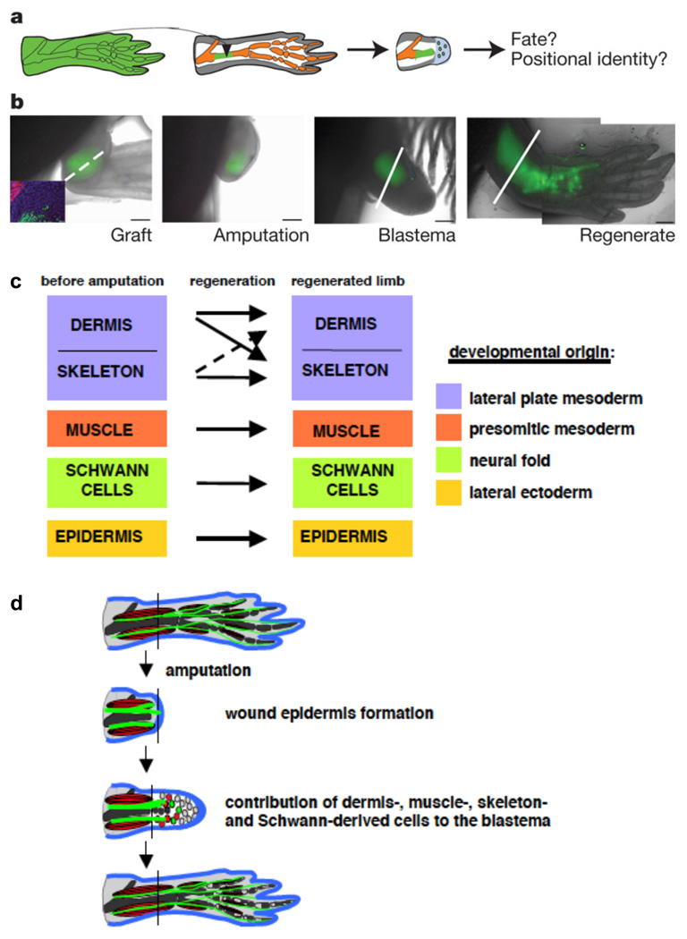
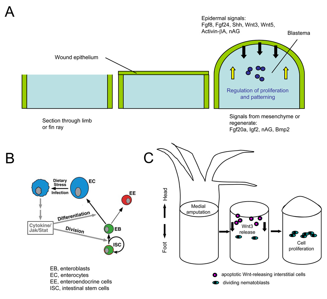
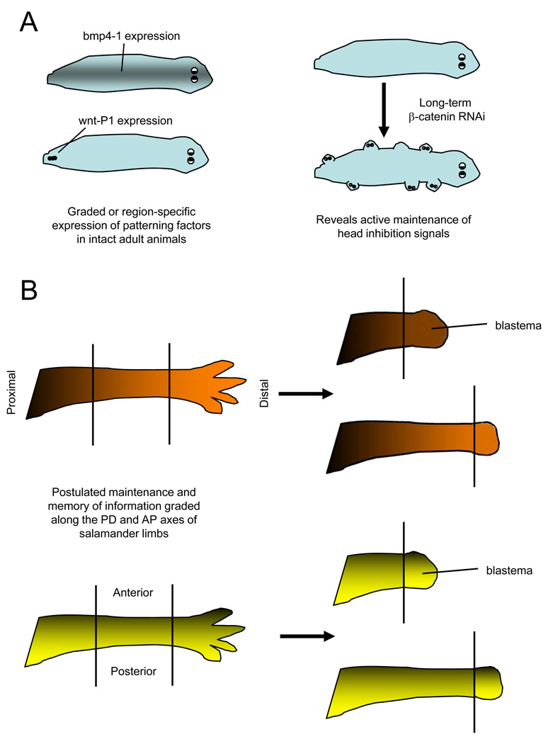
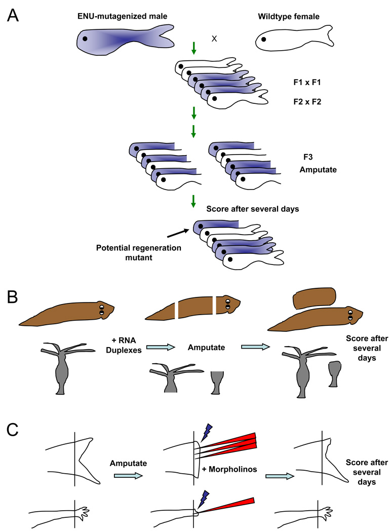
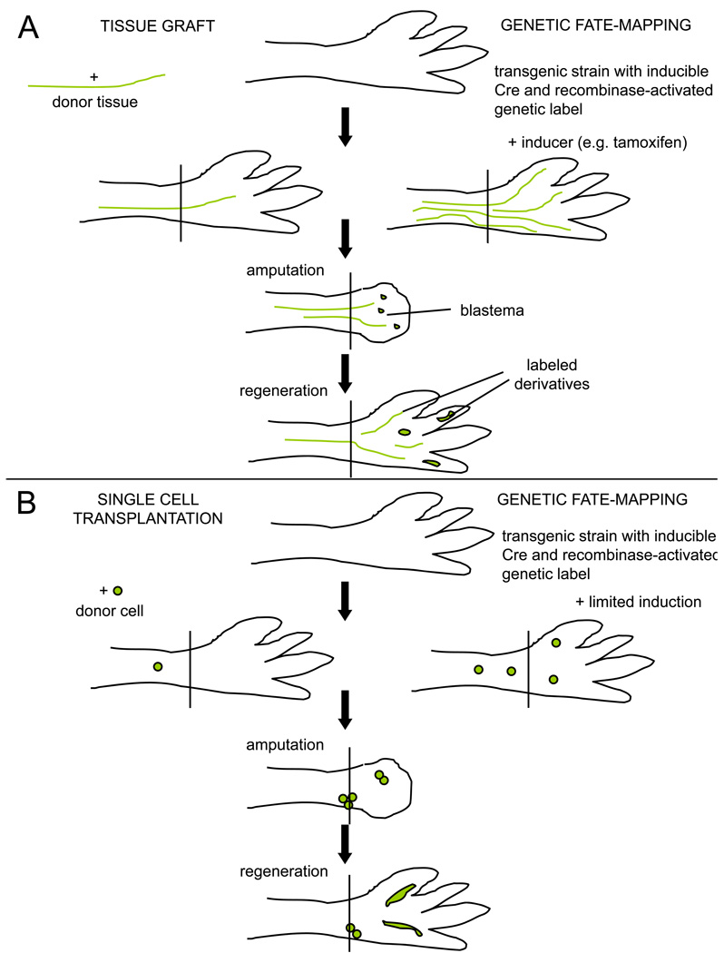
Questions about how and why tissue regeneration occurs have captured the attention of countless biologists, biomedical engineers and clinicians. Regenerative capacity differs greatly across organs and organisms, and a range of model systems that use different regenerative strategies and that offer different technical advantages have been studied to understand regeneration. Making use of this range of systems and approaches, recent advances have allowed progress to be made in understanding several key issues that are common to natural regenerative events. These issues include: the determination of regenerative capacity; the importance of stem cells, dedifferentiation and transdifferentiation; how regenerative signals are initiated and targeted; and the mechanisms that control regenerative proliferation and patterning.


Figures

Figure 1
Figure 1. Injured tissues retain lineages during axolotl limb regeneration
A. Schematic of cartilage grafting from transgenic EGFP-expressing axolotl to wild-type recipient. After amputation, the tissue contributions of the donor graft and location in regenerated structures can be assessed. B. Time course indicating the progression of EGFP-labeled donor tissue throughout the regeneration experiment. The amputation (dotted line) is made through an area containing a stable cartilage graft (green), and the graft and its derivatives are then visualized by whole-mount imaging throughout stages of regeneration. The Inset in the first image indicates that the EGFP-labeled cartilage graft does not co-label with a marker of skeletal muscle (red). C. Summary of results from tissue-grafting experiments by Kragl and colleagues. Key tissue lineages like muscle, cartilage, Schwann cells, and epidermis remain restricted to their developmental origin and do not transdifferentiate to other lineages during limb regeneration. D. Model of tissue contributions during blastema formation and limb regeneration, from ref 63. Blastemal cells arise from different tissue types but remain compartmentalized in the blastema. During regeneration, there is little lineage-switching, other than dermal cells; that is, blastemal cells retain their memory of origin as they are patterned into new limb structures. All figure panels reprinted with permission from .
Figure 2
Figure 2. Signals initiating regeneration
A. Signaling during blastema formation. Following amputation of a zebrafish fin or salamander limb, a wound epithelium quickly covers the appendage stump and matures into a key paracrine signaling structure for the blastema. Multiple factors are synthesized in the epidermis that are important for initiation and/or regulation of blastemal proliferation, while other factors appear to signal from underlying structures like the blastema or nerve to the epidermis. B. Model for regenerative signaling in the Drosophila midgut. Enterocytes (EC) encountering stress or undergoing cell death release Unpaired cytokines to activate production of replacement enterocytes and enteroendocrine cells (EE) by intestinal stem cells (ISC) and enteroblast (EB) progenitors, through activating Jak/Stat signaling in these progenitor cell types. Reprinted with permission from . C. Model for regenerative signaling by apoptotic cells after mid-gastric bisection in Hydra. Amputation causes apoptosis in cells near the plane of injury within 30 minutes, and concomitant Wnt3 release from those apoptotic cells. Wnt3 acts as a mitogen for interstial cells like nematoblasts, promoting regeneration by effects in addition to its role as a head organizer.
Figure 3
Figure 3. Models of positional memory in invertebrates and vertebrates
A. Maintenance of patterning signals in planarians. (Left) bmp4-1, which regulates midline and dorsoventral patterning, is expressed in the midline region of intact animals. wntP-1 regulates anteroposterior (AP) polarity during regeneration, and is normally expressed in a few cells in the tail of intact animals. (Right) Knockdown of β-catenin causes head-like protrusions in ectopic locations, revealing homeostatic maintenance of the AP axis by this signaling molecule, . B. Model for contributions to positional memory in highly regenerative vertebrate tissues. Graded distribution of morphogenetic factors along the proximodistal (PD; top) or anteroposterior (AP; bottom) axis might also assist in recognition of positional identity during amphibian limb regeneration. Such factors would have a graded or region-specific distribution in the intact limb that helps maintain cell identity, and that can be quickly recovered in the regeneration blastema to help pattern the regenerate.
Box 1
Box 1. Loss-of-function approaches in highly regenerative systems
A. A forward genetic screen in zebrafish. Point mutations are randomly induced by chemical treatment with ENU, followed by breeding mutations to homozygosity through the F3 generation. F3 animals have their fins clipped at 2–3 months of age, and are scored for regenerative defects after 1–2 weeks. This can also be performed as a temperature-sensitive screen for conditional mutations, with animals shifted from 25–26 degrees to 33 degrees during the regeneration period, . Robust mutants are chosen for genetic mapping and mutation identification. B. RNA interference in planarians and Hydra. RNA duplexes targeting a candidate gene are introduced by injection or by feeding of bacteria containing RNAi-generating expression cassettes, in various forms, to adult animals. Animals are then amputated and scored for regeneration, sometimes followed by an additional round of RNAi and regeneration tests. C. Morpholino electroporation. Fluorescently labeled antisense morpholinos are injected into the blastema of the regenerating zebrafish fin (top) or axolotl limb, followed by electroporation to allow nucleic acid entry into cells. The regenerate is assessed after several days; in this example, fin regeneration is delayed in morpholino-treated region.
Box 2
Box 2. Lineage tracing techniques used in regeneration studies
A. Tests of tissue contributions. (Left) Tissue grafts are accepted well in salamanders, when performed either at the embryo or adult stage. A tissue type from a genetically distinct animal, e.g. a triploid animal or a transgenic animal expressing a fluorescent reporter gene in all cell types, can be dissected surgically and implanted into the intact limb of an unlabeled host. After amputation through the host limb, labeled tissues in the blastema and in the regenerate represent the derivatives of the graft. If multiple tissue contributions are detected after a transplant, issues of initial graft purity arise. (Right) Transgenic fate-mapping approaches typically use a cell type-specific promoter driving an inducible recombinase, e.g. a tamoxifen-inducible Cre fused to a mutated estrogen receptor, to trace the progeny of cells activating that promoter. After irreversibly labeling a limb cell type by tamoxifen injection, the limb is amputated and the blastema and regenerate assessed for labeled derivatives. A downside to this approach is that it relies on the availability and fidelity of a presumed tissue-specific promoter, relying on one marker for conclusions. B. Clonal analyses can aid conclusions in lineage tracing experiments. (Left) A single labeled cell purified by flow cytometry is implanted into the intact host limb, or into the blastema (not shown). Labeled tissues in the regenerate can be traced back to that single cell, permitting rigorous tests of multipotentiality and self-renewal. (Right) A limited dose of tamoxifen will induce recombination in a small number of isolated cells, and presumably in those cells with relatively strong activation of the cell type-specific promoter driving the recombinase. Clones of cells in the regenerate can be assessed for cell type-specific markers to determine the tissue diversity of clonally related cells.

Similar articles

Cited by

References

    1. Morgan TH. Experimental studies of the regeneration of Planaria maculata. Arch. Entw. Mech. Org. 1898;7:364–397.
    1. Reddien PW, Sanchez Alvarado A. Fundamentals of planarian regeneration. Annu Rev Cell Dev Biol. 2004;20:725–757. - PubMed
    1. Gierer A, et al. Regeneration of hydra from reaggregated cells. Nat New Biol. 1972;239:98–101. - PubMed
    1. Bosch TC. Why polyps regenerate and we don't: towards a cellular and molecular framework for Hydra regeneration. Dev Biol. 2007;303:421–433. - PubMed
    1. Garza-Garcia AA, Driscoll PC, Brockes JP. Evidence for the local evolution of mechanisms underlying limb regeneration in salamanders. Integrative and Comparative Biology. 2010:1–8. - PubMed

Publication types