Review
. 2010 Nov;7(11):653-64.
doi: 10.1038/nrclinonc.2010.139. Epub 2010 Sep 14.

Delivering nanomedicine to solid tumors

Affiliations
Review

Delivering nanomedicine to solid tumors

Rakesh K Jain et al. Nat Rev Clin Oncol. 2010 Nov.

Abstract

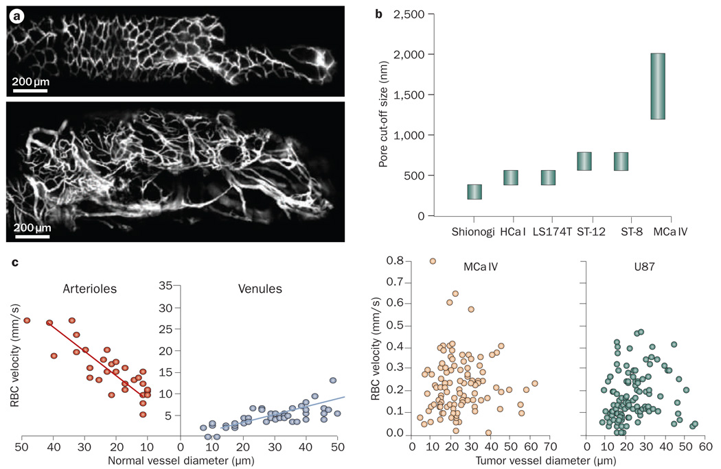
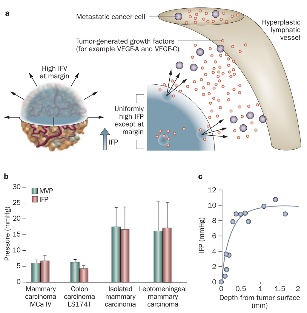
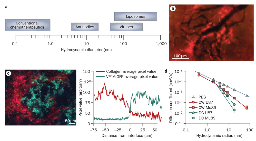
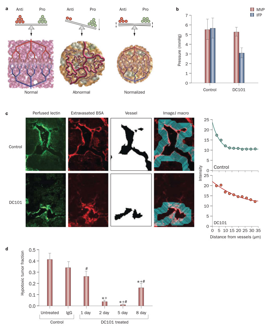
Recent advances in nanotechnology have offered new hope for cancer detection, prevention, and treatment. While the enhanced permeability and retention effect has served as a key rationale for using nanoparticles to treat solid tumors, it does not enable uniform delivery of these particles to all regions of tumors in sufficient quantities. This heterogeneous distribution of therapeutics is a result of physiological barriers presented by the abnormal tumor vasculature and interstitial matrix. These barriers are likely to be responsible for the modest survival benefit offered by many FDA-approved nanotherapeutics and must be overcome for the promise of nanomedicine in patients to be realized. Here, we review these barriers to the delivery of cancer therapeutics and summarize strategies that have been developed to overcome these barriers. Finally, we discuss design considerations for optimizing the delivery of nanoparticles to tumors.


Figures

Figure 1
Figure 1
Vascular structure and function in tumors. a | Longitudinal fluorescence imaging of normal (top) and tumor (bottom) colon tissue in a floxed Apc mouse. Permission obtained from Nature Publishing Group © Kim, P. et al. Nat. Methods 7, 303–305 (2010). b | Tumor vessels are leaky and have large pore sizes, which for some tumor types can be as large as a few micrometers in size. Permission obtained from the National Academy of Sciences, USA © Hobbs, S. K. et al. Proc. Natl Acad. Sci. USA 95, 4607–4612 (1998). c | Blood velocity in normal pial vessels (left) and tumors (right) as a function of vessel diameter. Unlike normal tissue, in tumors blood velocity does not depend on vessel diameter. Permission obtained from the American Association of Cancer Research © Yuan, F. et al. Cancer Res. 54, 4564–4568 (1994). Abbreviation: RBC, red blood cell.
Figure 2
Figure 2
Elevated IFP in tumors. a | The IFP is uniformly elevated in tumors except at the margin. The steep drop of IFP at the margin causes fluid, growth factors and cells to leak out of the tumor into the peritumoral tissue, which in turn might facilitate angiogenesis and metastasis, and inhibit drug delivery. Permission obtained from American Association of Cancer Research © Jain, R. K. et al. Cancer Res. 67, 2729–2735 (2007). b | The IFP and MVP for different tumor types. Permission obtained from the American Association of Cancer Research © Boucher, Y. & Jain, R. K. Cancer Res. 52, 5110–5114 (1992) and © Boucher, Y. et al. Cancer Res. 56, 4264–4266 (1996). c | IFP profile as a function of the distance from the tumor surface. Permission obtained from the American Association of Cancer Research © Boucher, Y. et al. Cancer Res. 50, 4478–4484 (1990). Abbreviations: IFP, interstitial fluid pressure; IFV, interstitial fluid velocity; MVP, microvascular pressure.
Figure 3
Figure 3
Barriers to interstitial transport of nanoparticles. a | The size of therapeutic agents can differ up to four orders of magnitude. b | The distribution of liposomes with a size of 90 nm in a tumor. The nanoparticles (bright red color) extravasate from some tumor blood vessels (black color) but because of their large size they cannot penetrate the tumor interstitial matrix and are concentrated in the perivascular region. Please note there is no extravasation from some vessels. Permission obtained from the American Association of Cancer Research © Yuan, F. et al. Cancer Res. 54, 3352–3356 (1994). c | Diffusion of nanoparticles in the tumor interstitium depends on collagen content. At high collagen regions (red color) the concentration of herpes simplex virus (green color; with a size of 150 nm) is low, while at low collagen regions the concentration of the virus increases. Permission obtained from the American Association of Cancer Research © McKee, T. D. et al. Cancer Res. 66, 2509–2513 (2006). d | Diffusion also depends on the implantation site. The plot depicts the diffusion coefficient of macromolecules in Mu89 melanomas and U87 glioblastomas implanted in the cranium and the dorsal skin. The diffusion coefficients in PBS solution are shown for comparison. Permission obtained from the National Academy of Sciences, USA © Pluen, A. et al. Proc. Natl Acad. Sci. USA 98, 4628–4633 (2001). Abbreviations: CW, cranial window; DC, dorsal chamber; PBS, phosphate buffered saline.
Figure 4
Figure 4
Effect of vascular normalization. a | Tumor angiogenesis results from an imbalance between antiangiogenic and proangiogenic factors. Normalization aims to restore this balance and bring the tumor vasculature to a more normal phenotype. Permission obtained from the American Association for the Advancement of Science © Jain, R. K. Science 307, 58–62 (2005). b | Use of an anti-VEGF antibody, DC101, reduced IFP and had no effect on MVP. Permission obtained from the American Association of Cancer Research © Tong, R. T. et al. Cancer Res. 64, 3731–3736 (2004). c | Frozen sections of murine mammary adenocarcinoma (MCa IV) tumors with perfused biotinylated lectin to indentify functional vessels, and extravasated tetramethyl rhodamine isothiocyanate–BSA to study interstitial penetration. Vessels were identified using the ImageJ software and the average intensity of BSA was quantified as a function of distance from the blood vessel wall. Tumors treated with the antiangiogenic agent DC101 exhibited increased penetration of BSA in the tumor interstitial space. Permission obtained from the American Association of Cancer Research © Tong, R. T. et al. Cancer Res. 64, 3731–3736 (2004). d | Normalization by DC101 decreased hypoxia in brain tumors in mice. Hypoxia reached a minimum at day 5, and a partial relapse occurred at day 8. *P <0.05, compared with untreated control; +P <0.05, compared with rat IgG-treated control (day 2); #P <0.05, compared with day 2 after initiation of DC101 therapy. Permission obtained from Elsevier Ltd. © Winkler, F. et al. Cancer Cell 6, 553–563 (2004). Abbreviations: BSA, bovine serum albumin; IFP, interstitial fluid pressure; MVP, microvascular pressure.
Figure 5
Figure 5
Effect of normalizing collagen matrix. a | Effect of interstitial matrix normalization on oncolytic viral therapy. Mu89 melanomas implanted in the dorsal skin-fold chamber were treated with the oncolytic vector MGH2 in combination with collagenase. Infection of tumor cells was detected by expression of the reporter gene GFP (encoded within the virus). Coinjection of MGH2 with collagenase increased cell infection and resulted in tumor regression (blue line). *P <0.05. Permission obtained from the American Association of Cancer Research © McKee, T. D. et al. Cancer Res. 66, 2509–2513 (2006). b | Effect of relaxin on collagen matrix in tumors. Second harmonic generation images of the collagen structure in a Mu89 melanoma during relaxin treatment. Maximum intensity projections of the same region of collagen fibers at different time points are shown. Relaxin treatment decreased collagen levels (white). Permission obtained from Nature Publishing Group © Brown, E. et al. Nat. Med. 9, 796–800 (2003). Abbreviation: GFP, green fluorescent protein.
Figure 6
Figure 6
Mean interstitial pH and pO2 as a function of the distance to the nearest blood vessel. Please note the low pH and low pO2 only becomes significantly low beyond 100 µm from a blood vessel wall. Thus, pH sensitive particles need to penetrate beyond 100 µm to take advantage of the low pH. Permission obtained from Nature Publishing Group © Helmlinger, G. et al. Nat. Med. 3, 177–182 (1997). Abbreviation: pO2, partial oxygen pressure.

References

    1. Torchilin VP. Targeted pharmaceutical nanocarriers for cancer therapy and imaging. AAPS J. 2007;9:E128–E147. - PMC - PubMed
    1. Schroeder A, Levins CG, Cortez C, Langer R, Anderson DG. Lipid-based nanotherapeutics for siRNA delivery. J. Intern. Med. 2010;267:9–21. - PMC - PubMed
    1. Wagner V, Dullaart A, Bock AK, Zweck A. The emerging nanomedicine landscape. Nat. Biotechnol. 2006;24:1211–1217. - PubMed
    1. Duncan R. Polymer conjugates as anticancer nanomedicines. Nat. Rev. Cancer. 2006;6:688–701. - PubMed
    1. Stroh M, et al. Quantum dots spectrally distinguish multiple species within the tumor milieu in vivo. Nat. Med. 2005;11:678–682. - PMC - PubMed

Publication types