. 2010 Sep 3;5(9):e12570.
doi: 10.1371/journal.pone.0012570.

Small-molecule antioxidant proteome-shields in Deinococcus radiodurans

Affiliations

Small-molecule antioxidant proteome-shields in Deinococcus radiodurans

Michael J Daly et al. PLoS One. .

Abstract

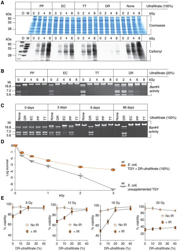
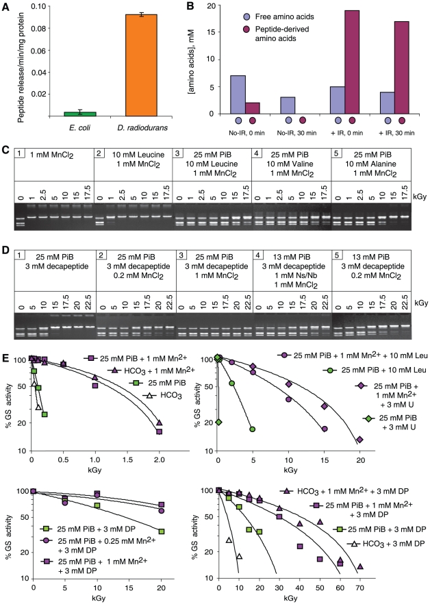
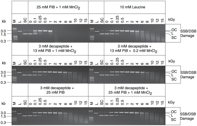
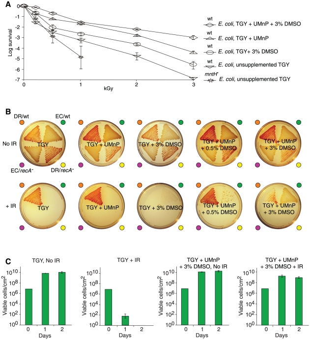
For Deinococcus radiodurans and other bacteria which are extremely resistant to ionizing radiation, ultraviolet radiation, and desiccation, a mechanistic link exists between resistance, manganese accumulation, and protein protection. We show that ultrafiltered, protein-free preparations of D. radiodurans cell extracts prevent protein oxidation at massive doses of ionizing radiation. In contrast, ultrafiltrates from ionizing radiation-sensitive bacteria were not protective. The D. radiodurans ultrafiltrate was enriched in Mn, phosphate, nucleosides and bases, and peptides. When reconstituted in vitro at concentrations approximating those in the D. radiodurans cytosol, peptides interacted synergistically with Mn(2+) and orthophosphate, and preserved the activity of large, multimeric enzymes exposed to 50,000 Gy, conditions which obliterated DNA. When applied ex vivo, the D. radiodurans ultrafiltrate protected Escherichia coli cells and human Jurkat T cells from extreme cellular insults caused by ionizing radiation. By establishing that Mn(2+)-metabolite complexes of D. radiodurans specifically protect proteins against indirect damage caused by gamma-rays delivered in vast doses, our findings provide the basis for a new approach to radioprotection and insight into how surplus Mn budgets in cells combat reactive oxygen species.


Conflict of interest statement

Competing Interests: The authors have declared that no competing interests exist.

Figures

Figure 1
Figure 1. In vitro and ex vivo protection by DR-ultrafiltrate.
(A) DR-ultrafiltrate prevents protein oxidation. The indicated ultrafiltrates were mixed with purified E. coli proteins and irradiated to the indicated doses of μ-radiation (kGy). Proteins were then separated by polyacrylamide gel electrophoresis and visualized by Coomassie staining. Duplicate gels were subjected to Western blot carbonyl analysis, which reveals the presence (black) or absence (no signal) of protein oxidation. PP, P. putida; EC, E. coli; TT, T. thermophilus; and DR, D. radiodurans. O and M, size-standards. (B) DR-ultrafiltrate preserves the activity of an irradiated enzyme. BamHI was irradiated in the indicated ultrafiltrates, then incubated with μ-DNA and subjected to agarose gel electrophoresis. (C) DR-ultrafiltrate preserves the activity of a desiccated enzyme. BamHI was desiccated from the indicated ultrafiltrates and stored in a desiccator for the indicated times, and then assayed for residual activity as in panel B. (D) DR-ultrafiltrate protects E. coli. Wild-type E. coli (MM1925) cells were grown in TGY medium supplemented with DR-ultrafiltrate and irradiated without change of broth to the indicated doses, then recovered on TGY medium. Colony forming unit (CFU) survival assays were in triplicate for each dose, with standard deviations shown. (E) DR-ultrafiltrate protects human Jurkat T cells. DR-ultrafiltrate was added to the growth medium 1 day before irradiation. The viability of irradiated cells was determined by trypan blue staining 2 days after irradiation. Viability assays were in triplicate, with standard deviations shown.
Figure 2
Figure 2. Composition of the DR-ultrafiltrate.
(A) Manganese (Mn) and phosphate (PO4) concentrations in bacterial ultrafiltrates (100%). (B) Nucleoside and base (Ns/Nb) concentrations in bacterial ultrafiltrates (100%) (Table S1). (C) Sum of free amino acids and those in peptide linkage in bacterial ultrafiltrates (100%) after acid hydrolysis. The total amino acid concentration of the DR-ultrafiltrate is 53 mM, of which 97% are in peptides (see also Figure S2). (D) Mn-complexes. The distribution of Mn bound to small molecules and peptides in aqueous-phase extracts of D. radiodurans homogenate was determined by size exclusion chromatography. The cell homogenate was prepared from cells disrupted in the presence of protease inhibitors. Note, gel filtration causes a significant dilution of the original sample, so the measured concentration in the fractions does not reflect the undiluted concentration in the sample. The 100% value for amino acids is 330 µM and that for Mn is 3.8 µM. As any unbound Mn or free amino acid-bound Mn would have eluted late in the chromatographic analysis, we concluded that Mn was bound to peptides. Proteins and large peptides >3,500 Da eluted together at the exclusion volume of the column; average molecular weight of an amino acid in peptide linkage is 115 Da.
Figure 3
Figure 3. Radioprotection by mixtures of Mn2+, orthophosphate, nucleosides and bases.
(A) BamHI activity. BamHI was irradiated in the indicated mixtures and then assayed for residual activity as in Figure 1B. Abbreviations: PiB, potassium phosphate buffer, pH 7.4; A, adenosine; U, uridine; Ns/Nb, nucleosides and bases (1 mM; see Table S1 for the Ns/Nb added). Irradiations were under aerobic conditions unless stated otherwise (gel 2). (B) Nucleosides prevent protein oxidation. Proteins purified from E. coli were mixed with adenosine (A) or uridine (U), phosphate buffer (PiB), pH 7.4 and Mn2+, and irradiated to the indicated doses (kGy). The irradiated proteins were separated by polyacrylamide gel electrophoresis and visualized by Coomassie staining as in Figure 1A. Note, the ability of the DR-ultrafiltrate to prevent in vitro ionizing radiation-induced protein carbonylation corresponds to the preservation of stainable (Coomassie) banding.
Figure 4
Figure 4. Role of amino acids and peptides in ionizing radiation resistance.
(A) Cytosolic protease activities in E. coli and D. radiodurans. (B) Cytosolic distribution and concentration of amino acids in D. radiodurans. No-IR, non-irradiated control cells held in 25 mM phosphate buffer, pH 7.4 on ice, then washed and held in 25 mM phosphate buffer, pH 7.4 (32°C) for 0 or 30 min. +IR, cells irradiated to 7 kGy in 25 mM phosphate buffer, pH 7.4 on ice, then washed and held in 25 mM phosphate buffer, pH 7.4 (32°C) for 0 or 30 min. Cells were harvested, resuspended in 20% TCA, and broken open. Aliquots of neutralized supernatant were analyzed for free amino acid and peptide-derived amino acid content. (C) Radioprotection of BamHI by amino acids. PiB, potassium phosphate buffer, pH 7.4. (D) Radioprotection of BamHI by the decapeptide (H-Asp-Glu-His-Gly-Thr-Ala-Val-Met-Leu-Lys-OH; 1,261 Da). Ns/Nb, nucleosides and bases (1 mM; see Table S1 for the Ns/Nb added). (E) Radioprotection of glutamine synthetase (GS) by Mn2+ and leucine (Leu), uridine (U), or the decapeptide (DP) in potassium phosphate buffer (PiB), pH 7.4 or sodium bicarbonate buffer (HCO3), pH 7.4. Adenosine could not be evaluated because it is an allosteric inhibitor of glutamine synthetase.
Figure 5
Figure 5. The HO-scavenging properties of Mn2+, orthophosphate, leucine and the decapeptide (H-Asp-Glu-His-Gly-Thr-Ala-Val-Met-Leu-Lys-OH).
Structural forms of the plasmid (pUC19): OC, open circular; L, linear; and SC, super-coiled. SSB, single-strand break; DSB, double-strand break. M, DNA size marker; PiB, phosphate buffer, pH 7.4. In the absence of HO-scavenging agents, approximately 80% of ionizing radiation-induced damage to purified DNA in aqueous solution is caused by HO, where one SSB in a SC circular plasmid molecule yields an OC form . In contrast to DNA damage, 3 mM decapeptide, 25 mM phosphate buffer, pH 7.4, and 1 mM Mn2+ preserved the activity of enzymes exposed to 50 kGy (Figure 4E).
Figure 6
Figure 6. Radioprotection of E. coli.
(A) Survival of E. coli exposed to acute ionizing radiation (IR). Cells were grown in, irradiated in, and recovered on the indicated medium (see also Figure S4B). wt, wild-type (MM1925); mntH , isogenic Mn-transport mutant (MM2115); UMnP contained 3 mM uridine/1 µM Mn2+/13 mM phosphate buffer, pH 7.4; DMSO, dimethyl sulfoxide. As the HO-scavenger uridine is a good growth substrate for E. coli (Figure S4A) and is not accumulated by the cells, we included DMSO, which E. coli does not metabolize (Figure S4A). Standard deviations shown. (B) Growth of E. coli under high-level chronic μ-radiation on solid medium (TGY). No IR, non-irradiated control plates incubated for 2 days at 25°C; +IR, plates incubated under 42 Gy/hour at the same temperature for the same time. Strain abbreviations: DR/wt, wild-type D. radiodurans; EC/wt, wild-type E. coli (MM1925); DR/recA (rec30), D. radiodurans DNA repair mutant; EC/recA (DH10B), E. coli DNA repair mutant. Each agar sector was inoculated with 1×10⁷ cells (see also Figure S4C). (C) Quantification of E. coli growth under chronic ionizing radiation. Cells on 4 cm2 agar sectors corresponding to those in panel B were harvested at 1 and 2 days after incubation under 42 Gy/hour at 25°C. Each agar sector was inoculated with 1×10⁷ cells. The number of viable cells per sector after 1 or 2 days was enumerated in triplicate, with standard deviations shown. Note, E. coli cells harvested from TGY+UMnP+3% DMSO+IR did not grow when transferred to non-supplemented TGY+IR.

References

    1. Daly MJ, Gaidamakova EK, Matrosova VY, Vasilenko A, Zhai M, et al. Protein oxidation implicated as the primary determinant of bacterial radioresistance. PLoS Biol. 2007;5:e92. - PMC - PubMed
    1. Kriško A, Radman M. Protein damage and death by radiation in Escherichia coli and Deinococcus radiodurans. Proc Natl Acad Sci USA. 2010 ( www.pnas.org/cgi/doi/10.1073/pnas.1009312107) - DOI - PMC - PubMed
    1. Daly MJ, Gaidamakova EK, Matrosova VY, Vasilenko A, Zhai M, et al. Accumulation of Mn(II) in Deinococcus radiodurans facilitates gamma-radiation resistance. Science. 2004;306:1025–1028. - PubMed
    1. Qiu X, Daly MJ, Vasilenko A, Omelchenko MV, Gaidamakova EK, et al. Transcriptome analysis applied to survival of Shewanella oneidensis MR-1 exposed to ionizing radiation. J Bacteriol. 2006;188:1199–1204. - PMC - PubMed
    1. Daly MJ. A new perspective on radiation resistance based on Deinococcus radiodurans. Nat Rev Microbiol. 2009;7:237–245. - PubMed

Publication types

LinkOut - more resources