. 2010 Sep 9;6(9):e1001112.
doi: 10.1371/journal.pgen.1001112.

Preferential re-replication of Drosophila heterochromatin in the absence of geminin

Affiliations

Preferential re-replication of Drosophila heterochromatin in the absence of geminin

Queying Ding et al. PLoS Genet. .

Abstract

To ensure genomic integrity, the genome must be duplicated exactly once per cell cycle. Disruption of replication licensing mechanisms may lead to re-replication and genomic instability. Cdt1, also known as Double-parked (Dup) in Drosophila, is a key regulator of the assembly of the pre-replicative complex (pre-RC) and its activity is strictly limited to G1 by multiple mechanisms including Cul4-Ddb1 mediated proteolysis and inhibition by geminin. We assayed the genomic consequences of disregulating the replication licensing mechanisms by RNAi depletion of geminin. We found that not all origins of replication were sensitive to geminin depletion and that heterochromatic sequences were preferentially re-replicated in the absence of licensing mechanisms. The preferential re-activation of heterochromatic origins of replication was unexpected because these are typically the last sequences to be duplicated in a normal cell cycle. We found that the re-replication of heterochromatin was regulated not at the level of pre-RC activation, but rather by the formation of the pre-RC. Unlike the global assembly of the pre-RC that occurs throughout the genome in G1, in the absence of geminin, limited pre-RC assembly was restricted to the heterochromatin by elevated cyclin A-CDK activity. These results suggest that there are chromatin and cell cycle specific controls that regulate the re-assembly of the pre-RC outside of G1.


Conflict of interest statement

The authors have declared that no competing interests exist.

Figures

Figure 1
Figure 1. Geminin depletion results in increased ploidy.
(A) Histogram of DNA content determined by flow cytometry for cells treated with nonspecific pUC control dsRNA for 24 (gray; solid outline) and 48 hours (gray; dashed outline) or geminin dsRNA for 24 (blue; solid outline) and 48 (green; dashed outline) hours. (B) Boxplot of the relative copy number of DNA from geminin depleted cells at 48 hours versus DNA from control cells for individual array probes grouped by chromosome. The heterochromatic 4th chromosome is at a significantly elevated copy number (p<1×10⁻¹⁶). (C) The relative copy number for array probes following 48 hours of geminin depletion (black) as a function of chromosomal position for the left and right arms of Drosophila chromosome 2. The relative copy number comparing two independent populations of control cells is shown in gray. Transposon density (fraction of sequence covered by transposable elements) is indicated by the red histogram at the bottom of the plot.
Figure 2
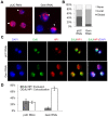
Figure 2. Re-replication is specific to pericentromeric heterochromatin.
(A) Fluorescence microscopy of cells after a 4 hour BrdU pulse at 18 hours post RNAi treatment with control (pUC) or geminin dsRNA. BrdU labeled sequences were detected with a rat anti-BrdU antibody (red) and DNA was counterstained with DAPI (blue). BrdU staining patterns were classified as ‘global’ (arrow), ‘local’ (arrowhead) or ‘none’. (B) Distribution of the different BrdU incorporation patterns. At least 200 cells were counted from three independent experiments. The distribution of BrdU incorporation patterns was significantly different between control cells and geminin depleted cells (p<1×10⁻¹⁶). (C) Immunofluorescence of control and geminin depleted cells following a 30 min pulse of EdU at 24 hours post RNAi. EdU and HP1 were detected with Alexa Fluor 488-azide (green) and rabbit anti-HP1 antibody (red), respectively, and DNA was counterstained with DAPI (blue). (D) Quantification of EdU and HP1 localization patterns in control and geminin depleted cells with error bars indicating standard error. At least 200 cells were counted in three independent experiments. The distribution of EdU/HP1 localization patterns was significantly different between control cells and geminin depleted cells (p<1×10⁻¹⁶).
Figure 3
Figure 3. Re-replication is dependent on chromatin environment not time of replication.
(A) Boxplot of the relative copy number between geminin depleted and control cells for array probes in the euchromatin or heterochromatin. The heterochromatin is at a signficantly higher copy number (p<1×10⁻¹⁶). (B) The relative copy number for array probes in the vicinity of pericentromeric heterochromatin on the right arm of chromosome 2 following geminin RNAi treatment for 48 hours (euchromatin, black; heterochromatin, red). (C) Boxplot of the relative time of replication for sequences in the euchromatin or heterochromatin from normal mitotic cells. The heterochromatin is signficantly later replicating than the euchromatin (p<1×10⁻¹⁶). (D) Replication timing values as a function of chromosomal position for unique sequence probes in the vicinity of the pericentric heterochromatin on the right arm of chromosome 2 (euchromatin, black; heterochromatin, red).
Figure 4
Figure 4. Limited pre-RC re-assembly occurs in the absence of geminin.
(A) Chromatin from control RNAi (pUC), G1/S arrested (HU), and geminin depleted re-replicating cells (Gem) was fractionated biochemically and immunoblotted for MCMs and ORC2. Levels of ORC and MCMs are also shown for whole cell extracts (WCE). (B) Fraction of control or geminin depleted cells exhibiting active DNA synthesis (EdU incorporation), with error bars indicating standard error. DNA synthesis was determined by pulsing the cells for 30 minutes with EdU at the given time points. At least 200 cells were counted in three independent experiments. (C) Time course of the fraction of control (open diamond) or geminin depleted (open square) cells exhibiting nuclear MCM localization following RNAi treatment for the indicated duration, with error bars indicating standard error. At least 200 cells were counted in three independent experiments.
Figure 5
Figure 5. MCMs are preferentially loaded onto heterochromatin in the absence of geminin.
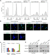
(A) Flow cytometry analysis of DNA content for cells treated with nonspecific pUC or geminin RNAi for 24 hours (dotted outline; gray), 48 hours (solid outline; gray) or 48 hours with aphidicolin (APH) added at 24 hours (solid outline; blue). (B) Quantification of MCM localization patterns. MCM staining was classified into 3 types: type I - MCM localization throughout the nucleus, type II - MCM localization to a small portion of the nucleus (heterochromatin), and type III - no significant MCM localization observed. The numbers under each staining pattern represent the percentage of nuclei with that particular pattern. At least 200 cells were counted in three independent experiments. The distribution of MCM localization patterns in geminin depleted cells treated with aphidicolin was signficantly different from control cells treated with aphidcolin (p<1×10⁻¹⁶) and similarily, geminin depleted cells were significantly different from geminin depleted cells treated with aphidicolin (p<1×10⁻¹⁶). (C) Low and high magnification examples of type I (arrow) and II (arrow head) MCM staining patterns in control and geminin depleted cells treated with aphidicolin. The MCMs, HP1 and DAPI are shown in green, red and blue, respectively. The type II pattern of MCM localization in geminin depleted cells often exhibited heterogeneous staining with faint signal over the entire HP1 region (open arrowhead) or partial overlap with HP1 (open arrow).
Figure 6
Figure 6. Cyclin A - CDK activity restricts pre-RC formation to the heterochromatin.
(A) Histogram of DNA content for cells treated with pUC dsRNA, cyclin A dsRNA or cyclin A and geminin dsRNAs for 24 hours and 48 hours. (B) Immunostaining of chromatin-associated MCMs at 24 hours post cyclin A, cyclin A and geminin, or geminin RNAi treatment. (C) Quantification of the different MCM localization patterns as described in Figure 5, with error bars indicating standard error. At least 200 cells were counted from three independent experiments. The distribution of MCM localization patterns in geminin depleted cells was significantly different from cyclin A depleted cells (p<1×10⁻¹⁶) and cells simultaneously depleted of geminin and cyclin A (p<1×10⁻¹⁶). (D) Immunoblot analysis of cyclin A and geminin levels following the individual and co-RNAi depletion experiments.

References

    1. Diffley JF. Regulation of early events in chromosome replication. Curr Biol. 2004;14(18):778–786. - PubMed
    1. Arias EE, Walter JC. Strength in numbers: preventing rereplication via multiple mechanisms in eukaryotic cells. Genes Dev. 2007;21:497–518. - PubMed
    1. Drury LS, Perkins G, Diffley JF. The Cdc4/34/53 pathway targets Cdc6p for proteolysis in budding yeast. EMBO J. 1997;16:5966–5976. - PMC - PubMed
    1. Jallepalli PV, Brown GW, Muzi-Falconi M, Tien D, Kelly TJ. Regulation of the replication initiator protein p65cdc18 by CDK phosphorylation. Genes Dev. 1997;11(21):2767–2779. - PMC - PubMed
    1. Nguyen V, Co C, Irie K, Li J. Clb/Cdc28 kinases promote nuclear export of the replication initiator proteins Mcm2-7. Curr Biol. 2000;10:195–205. - PubMed

Publication types

Associated data

LinkOut - more resources