. 2010 Aug 23:4:39.
doi: 10.3389/fnsys.2010.00039. eCollection 2010.

Rewiring neural interactions by micro-stimulation

Affiliations

Rewiring neural interactions by micro-stimulation

James M Rebesco et al. Front Syst Neurosci. .

Abstract

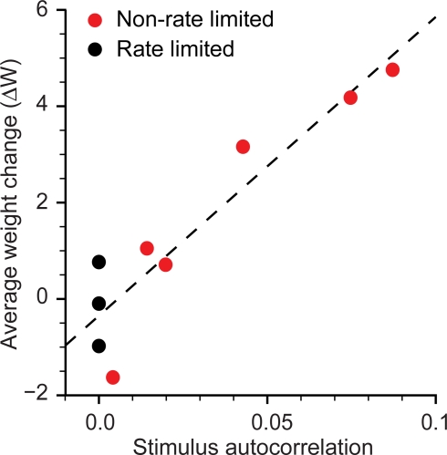
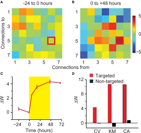
Plasticity is a crucial component of normal brain function and a critical mechanism for recovery from injury. In vitro, associative pairing of presynaptic spiking and stimulus-induced postsynaptic depolarization causes changes in the synaptic efficacy of the presynaptic neuron, when activated by extrinsic stimulation. In vivo, such paradigms can alter the responses of whole groups of neurons to stimulation. Here, we used in vivo spike-triggered stimulation to drive plastic changes in rat forelimb sensorimotor cortex, which we monitored using a statistical measure of functional connectivity inferred from the spiking statistics of the neurons during normal, spontaneous behavior. These induced plastic changes in inferred functional connectivity depended on the latency between trigger spike and stimulation, and appear to reflect a robust reorganization of the network. Such targeted connectivity changes might provide a tool for rerouting the flow of information through a network, with implications for both rehabilitation and brain-machine interface applications.

Keywords: brain machine interface; functional connectivity; hebbian association; plasticity; rat; sensorimotor cortex.


Figures

Figure 1
Figure 1
Schematic diagram of the inferred functional connectivity (IFC) algorithm. (A) The discharge of each neuron is driven by input spike trains from itself and from the other neuron, each convolved with a temporal kernel. The convolved inputs are summed, a static non-linearity transforms them into firing rates, and spikes are generated from the resulting Poisson distribution. (B) Recorded spikes are used by the model to estimate the time-dependent pairwise kernels. (C) Each kernel is integrated to produce the connection weight Wij that represents the net effect of neuron j on neuron i. A color scale represents the strength of the connections.
Figure 2
Figure 2
Functional connectivity of subnetworks within large simulated networks. (A) A representative example of the true (top, left) and inferred (bottom, left) connectivity matrix W for a subnetwork of six neurons embedded in a 10,000 neuron network. Plasticity experiment on the same network; true (top, right) and inferred (bottom, right) matrix of connectivity changes ΔW. Units for the true network are defined through the Izhikevich model of neuron spiking, while the units of the inferred network are given by the IFC model. (B) Comparison of R2 for inferred weights W and weight changes ΔW for 80 experiments. Lines join the paired values resulting from a given experiment. Difference in means was highly significant (n = 80, KS-test, p < 10−9).
Figure 3
Figure 3
Timecourse of trigger-to-target ΔW and ΔR. Stimulation took place during the 48-h window shown in yellow. (A) Latency between trigger spike and target stimulation was 5 ms. (B) Stimulation rate (binned at 1 min) for the experiment shown in (A). (C) Latency between trigger spike and target stimulation was 500 ms. (D) Stimulation rate for the experiment shown in (C).
Figure 4
Figure 4
Sensitivity of R and W. Comparison of fluctuations of R and W in prestimulation periods enabled an estimate of the sensitivity of the two measures. A mean-normalized estimate of the standard deviation is shown for R (left) as well as W (right).
Figure 5
Figure 5
Observed changes in cross-correlations and IFC kernels. IFC kernels (left) and cross-correlation functions (right) for animal BT. These functions are shown before (black) and after (red) the stimulation period. Autocorrelation scale reflecting self connections is 10 times that of the cross-correlations.
Figure 6
Figure 6
Connectivity changes from combined stimulation. Two target electrodes were stimulated in a single experiment, each at a different latency with respect to the trigger spikes. Red indicates the connection stimulated at 5-ms latency; green indicates the connection stimulated at 500-ms latency. (A) Timecourse of trigger-to-target ΔW. (B) Matrix and histogram of connectivity changes ΔW for the 24-h period prior to the onset of stimulation. (C) Matrix and histogram of connectivity changes ΔW for the 48-h stimulation period.
Figure 7
Figure 7
Network-wide potentiation effects. Matrix of connectivity changes ΔW for the two stimulation experiments of Figure 3. In all plots, the trigger-to-target connection is highlighted. (A) ΔW matrix and histogram for the period from −24 to 0 h for the 5-ms latency stimulation. (B) Corresponding results for the period from −24 to 0 h for the 500-ms latency stimulation. (C) Results for the period from 0 to 48 h after the onset of stimulation at 5-ms latency. (D) Corresponding results for the period from 0 to 48 h after the onset of stimulation at 500-ms latency.
Figure 8
Figure 8
Effect of stimulus autocorrelation on network-wide potentiation. For all experiments at 5-ms latency (red), the average connectivity change ΔW for non-targeted connections had a linear dependence on the autocorrelation of the stimulus train averaged over lags from 20 to 100 ms. Average weight changes for the rate limited experiments with an enforced autocorrelation of 0 (black) are clustered around zero.
Figure 9
Figure 9
Network-wide potentiation effects in rate-limited experiments. (A) ΔW matrix for the period from −24 to 0 h for the rate-limited, 5-ms latency stimulation. (B) Results for the period from 0 to 48 h after the onset of rate-limited stimulation at 5-ms latency. (C) Timecourse of trigger-to-target ΔW. (D) Summary of ΔW for targeted (red) and the average of all non-targeted connections (black).
Figure 10
Figure 10
Potentiation of targeted and non-targeted connections at short and long latencies. In all panels, red refers to 5-ms latency, and green refers to 500-ms latency. (A) Mean weight change of targeted connections. Experiments on animals Br, Ti, and Pt involved stimulation of two electrodes at different latencies, as in Figure 6. (B) ΔW of the targeted connection as a function of the average ΔW for all non-targeted connections; equal values lie on the diagonal dashed line. Paired results for experiments with two stimulation electrodes are identified by vertical dashed lines. (C) Average connectivity changes ΔW for targeted and non-targeted connections, at 5- and 500-ms latency. The upper error bound for the targeted, 5-ms connections has been truncated.

References

    1. Abbott L. F., Blum K. I. (1996). Functional significance of long-term potentiation for sequence learning and prediction. Cereb. Cortex 6, 406–41610.1093/cercor/6.3.406 - DOI - PubMed
    1. Ahissar E., Abeles M., Ahissar M., Haidarliu S., Vaadia E. (1998). Hebbian-like functional plasticity in the auditory cortex of the behaving monkey. Neuropharmacology 37, 633–65510.1016/S0028-3908(98)00068-9 - DOI - PubMed
    1. Baccala L. A., Sameshima K. (2001). “Overcoming the limitations of correlation analysis for many simultaneously processed neural structures,” in Progress in Brain Research, Vol. 130 ed. Nicolelis M. A. (New York: Elsevier; ), 33–47 - PubMed
    1. Bi G., Poo M. (2001). Synaptic modification by correlated activity: Hebb's postulate revisited. Annu. Rev. Neurosci. 24, 139–16610.1146/annurev.neuro.24.1.139 - DOI - PubMed
    1. Bi G. Q., Poo M. M. (1998). Synaptic modifications in cultured hippocampal neurons: dependence on spike timing, synaptic strength, and postsynaptic cell type. J. Neurosci. 18, 10464–10472 - PMC - PubMed