. 2010 Dec;15(12):1425-34.
doi: 10.1007/s10495-010-0539-z.

Drosophila Chk2 and p53 proteins induce stage-specific cell death independently during oogenesis

Affiliations

Drosophila Chk2 and p53 proteins induce stage-specific cell death independently during oogenesis

Anna Bakhrat et al. Apoptosis. 2010 Dec.

Abstract

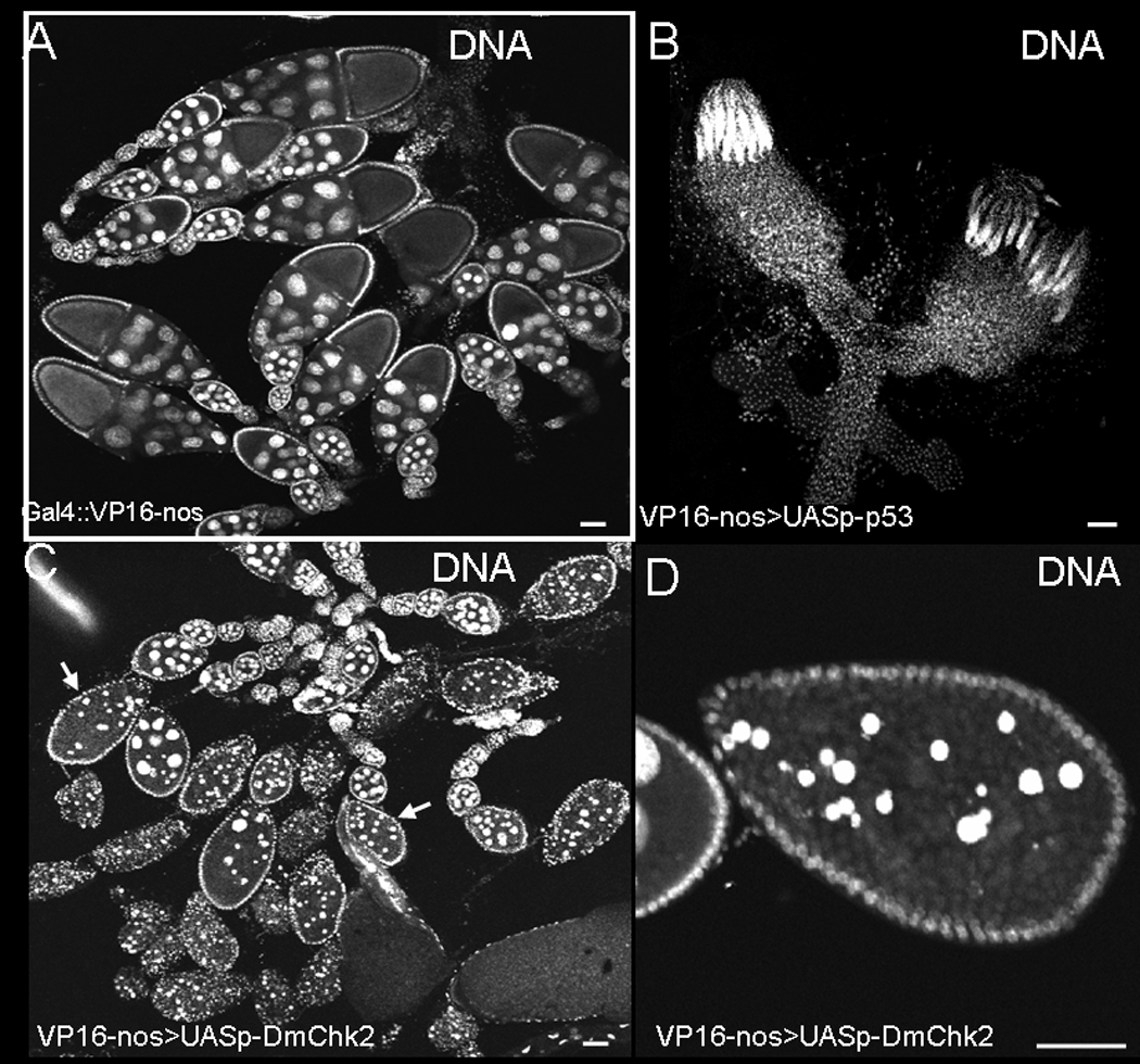
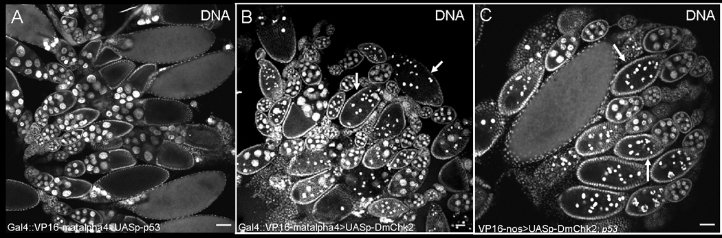
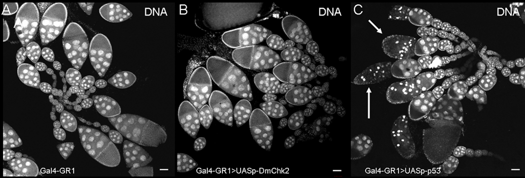
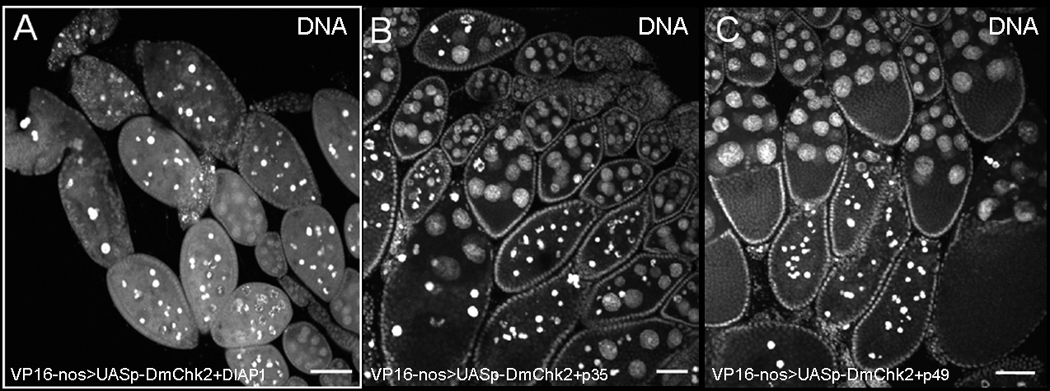
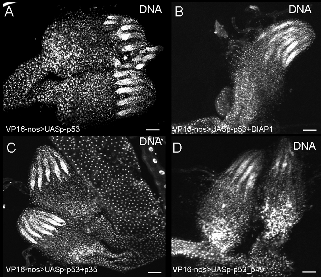
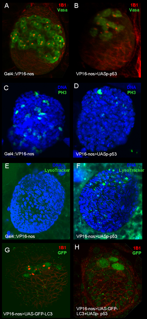
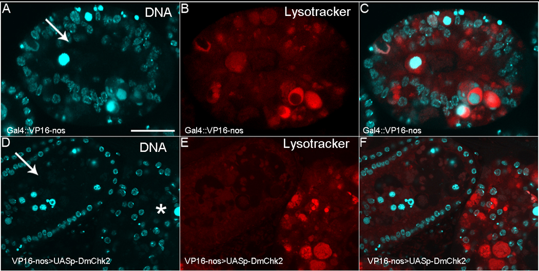
In Drosophila, the checkpoint protein-2 kinase (DmChk2) and its downstream effector protein, Dmp53, are required for DNA damage-mediated cell cycle arrest, DNA repair and apoptosis. In this study we focus on understanding the function of these two apoptosis inducing factors during ovarian development. We found that expression of Dmp53, but not DmChk2, led to loss of ovarian stem cells. We demonstrate that expression of DmChk2, but not Dmp53, induced mid-oogenesis cell death. DmChk2 induced cell death was not suppressed by Dmp53 mutant, revealing for the first time that in Drosophila, over-expression of DmChk2 can induce cell death which is independent of Dmp53. We found that over-expression of caspase inhibitors such as DIAP1, p35 and p49 did not suppress DmChk2- and Dmp53-induced cell death. Thus, our study reveals stage-specific effects of Dmp53 and DmChk2 in oogenesis. Moreover, our results demonstrate that although DmChk2 and Dmp53 affect different stages of ovarian development, loss of ovarian stem cells by p53 expression and mid-oogenesis cell death induced by DmChk2 do not require caspase activity.


Figures

Figure 1
Figure 1. Effects of Dmp53 and DmChk2 over-expression using nanos-Gal4VP16 in germline
Egg chambers were stained with Hoechst. A) nanos-Gal4VP16 Drosophila ovaries. B) Over-expression of Dmp53 in the germline using the above Gal4 driver resulted in small ovaries probably due to loss of germline stem cells. C) Over-expression of DmChk2 in the germline leads to massive egg chamber death; degenerating egg chambers are indicated by arrows. D) Closer examination of a degenerating egg chamber over-expressing DmChk2. Scale bar is 50µm.
Figure 2
Figure 2. Effects of Dmp53 and DmChk2 over-expression using P{matalpha4-GAL-VP16} V2H in the germline
A) DNA staining of egg chambers from P{matalpha4-GAL-VP16}V2H:UASp-p53 fly. B) DNA staining of egg chambers from P{matalpha4-GAL-VP16}V2H : UASp-HA-Chk2 fly. Degenerating egg chambers of stages 8–9 are indicated by arrows. C) DNA staining of egg chambers from P{matalpha4-GAL-VP16}V2H/ UASp-HA-Chk2; p53 flies. Degenerating egg chambers of stages 8–9 are indicated by arrows. Scale bar is 50µm.
Figure 3
Figure 3. Over-expression of Dmp53 but not of DmChk2 in the follicle somatic cells using GR1-Gal4 driver induced germline cell death
Egg chambers were stained with Hoechst. A) Egg chambers from GR1-GAL4 flies. B) Expressing DmChk2 in the follicle cell does not affect egg chamber survival. C) Expressing Dmp53 in the follicle cells lead to egg chamber death (marked with arrows).
Figure 4
Figure 4. DmChk2-mid oogenesis cell death is caspase-independent
Egg chambers were stained with Hoechst. A) Egg chambers from flies over-expressing DmChk2 and DIAP1 undergo cell death similar to egg chambers expressing DmChk2 alone. Over-expressing p35 (B) or p49 (C) does not suppress DmChk2-mid oogenesis cell death. Scale bar is 50µm.
Figure 5
Figure 5. Expression of caspase inhibitors do not suppress p53-mediated stem cell loss
Ovaries were stained with Hoechst. A) ovaries from nanos-Gal4VP16; pUASp53. Expression of p53 led to loss of stem cells. Over-expressing DIAP1 (B) or p35 (C) or p49 (D) does not suppress Dmp53-stem cell loss.
Figure 6
Figure 6. Expression of Dmp53 in the germline lead to loss of primordial germ cells (PGC's)
Gonads from third instar larvae were stained with germ cell marker, vasa (A–B; Green), anti-Adducin-like antibodies (A–B and G–H; Red), anti-phospho-histone 3 (C–D, Green), Lysotracker (E–F, Green), anti-GFP (G–H) and DAPI (C–F, Blue). A) nanos-Gal4VP16 ovary. B) Ovary from nanos-Gal4VP16; UASp- p53 fly, over-expressing of Dmp53 lead to loss pf PGC's. C) nanos-Gal4VP16. D) Ovary from nanos-Gal4VP16; UASp- p53 fly, over-expressing of Dmp53 lead to decrease in numbers of proliferating PGC's. E) Ovary from nanos-Gal4VP16. F) Ovary from nanos-Gal4VP16; UASp- p53 fly, LysoTracker positive puncta (arrows) have begun to accumulate in the ovary. G) Ovary from nanos-Gal4VP16; GFP-LC3 stained with anti-GFP. H) Ovary from nanos-Gal4VP16/UASp-p53; GFP-LC3 stained with anti-GFP.
Figure 7
Figure 7. DmChk2-mediated cell death is characterized by DNA fragmentation
All egg chambers were stained with DAPI (Blue) and TUNEL (Green). A) A control degenerating egg chamber from a nutrient deprived fly has condensed and fragmented nurse cell nuclei. B) TUNEL-positive puncta are detected in the same egg chamber. C) Merge of A–B. D) A degenerating egg chamber from a female over-expressing DmChk2 has condensed and fragmented nurse cell nuclei. E) The degenerating egg chamber over-expressing DmChk2 also has TUNEL-positive puncta. F) Merge of D–E. Scale bar is 50µm. All images were taken at the same magnification.
Figure 8
Figure 8. Lysosome activity is delayed in flies over-expressing DmChk2
All egg chambers were stained with DAPI (Blue) and LysoTracker (Red). A) Control NGT;nosGAL4 egg chamber from a nutrient deprived fly with condensed nurse cell nuclei that have fragmented. B) LysoTracker positive puncta have begun to accumulate in this egg chamber. C) Merge of A–B. D) Egg chambers over-expressing DmChk2 at different stages of degeneration (arrow, middle and *, late) E) LysoTracker staining can only be seen in the later degenerating egg chamber (*). F) Merge of D–E. Scale bar is 20µm. All egg chambers were taken at the same magnification.

References

    1. Stracker TH, Usui T, Petrini JH. Taking the time to make important decisions: the checkpoint effector kinases Chk1 and Chk2 and the DNA damage response. DNA Repair (Amst) 2009;8:1047–1054. - PMC - PubMed
    1. Hirao A, Cheung A, Duncan G, Girard PM, Elia AJ, Wakeham A, et al. Chk2 is a tumor suppressor that regulates apoptosis in both an ataxia telangiectasia mutated (ATM)-dependent and an ATM-independent manner. Mol Cell Biol. 2002;22:6521–6532. - PMC - PubMed
    1. Takai H, Naka K, Okada Y, Watanabe M, Harada N, Saito S, et al. Chk2-deficient mice exhibit radioresistance and defective p53-mediated transcription. EMBO J. 2002;21:5195–5205. - PMC - PubMed
    1. Chen L, Gilkes DM, Pan Y, Lane WS, Chen J. ATM and Chk2-dependent phosphorylation of MDMX contribute to p53 activation after DNA damage. EMBO J. 2005;24:3411–3422. - PMC - PubMed
    1. LeBron C, Chen L, Gilkes DM, Chen J. Regulation of MDMX nuclear import and degradation by Chk2 and 14-3-3. EMBO J. 2006;25:1196–1206. - PMC - PubMed

Publication types

MeSH terms