. 2011 Apr;226(4):1082-1089.
doi: 10.1002/jcp.22423.

The role of calcium release activated calcium channels in osteoclast differentiation

Affiliations

The role of calcium release activated calcium channels in osteoclast differentiation

Yandong Zhou et al. J Cell Physiol. 2011 Apr.

Abstract

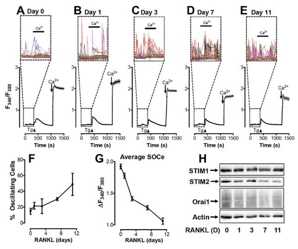
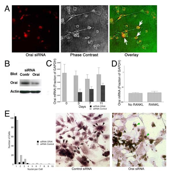
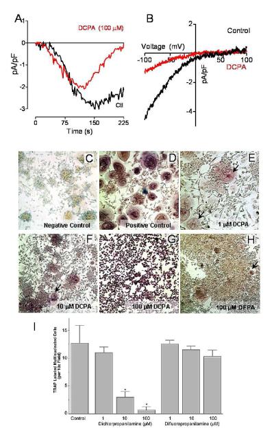
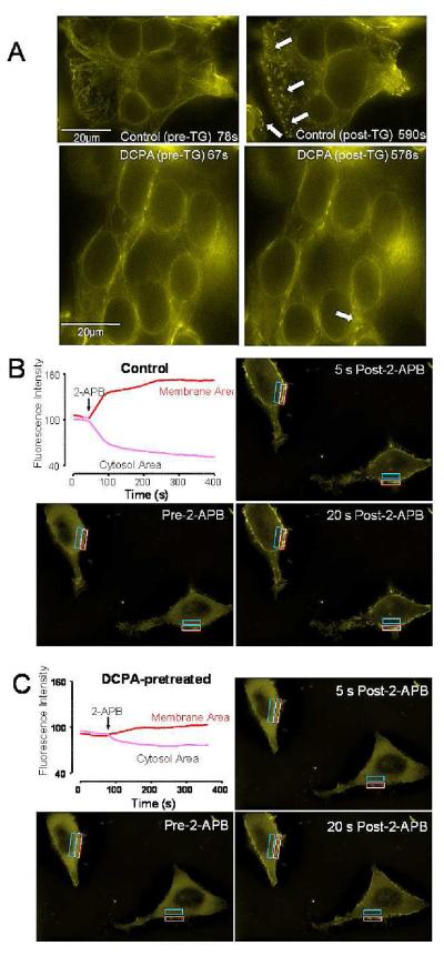
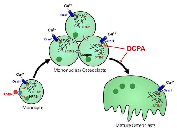
Osteoclasts are specialized macrophage derivatives that secrete acid and proteinases to mobilize bone for mineral homeostasis, growth, and replacement or repair. Osteoclast differentiation generally requires the monocyte growth factor m-CSF and the TNF-family cytokine RANKL, although differentiation is regulated by many other cytokines and by intracellular signals, including Ca(2+). Studies of osteoclast differentiation in vitro were performed using human monocytic precursors stimulated with m-CSF and RANKL, revealing significant loss in both the expression and function of the required components of store-operated Ca(2+) entry over the course of osteoclast differentiation. However, inhibition of CRAC using either the pharmacological agent 3,4-dichloropropioanilide (DCPA) or by knockdown of Orai1 severely inhibited formation of multinucleated osteoclasts. In contrast, no effect of CRAC channel inhibition was observed on expression of the osteoclast protein tartrate resistant acid phosphatase (TRAP). Our findings suggest that despite the fact that they are down-regulated during osteoclast differentiation, CRAC channels are required for cell fusion, a late event in osteoclast differentiation. Since osteoclasts cannot function properly without multinucleation, selective CRAC inhibitors may have utility in management of hyperresorptive states.


Figures

Figure 1
Figure 1. Modulation of store-operated calcium channels during osteoclast differentiation
A-E. Store operated calcium entry (SOCe) was measured in monocytes maintained in m-CSF (A; Day 0) supplemented with RANKL for 1 (B), 3 (C), 7 (D) or 11 (E) days. ER Ca2+ depletion via the addition of thapsigargin (Tg; 2 μM) in nominally Ca2+-free medium. Extracellular Ca2+ concentration was increased from 0 to 1 mM either before or after store depletion where indicated to differentiate between store-independent (before Tg) and store-dependent (after Tg) Ca2+ entry. Each trace represents 30 to 40 cells, with the shaded areas indicating standard error (SEM). Insets: Each individual cell is depicted within the boxed regions to reveal Ca2+ fluxes during the period prior to the addition of Tg. F. The percentage of cells exhibiting Ca2+ fluxes as depicted in panels A-E were determined in 4 experiments and averaged. Error bars show SEM. G. The total amount of store operated Ca2+ entry at each time point is depicted. Averages are based on 125 to 175 cells collected during 4 experiments performed as depicted above (A-E). Error bars show SEM. H. Western blots for STIM1, STIM2, Orai1 and Actin in isolated monocytes maintained in m-CSF (day 0) and supplemented with RANKL for 1, 3, 7 or 11 days.
Figure 2
Figure 2. siRNA knockdown of Orai inhibits human osteoclast development in vitro
A. Transfection of a mixture of four siRNAs to reduce Orai1 expression was performed. Transfection with fluorescently tagged siRNA was used to allow efficiency to be monitored; this is shown one day after transfection (red signal, left) compared with phase to show the cells (middle frame) and the two overlain (phase in the red channel in this case, right). Approximately 75% of cells were transfected with detectable amounts of siRNA. B. Three days after transfection of siRNAs Orai1 protein was determined by Western blot, relative to controls transfected with scrambled siRNA. The primary antibody was diluted 1:200 and the secondary anti-antibody was used at a 1:1000 dilution. The siRNA reduced Orai1 by ~80%. C. Orai1 mRNA was quantified in transfected and control cells relative to GAPDH by quantitative real-time PCR as a function of time. After three days, mRNA is reduced ~60% but the siRNA was then progressively lost. D. Treatment of cell cultures for seven days with RANKL relative to the same medium without RANKL did not affect Orai1 mRNA level relative to GAPDH, suggesting that expression is not down regulated by osteoclast differentiation. E. Cells with Orai1 knocked down produce few multinucleated cells; the graph in the left frame shows summaries of cell number versus nuclei per cell from high power fields from four separate cultures of control or Orai1 knockdown cells, each field containing each ~30 cells. Cells were maintained in osteoclast differentiation medium, with RANKL and m-CSF, for seven days after transfection. Multinucleated cells are reduced ~70% by knockdown and very few cells with more than three nuclei were present (black bars) relative to controls (grey bars). Representative fields from control and Orai1 knockdown cells stained for TRAP activity are shown in the middle and right frames. Features include that there are mononuclear cells with TRAP expression in the Orai1 knockdown (green arrows), but very few multinucleated cells are present relative to the control (black arrows, middle frame, indicate some nuclei in multinucleated cells).
Figure 3
Figure 3. Pharmacological CRAC channel inhibition blocks osteoclast differentiation
(A) Time-course of whole-cell current measurement in 10 mM Ca2+ in HEK293 cells stably overexpressing Orai1 and transfected with STIM1 (-100 mV holding potential). Cells were pretreated for ~10 min with DCPA (100 μM) or DMSO (Control). Traces represent the average of 3 separate experiments. (B) Current-voltage (I/V) relationships of CRAC currents extracted from representative cells shown in panel A at maximal current density revealing typical CRAC channel properties. Data represent leak-subtracted currents evoked by 50 ms voltage ramps from −100 to +100 mV, normalized to cell capacitance (pA/pF). (C-G) Human monocytes were induced to differentiate by the addition of RANKL and m-CSF as described in the Materials and Methods. (C) A representative photomicrograph of cells after 8 days of culture without RANKL/m-CSF (negative control) and (D) with optimal concentrations of RANKL/m-CSF (positive control). Photomicrographs E through F shows the effect of blocking CRAC channel Ca2+ influx by the addition of graded concentrations of 3,4-dichloropropioanilide (DCPA). (E) DCPA, 1 μM had no effect on osteoclast differentiation, (F) 10 μM showed some inhibition of osteoclast differentiation and (G) 100 μM completely inhibited multinucleation of osteoclasts, while TRAP production was similar to control cells. (H) The inactive congener DFPA (100 μM) did not affect osteoclast differentiation, consistent with its inability to affect store operated calcium entry (Lewis and Barnett, unpublished data). (I) Numbers of TRAP positive, multinucleated cells (per 10× field) in monocyte cultures treated with RANKL, m-CSF and either DCPA or DFPA.
Figure 4
Figure 4. Effects of pharmacological inhibitors on STIM1 puncta formation and calcium currents in HEK293 cells
A. The store-operated calcium inhibitor DCPA inhibits puncta formation by the CRAC channel component, STIM1. The top two photomicrographs, show HEK293 cells, transfected with YFP-STIM1, that were stimulated with 2 μM thapsigargin (TG) at 78 seconds and images collected for 963 seconds and an average of 35 cells were observed. A representative image (60X) from a single focal plan for all images is shown. In the top right panel, the arrows point to areas of puncta formation within a single representative field. The bottom photomicrographs shows the effect of pretreating the cells with DCPA on STIM1 puncta formation. These cells were treated with 100 μM DCPA and were stimulated with 2 μM TG at 67 seconds and images were collected for 968 seconds. The bottom left photomicrograph was taken just before stimulating the cells with TG (67s). The bottom right photomicrograph taken at 578s, shows a diffuse pattern of fluorescence in the areas of the membrane where the development of puncta would be expected, as indicated by the arrows. B-C. Time-course of redistribution of STIM1-CT-YFP fluorescence induced by a second pharmacological inhibitor of SOCe, 2-APB (50 μM) in HEK293 cells stably expressing Orai1 and transfected with STIM1-CT-YFP. (B) Changes induced by 2-APB in STIM1-CT-YFP fluorescence at the membrane (red trace; outlined in white on the photomicrograph) or within the cytosol (blue trace; outlined in blue on the photomicrograph). The three photomicrographs were taken just prior to the addition of 2ABP and then 5 and 20s later. Beginning with the addition of 2-ABP, STIM1-CT-YFP fluorescence moved from an approximate equal distribution between cytosol and membrane to a predominantly membrane associated position. (C) Inhibition of 2-APB-induced association of the C-terminal YFP-tagged STIM fragment by DCPA. After an ~10 minute incubation in DCPA (100 μM), 2-APB induced redistribution of STIM1-CT YFP fluorescence was determined. As shown by the graph (Figure 4C, top left) DCPA inhibited the redistribution on the fluorescence to the membrane area. Three photomicrographs were taken as the same time points described in B, i.e., just prior to the addition of 2ABP and then 5 and 20s later. The redistribution of STIM1-CT-YFP fluorescence, beginning with the addition of 2-ABP, showed substantially less redistribution between cytosol and membrane than the control group shown in (B).
Figure 5
Figure 5. Model of the role of CRAC channels in osteoclast differentiation
Two store-operated Ca2+ entry (SOCe) events are required for m-CSF plus RANKL stimulation of osteoclast differentiation. The early SOCe through CRAC channels is postulated to activate NFATc1 through Ca2+-dependent signaling pathways. The second (later) SOCe via CRAC channels (Orai1/STIM1) occurs at the time when mononuclear osteoclasts merge into multinucleated forms characteristic of fully differentiated osteoclasts. The role of SOCe at the later stage is postulated to be required to activate Ca2+-dependent fusogens. These fusogens cause the formation of multinucleation required for osteoclast function. DCPA is a novel pharmacological inhibitor of STIM1 puncta formation as shown. The inhibition of STIM1 puncta formation prevents the formation of an active CRAC channel, thus, inhibiting terminal differentiation of the mononuclear osteoclasts into mature multinuclear osteoclasts.

References

    1. Blair HC, Yaroslavskiy BB, Robinson LJ, Mapara MY, Pangrazio A, Guo L, Chen K, Vezzoni P, Tolar J, Orchard PJ. Osteopetrosis with micro-lacunar resorption because of defective integrin organization. Lab Invest. 2009;89:1007–1017. - PMC - PubMed
    1. Blair HC, Schlesinger PH, Huang CL, Zaidi M. Calcium signalling and calcium transport in bone disease. Sub-cellular biochemistry. 2007;45:539–562. - PMC - PubMed
    1. Chang EJ, Ha J, Huang H, Kim HJ, Woo JH, Lee Y, Lee ZH, Kim JH, Kim HH. The JNK-dependent CaMK pathway restrains the reversion of committed cells during osteoclast differentiation. Journal of Cell Science. 2008;121:2555–2564. - PubMed
    1. Day CJ, Kim MS, Lopez CM, Nicholson GC, Morrison NA. NFAT expression in human osteoclasts. J Cell Biochem. 2005;95:17–23. - PubMed
    1. Deng X, Wang Y, Zhou Y, Soboloff J, Gill DL. STIM and Orai: Dynamic Intermembrane Coupling to Control Cellular Calcium Signals. J Bio Chem. 2009;284:22501–22505. - PMC - PubMed

Publication types

MeSH terms