

The adult human pubic symphysis: a systematic review
- PMID: 20840351
- PMCID: PMC3035856
- DOI: 10.1111/j.1469-7580.2010.01300.x
The adult human pubic symphysis: a systematic review
Abstract
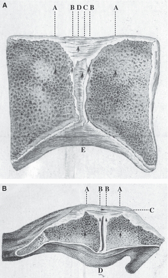
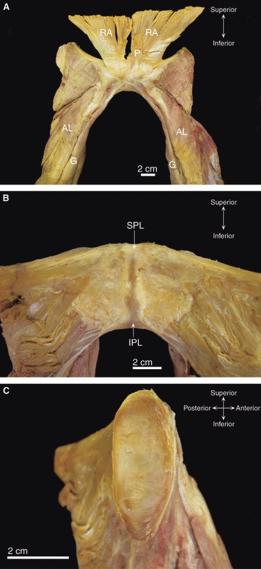
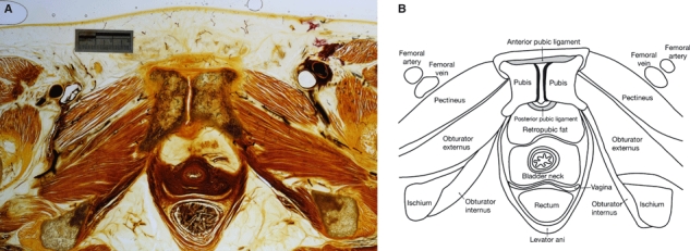
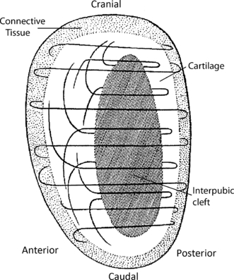
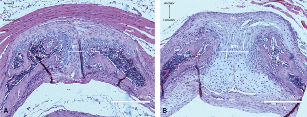
The pubic symphysis is a unique joint consisting of a fibrocartilaginous disc sandwiched between the articular surfaces of the pubic bones. It resists tensile, shearing and compressive forces and is capable of a small amount of movement under physiological conditions in most adults (up to 2 mm shift and 1° rotation). During pregnancy, circulating hormones such as relaxin induce resorption of the symphyseal margins and structural changes in the fibrocartilaginous disc, increasing symphyseal width and mobility. This systematic review of the English, German and French literature focuses on the normal anatomy of the adult human pubic symphysis. Although scientific studies of the joint have yielded useful descriptive data, comparison of results is hampered by imprecise methodology and/or poorly controlled studies. Several aspects of the anatomy of the pubic symphysis remain unknown or unclear: the precise attachments of surrounding ligaments and muscles; the arrangement of connective tissue fibres within the interpubic disc and the origin, structure and function of its associated interpubic cleft; the biomechanical consequences of sexual dimorphism; potential ethnic variations in morphology; and its precise innervation and blood supply. These deficiencies hinder our understanding of the normal form and function of the joint, which is particularly relevant when attempting to understand the mechanisms underlying pregnancy-related pubic symphyseal pain, a neglected and relatively common cause of pubic pain. A better understanding of the normal anatomy of the human pubic symphysis should improve our understanding of such problems and contribute to better treatments for patients suffering from symphyseal pain and dysfunction.
© 2010 The Authors. Journal of Anatomy © 2010 Anatomical Society of Great Britain and Ireland.
Figures
References
-
- Abramson D, Roberts SM, Wilson PD. Relaxation of the pelvic joints in pregnancy. Surg Gynecol Obstet. 1934;58:595–613.
-
- Aeby C. Über die Symphysis Ossium Pubis des Menschen nebst Beiträgen zur Lehre vom hyalinen Knorpel und seiner Verknöcherung. Z Ration Med. 1858;4:1–25.
-
- Albert H, Godskesen M, Westergaard JG, et al. Circulating levels of relaxin are normal in pregnant women with pelvic pain. Eur J Obstet Gynecol. 1997;74:19–22. - PubMed
-
- Alicioglu B, Kartal O, Gurbuz H, et al. Symphysis pubis distance in adults: a retrospective computed tomography study. Surg Radiol Anat. 2008;30:153–157. - PubMed
-
- Bahlmann F, Merz E, Macchiella D, et al. Sonographische Darstellung des Symphysenspaltes zur Beurteilung eines Symphysenschadens in der Schwangerschaft und Post Partum. Z Geburtsh u Perinat. 1993;197:27–30. - PubMed
Publication types
MeSH terms
LinkOut - more resources
Full Text Sources
Other Literature Sources
Medical
