

Expression of proliferative and inflammatory markers in a full-thickness human skin equivalent following exposure to the model sulfur mustard vesicant, 2-chloroethyl ethyl sulfide
- PMID: 20840853
- PMCID: PMC2996832
- DOI: 10.1016/j.taap.2010.09.005
Expression of proliferative and inflammatory markers in a full-thickness human skin equivalent following exposure to the model sulfur mustard vesicant, 2-chloroethyl ethyl sulfide
Abstract
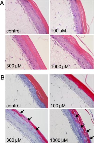
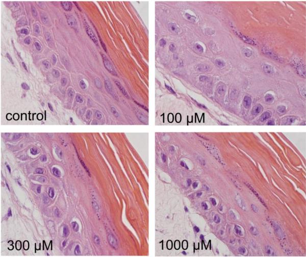
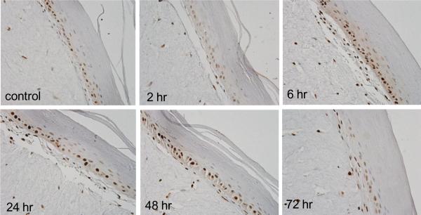
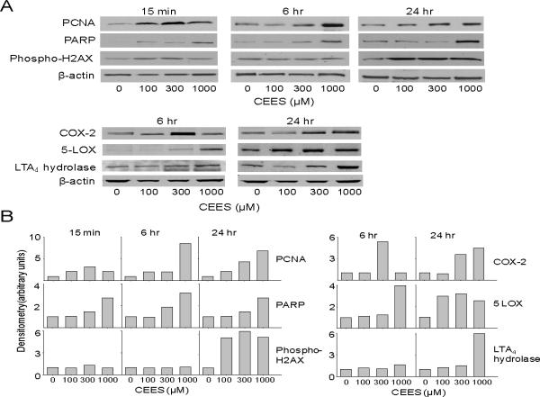
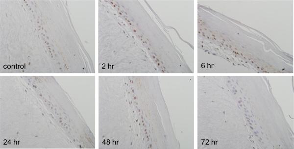
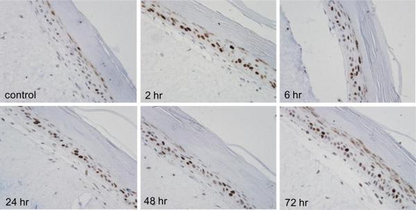
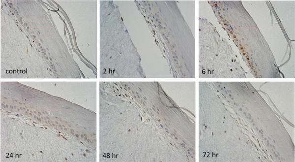
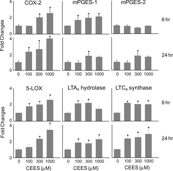
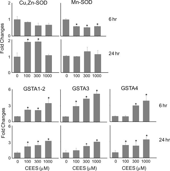
Sulfur mustard is a potent vesicant that induces inflammation, edema and blistering following dermal exposure. To assess molecular mechanisms mediating these responses, we analyzed the effects of the model sulfur mustard vesicant, 2-chloroethyl ethyl sulfide, on EpiDerm-FT™, a commercially available full-thickness human skin equivalent. CEES (100-1000 μM) caused a concentration-dependent increase in pyknotic nuclei and vacuolization in basal keratinocytes; at high concentrations (300-1000 μM), CEES also disrupted keratin filament architecture in the stratum corneum. This was associated with time-dependent increases in expression of proliferating cell nuclear antigen, a marker of cell proliferation, and poly(ADP-ribose) polymerase (PARP) and phosphorylated histone H2AX, markers of DNA damage. Concentration- and time-dependent increases in mRNA and protein expression of eicosanoid biosynthetic enzymes including COX-2, 5-lipoxygenase, microsomal PGE₂ synthases, leukotriene (LT) A₄ hydrolase and LTC₄ synthase were observed in CEES-treated skin equivalents, as well as in antioxidant enzymes, glutathione S-transferases A1-2 (GSTA1-2), GSTA3 and GSTA4. These data demonstrate that CEES induces rapid cellular damage, cytotoxicity and inflammation in full-thickness skin equivalents. These effects are similar to human responses to vesicants in vivo and suggest that the full thickness skin equivalent is a useful in vitro model to characterize the biological effects of mustards and to develop potential therapeutics.
Copyright © 2010 Elsevier Inc. All rights reserved.
Figures
References
-
- Babin MC, Ricketts K, Skvorak JP, Gazaway M, Mitcheltree LW, Casillas RP. Systemic administration of candidate antivesicants to protect against topically applied sulfur mustard in the mouse ear vesicant model (MEVM) J Appl Toxicol. 2000;20(Suppl 1):S141–144. - PubMed
-
- Bhat KR, Benton BJ, Ray R. Poly (ADP-ribose) polymerase (PARP) is essential for sulfur mustard-induced DNA damage repair, but has no role in DNA ligase activation. J Appl Toxicol. 2006;26:452–457. - PubMed
-
- Bhat KR, Benton BJ, Rosenthal DS, Smulson ME, Ray R. Role of poly(ADP-ribose) polymerase (PARP) in DNA repair in sulfur mustard-exposed normal human epidermal keratinocytes (NHEK) J Appl Toxicol. 2000;20(Suppl 1):S13–17. - PubMed
-
- Black AT, Joseph LB, Casillas RP, Heck DE, Gerecke DR, Sinko PJ, Laskin DL, Laskin JD. Role of MAP kinases in regulating expression of antioxidants and inflammatory mediators in mouse keratinocytes following exposure to the half mustard, 2-chloroethyl ethyl sulfide. Toxicol Appl Pharmacol. 2010;245:352–360. - PMC - PubMed
-
- Blaha M, Bowers W, Jr., Kohl J, DuBose D, Walker J. IL-1-related cytokine responses of nonimmune skin cells subjected to CEES exposure with and without potential vesicant antagonists. In Vitr Mol Toxicol. 2000a;13:99–111. - PubMed
Publication types
MeSH terms
Substances
Grants and funding
- R01 CA132624/CA/NCI NIH HHS/United States
- P30 ES005022/ES/NIEHS NIH HHS/United States
- R01 CA100994/CA/NCI NIH HHS/United States
- CA093798/CA/NCI NIH HHS/United States
- R01 CA093798/CA/NCI NIH HHS/United States
- AI51214/AI/NIAID NIH HHS/United States
- ES005022/ES/NIEHS NIH HHS/United States
- GM034310/GM/NIGMS NIH HHS/United States
- AI084138/AI/NIAID NIH HHS/United States
- CA132624/CA/NCI NIH HHS/United States
- R01 AI084137/AI/NIAID NIH HHS/United States
- AR055073/AR/NIAMS NIH HHS/United States
- R37 AI051214/AI/NIAID NIH HHS/United States
- R01 GM034310/GM/NIGMS NIH HHS/United States
- CA100994/CA/NCI NIH HHS/United States
- R01 AI051214/AI/NIAID NIH HHS/United States
- U54 AR055073/AR/NIAMS NIH HHS/United States
- ES004738/ES/NIEHS NIH HHS/United States
- U54AR055073/AR/NIAMS NIH HHS/United States
- R01 ES004738/ES/NIEHS NIH HHS/United States
- R55 CA093798/CA/NCI NIH HHS/United States
LinkOut - more resources
Full Text Sources
Research Materials
Miscellaneous
