Low-level gestational lead exposure increases retinal progenitor cell proliferation and rod photoreceptor and bipolar cell neurogenesis in mice
- PMID: 20840909
- PMCID: PMC3018503
- DOI: 10.1289/ehp.1002524
Low-level gestational lead exposure increases retinal progenitor cell proliferation and rod photoreceptor and bipolar cell neurogenesis in mice
Abstract
Background: Gestational lead exposure (GLE) produces novel and persistent rod-mediated electroretinographic (ERG) supernormality in children and adult animals.
Objectives: We used our murine GLE model to test the hypothesis that GLE increases the number of neurons in the rod signaling pathway and to determine the cellular mechanisms underlying the phenotype.
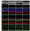
Results: Blood lead concentrations ([BPb]) in controls and after low-, moderate-, and high-dose GLE were ≤ 1, ≤ 10, approximately 25, and approximately 40 µg/dL, respectively, at the end of exposure [postnatal day 10 (PND10)]; by PND30 all [BPb] measures were ≤ 1 µg/dL. Epifluorescent, light, and confocal microscopy studies and Western blots demonstrated that late-born rod photoreceptors and rod and cone bipolar cells (BCs), but not Müller glial cells, increased in a nonmonotonic manner by 16-30% in PND60 GLE offspring. Retinal lamination and the rod:cone BC ratio were not altered. In vivo BrdU (5-bromo-2-deoxyuridine) pulse-labeling and Ki67 labeling of isolated cells from developing mice showed that GLE increased and prolonged retinal progenitor cell proliferation. TUNEL (terminal deoxynucleotidyl transferase dUTP nick end labeling) and confocal studies revealed that GLE did not alter developmental apoptosis or produce retinal injury. BrdU birth-dating and confocal studies confirmed the selective rod and BC increases and showed that the patterns of neurogenesis and gliogenesis were unaltered by GLE.
Conclusions: Our findings suggest two spatiotemporal components mediated by dysregulation of different extrinsic/intrinsic factors: increased and prolonged cell proliferation and increased neuronal (but not glial) cell fate. These findings have relevance for neurotoxicology, pediatrics, public health, risk assessment, and retinal cell biology because they occurred at clinically relevant [BPb] and correspond with the ERG phenotype.
Figures


Comment in
-
Lead doesn't spare the rod: low-level exposure supercharges retinal cell production in mice.Environ Health Perspect. 2011 Jan;119(1):A35. doi: 10.1289/ehp.119-a35b. Environ Health Perspect. 2011. PMID: 21196149 Free PMC article. No abstract available.
-
Redefining low lead levels.Environ Health Perspect. 2011 May;119(5):A202. doi: 10.1289/ehp.1103489. Environ Health Perspect. 2011. PMID: 21531660 Free PMC article. No abstract available.
Similar articles
-
Redefining low lead levels.Environ Health Perspect. 2011 May;119(5):A202. doi: 10.1289/ehp.1103489. Environ Health Perspect. 2011. PMID: 21531660 Free PMC article. No abstract available.
-
Increased proliferation of late-born retinal progenitor cells by gestational lead exposure delays rod and bipolar cell differentiation.Mol Vis. 2016 Dec 24;22:1468-1489. eCollection 2016. Mol Vis. 2016. PMID: 28050121 Free PMC article.
-
Low-level human equivalent gestational lead exposure produces supernormal scotopic electroretinograms, increased retinal neurogenesis, and decreased retinal dopamine utilization in rats.Environ Health Perspect. 2008 May;116(5):618-25. doi: 10.1289/ehp.11268. Environ Health Perspect. 2008. PMID: 18470321 Free PMC article.
-
The rod photoreceptor lineage of teleost fish.Prog Retin Eye Res. 2011 Nov;30(6):395-404. doi: 10.1016/j.preteyeres.2011.06.004. Epub 2011 Jun 30. Prog Retin Eye Res. 2011. PMID: 21742053 Free PMC article. Review.
-
Need rods? Get glycine receptors and taurine.Neuron. 2004 Mar 25;41(6):839-41. doi: 10.1016/s0896-6273(04)00153-9. Neuron. 2004. PMID: 15046714 Review.
Cited by
-
Lead exposure disrupts global DNA methylation in human embryonic stem cells and alters their neuronal differentiation.Toxicol Sci. 2014 May;139(1):142-61. doi: 10.1093/toxsci/kfu028. Epub 2014 Feb 11. Toxicol Sci. 2014. PMID: 24519525 Free PMC article.
-
Retinal toxicity of heavy metals and its involvement in retinal pathology.Food Chem Toxicol. 2024 Jun;188:114685. doi: 10.1016/j.fct.2024.114685. Epub 2024 Apr 23. Food Chem Toxicol. 2024. PMID: 38663763 Free PMC article. Review.
-
Late Effects of 1H + 16O on Short-Term and Object Memory, Hippocampal Dendritic Morphology and Mutagenesis.Front Behav Neurosci. 2020 Jun 26;14:96. doi: 10.3389/fnbeh.2020.00096. eCollection 2020. Front Behav Neurosci. 2020. PMID: 32670032 Free PMC article.
-
Redefining low lead levels.Environ Health Perspect. 2011 May;119(5):A202. doi: 10.1289/ehp.1103489. Environ Health Perspect. 2011. PMID: 21531660 Free PMC article. No abstract available.
-
The cellular and compartmental profile of mouse retinal glycolysis, tricarboxylic acid cycle, oxidative phosphorylation, and ~P transferring kinases.Mol Vis. 2016 Jul 23;22:847-85. eCollection 2016. Mol Vis. 2016. PMID: 27499608 Free PMC article.
References
-
- Alexiades MR, Cepko C. Quantitative analysis of proliferation and cell cycle length during development of the rat retina. Dev Dyn. 1996;205:293–307. - PubMed
-
- Alkondon M, Costa AC, Radhakrishnan V, Aronstam RS, Albuquerque EX. Selective blockade of NMDA-activated channel currents may be implicated in learning deficits caused by lead. FEBS Lett. 1990;261:124–130. - PubMed
-
- Barton KM, Levine EM. Expression patterns and cell cycle profiles of PCNA, MCM6, cyclin D1, cyclin A2, cyclin B1, and phosphorylated histone H3 in the developing mouse retina. Dev Dyn. 2008;237:672–682. - PubMed
-
- CDC (Centers for Disease Control and Prevention) Preventing Lead Poisoning in Young Children: A Statement by the Centers for Disease Control and Prevention. Atlanta, GA: CDC; 1991.
-
- Chetty CS, Reddy GR, Murthy KS, Johnson J, Sajwan K, Desiah D. Perinatal lead exposure alters the expression of neuronal nitric oxide synthase in rat brain. Int J Toxicol. 2001;20:113–120. - PubMed
Publication types
MeSH terms
Substances
Grants and funding
LinkOut - more resources
Full Text Sources
Medical

