. 2010 Oct 1;70(19):7421-30.
doi: 10.1158/0008-5472.CAN-10-0232. Epub 2010 Sep 14.

ERBB receptor activation is required for profibrotic responses to transforming growth factor beta

Affiliations

ERBB receptor activation is required for profibrotic responses to transforming growth factor beta

Mahefatiana Andrianifahanana et al. Cancer Res. .

Abstract

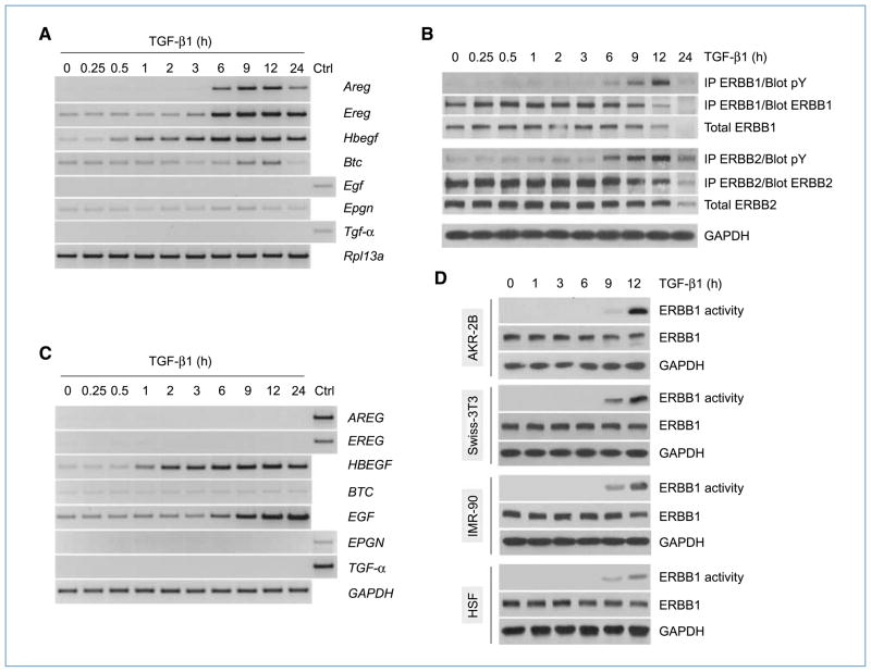
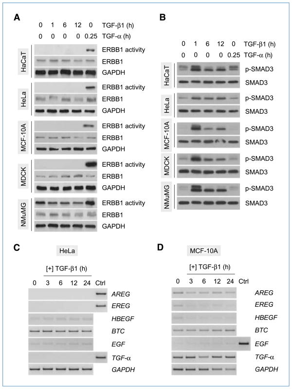
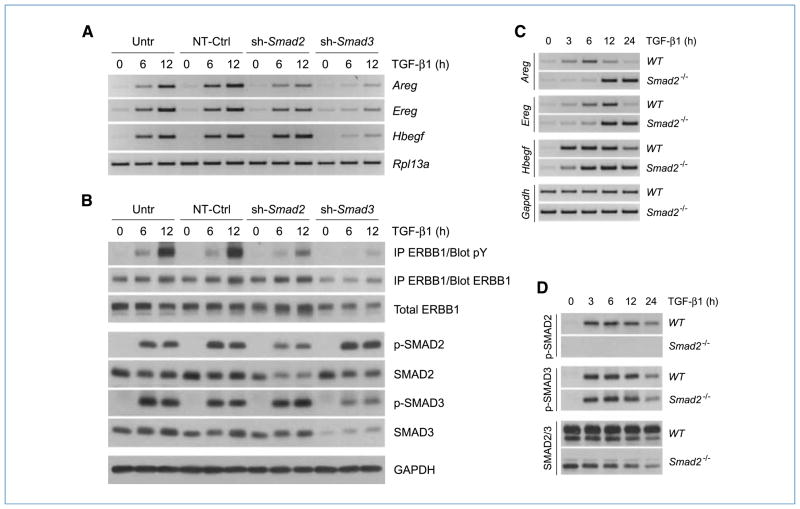
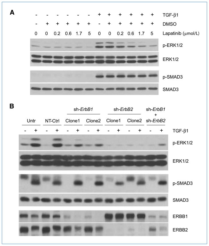
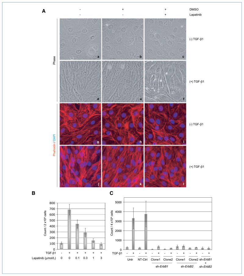
Engagement of the transforming growth factor-β (TGF-β) receptor complex activates multiple signaling pathways that play crucial roles in both health and disease. TGF-β is a key regulator of fibrogenesis and cancer-associated desmoplasia; however, its exact mode of action in these pathologic processes has remained poorly defined. Here, we report a novel mechanism whereby signaling via members of the ERBB or epidermal growth factor family of receptors serves as a central requirement for the biological responses of fibroblasts to TGF-β. We show that TGF-β triggers upregulation of ERBB ligands and activation of cognate receptors via the canonical SMAD pathway in fibroblasts. Interestingly, activation of ERBB is commonly observed in a subset of fibroblast but not epithelial cells from different species, indicating cell type specificity. Moreover, using genetic and pharmacologic approaches, we show that ERBB activation by TGF-β is essential for the induction of fibroblast cell morphologic transformation and anchorage-independent growth. Together, these results uncover important aspects of TGF-β signaling that highlight the role of ERBB ligands/receptors as critical mediators in fibroblast responses to this pleiotropic cytokine.


Conflict of interest statement

Disclosure of Potential Conflicts of Interest

No potential conflicts of interest were disclosed.

Figures

Figure 1
Figure 1
TGF-β induces ERBB ligand expression and receptor activation in mesenchymal cells. A, AKR-2B fibroblast cells were treated with TGF-β (10 ng/mL) and harvested at various time points, and total RNA (2 μg) was subjected to RT-PCR analysis using primers specific for the indicated target genes. Control (Ctrl) sample consisted of mouse heart RNA. B, AKR-2B cells were treated as in A, and total proteins (400 μg) were subjected to immunoprecipitation (IP) using ERBB1- or ERBB2-specific antibodies. Immunoprecipitates were analyzed by Western blotting using phosphotyrosine (pY)–specific antibodies. Membranes were stripped and reprobed with ERBB1 or ERBB2 antibodies. The expression levels of total ERBB1, ERBB2, and GAPDH (internal control) were assessed by Western blot analysis (50 μg). C, human fibroblast cells (IMR-90) were treated as in A, and total RNA was subjected to RT-PCR analysis for ERBB ligands. Control (Ctrl) sample consisted of human heart RNA. D, murine (AKR-2B and Swiss-3T3) and human (IMR-90 and HSF) fibroblast cells were treated as in A. ERBB1 was immunoprecipitated from total protein extracts (400–600 μg) and subjected to in vitro kinase assays using MBP as a substrate. Total protein aliquots (50 μg) were subjected to Western blot analysis to evaluate the expression levels of total ERBB1 and GAPDH (internal control).
Figure 2
Figure 2
Cell type–specific activation of ERBB by TGF-β. A, various epithelial lines from different species were treated with TGF-β (10 ng/mL) and harvested at the indicated times. Cells stimulated with TGF-α (20 ng/mL) for 15 min were used as positive controls for ERBB1 activation. ERBB1 was immunoprecipitated from total protein extracts (400–600 μg) and subjected to in vitro kinase assays using MBP as a substrate. Total protein aliquots (50 μg) were subjected to Western blot analysis to evaluate the expression levels of total ERBB1 and GAPDH (internal control). B, cells were treated with TGF-β or TGF-α as in A, and total proteins (50 μg) were analyzed by Western blotting using antibodies specific for phosphorylated SMAD3 (p-SMAD3) or total SMAD3. C and D, cells were treated with TGF-β (10 ng/mL) and harvested at various time points. Total RNA (2 μg) was subjected to RT-PCR analysis using primers specific for the indicated target genes. Control (Ctrl) sample consisted of human heart RNA.
Figure 3
Figure 3
Activation of the ERBB axis by TGF-β is SMAD dependent. A, AKR-2B cells stably expressing shRNA targeting Smad2 or Smad3 were treated with TGF-β (10 ng/mL) for 6 or 12 h and harvested for total RNA extraction. Untransduced AKR-2B cells (Untr) and cells transduced with nontargeting sequences (NT-Ctrl) were used as controls. Samples were subjected to RT-PCR analysis using primers specific for Areg, Ereg, or Hbegf. Rpl13a was used as an internal control. B, cells were treated as in A, and total proteins (500 μg) were subjected to immunoprecipitation (IP) using ERBB1-specific antibodies. Immunoprecipitates were analyzed by Western blotting using phosphotyrosine (pY)–specific antibodies. Membranes were stripped and reprobed with ERBB1 antibodies. Equivalent protein aliquots were subjected to Western blot analysis using antibodies specific for ERBB1, phosphorylated SMAD2 (p-SMAD2), and phosphorylated SMAD3 (p-SMAD3). SMAD2 and SMAD3 antibodies were used to determine silencing efficiency for both genes. GAPDH served as an internal control. C, murine embryonic fibroblast cells, lacking expression of Smad2 (Smad2−/−) as well as the WT counterpart, were treated with TGF-β (10 ng/mL), harvested at various time points, and subjected to RT-PCR analysis using primers specific for Areg, Ereg, or Hbegf. Gapdh was used as an internal control. D, cells were treated with TGF-β as in C, and total proteins (50 μg) were analyzed by Western blotting using antibodies specific for phosphorylated SMAD2 (p-SMAD2), phosphorylated SMAD3 (p-SMAD3), or total SMAD2/3.
Figure 4
Figure 4
Activation of ERBB by TGF-β elicits downstream signaling. A, AKR-2B fibroblast cells were left untreated or treated with TGF-β (10 ng/mL) for 12 h following a 45-min preincubation with different concentrations of the dual ERBB1/2-specific kinase inhibitor lapatinib or DMSO (0.025%, vehicle). Total proteins (50 μg) were analyzed by Western blotting using antibodies specific for phosphorylated ERK1/2 (p-ERK1/2), total ERK1/2, phosphorylated SMAD3 (p-SMAD3), or total SMAD3. B, AKR-2B cells stably transduced with lentivirus-based shRNA targeting ErbB1 (sh-ErbB1; clones 1 and 2), ErbB2 (sh-ErbB2; clones 1 and 2), or both (sh-ErbB1 + sh-ErbB2) were treated with TGF-β (10 ng/mL) for 6 or 12 h and harvested for total protein extraction. Untransduced AKR-2B cells (Untr) and cells transduced with nontargeting sequences (NT-Ctrl) were used as controls. Equivalent protein aliquots (50 μg) were subjected to Western blot analysis using antibodies specific for phosphorylated ERK1/2 (p-ERK1/2), total ERK1/2, phosphorylated SMAD3 (p-SMAD3), or total SMAD3. ERBB1 and ERBB2 antibodies were used to determine silencing efficiency for both genes. GAPDH served as an internal control. Note: the 6-h samples were used to evaluate SMAD3 phosphorylation, whereas the remaining study was performed on 12-h samples.
Figure 5
Figure 5
TGF-β induces morphologic transformation and AIG of fibroblasts via ERBB. A, AKR-2B fibroblast cells were grown to confluence on coverslips in six-well tissue culture plates and serum starved for 24 h. TGF-β was added (10 ng/mL) to the cells in the presence/absence of the ERBB1/2-specific kinase inhibitor lapatinib (5 μmol/L) or its vehicle (DMSO, 0.025%) and incubated for an additional 48 h. Following labeling of F-actin with TRITC-conjugated phalloidin (red), photomicrographs (original magnification, 60×) were produced under phase contrast (a–f) or fluorescent light (g–l). DAPI (blue) was used as a nuclear stain. B, AKR-2B cells were grown on soft agar in six-well plates (2 × 10⁴ per well) and left untreated or treated with TGF-β (10 ng/mL) in the presence or absence of different concentrations of lapatinib. Colonies (>50 μm in diameter) were counted at day 14 postseeding. Columns, means of triplicate wells from two separate experiments; bars, SDs. C, AKR-2B cells stably transduced with lentivirus-based shRNA targeting ErbB1 (sh-ErbB1; clones 1 and 2), ErbB2 (sh-ErbB2; clones 1 and 2), or both (sh-ErbB1 + sh-ErbB2) were grown on soft agar in six-well plates (4 × 10⁴ per well) and left untreated or treated with TGF-β (10 ng/mL) for 7 days. Untransduced AKR-2B cells (Untr) and cells transduced with nontargeting sequences (NT-Ctrl) were used as controls. Colonies were counted as in B. Columns, means of three replicates from three separate experiments; bars, SDs.

References

    1. Derynck R, Akhurst RJ. Differentiation plasticity regulated by TGF-β family proteins in development and disease. Nat Cell Biol. 2007;9:1000–4. - PubMed
    1. Massague J, Blain SW, Lo RS. TGFβ signaling in growth control, cancer, and heritable disorders. Cell. 2000;103:295–309. - PubMed
    1. Prud’homme GJ. Pathobiology of transforming growth factor-β in cancer, fibrosis and immunologic disease, and therapeutic considerations. Lab Invest. 2007;87:1077–91. - PubMed
    1. Rahimi RA, Leof EB. TGF-β signaling: a tale of two responses. J Cell Biochem. 2007;102:593–608. - PubMed
    1. Feng XH, Derynck R. Specificity and versatility in TGF-β signaling through Smads. Annu Rev Cell Dev Biol. 2005;21:659–93. - PubMed

Publication types

MeSH terms

Substances