. 2010 Nov 1;70(21):8642-50.
doi: 10.1158/0008-5472.CAN-10-1345. Epub 2010 Sep 14.

Heat shock protein 90 inhibition depletes LATS1 and LATS2, two regulators of the mammalian hippo tumor suppressor pathway

Affiliations

Heat shock protein 90 inhibition depletes LATS1 and LATS2, two regulators of the mammalian hippo tumor suppressor pathway

Catherine J Huntoon et al. Cancer Res. .

Abstract

Heat shock protein 90 (HSP90), which regulates the functions of multiple oncogenic signaling pathways, has emerged as a novel anticancer therapeutic target, and multiple small-molecule HSP90 inhibitors are now in clinical trials. Although the effects of HSP90 inhibitors on oncogenic signaling pathways have been extensively studied, the effects of these agents on tumor suppressor signaling pathways are currently unknown. Here, we have examined how HSP90 inhibitors affect LATS1 and the related protein LATS2, two kinases that relay antiproliferative signals in the Hippo tumor suppressor pathway. Both LATS1 and LATS2 were depleted from cells treated with the HSP90 inhibitors 17-allylamino-17-demethoxygeldanamycin (17-AAG), radicicol, and PU-H71. Moreover, these kinases interacted with HSP90, and LATS1 isolated from 17-AAG-treated cells had reduced catalytic activity, thus showing that the kinase is a bona fide HSP90 client. Importantly, LATS1 signaling was disrupted by 17-AAG in tumor cell lines in vitro and clinical ovarian cancers in vivo as shown by reduced levels of LATS1 and decreased phosphorylation of the LATS substrate YAP, an oncoprotein transcriptional coactivator that regulates genes involved in cell and tissue growth, including the CTGF gene. Consistent with the reduced YAP phosphorylation, there were increased levels of CTGF, a secreted protein that is implicated in tumor proliferation, metastasis, and angiogenesis. Taken together, these results identify LATS1 and LATS2 as novel HSP90 clients and show that HSP90 inhibitors can disrupt the LATS tumor suppressor pathway in human cancer cells.


Figures

Figure 1
Figure 1
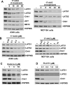
LATS1 and LATS2 are depleted by HSP90 inhibition. (A, B) Subconfluent A549 and MCF10A cells were treated with 1 μM 17-AAG (A) for the indicated times or with the indicated concentrations of 17-AAG for 24 h (B). (C, D) A549 cells were treated with the indicated concentrations of radicicol (C) or PU-H71 (D) for 24 h. Following treatment, cell lysates were analyzed by immunoblotting for the indicated antigens.
Figure 2
Figure 2
LATS1 is an HSP90 client. (A, B) Subconfluent A549 cells were treated with vehicle or 10 mM hydroxyurea (HU) for 24 h to induce arrest at the G1/S phase border. The HU-arrested cells were then incubated with vehicle (0.1% DMSO) or 1 μM 17-AAG in the continued presence of HU for the indicated times. Following treatment, the cell samples were divided; one portion was used to assess cycle by staining the DNA with propidium iodide (A), and the remaining cells were lysed and immunoblotted for the indicated antigens (B). (C, D) A549 cell lysates were immunoprecipitated (IP) with non-reactive rabbit serum (NRS) or anti-LATS2 rabbit antiserum (C); or subconfluent A549 cells were treated with vehicle (-) or 1 μM 17-AAG (+) for 24 h, and cell lysates were then immunoprecipitated with NRS or anti-LATS1 rabbit antiserum (D). The immunoprecipitates were then washed and sequentially immunoblotted for HSP90ß and LATS1 or LATS2. Cell lysates were immunoblotted to show equal amounts of HSP90ß in all lysates.
Figure 3
Figure 3
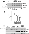
MOB1B stabilizes LATS1 but does not prevent 17-AAG from reducing LATS1 kinase activity. (A, B) A549 cells were transfected with empty vector (EV) or plasmid encoding SFB-LATS1 alone or combination with vectors that express FLAG-MST2 and FLAG-MOB1B. Twenty-four h after transfection, cells were treated with 1 μM 17-AAG for 24 h and SFB-LATS1 was recovered from cell lysates by reaction with streptavidin-agarose beads (which bind the streptavidin-binding peptide sequence of the SFB tag). Bead-bound SFB-LATS1 was subjected to in vitro kinase assays with GST-YAP as the substrate. Reactions were stopped with SDS-PAGE sample buffer and fractionated by SDS-PAGE, and transferred to a membrane. After membrane-bound radioactivity was detected and quantitiated by phosphorimaging, the membrane was blotted with anti-FLAG antibody to detect precipitated LATS1. (A) Representative experiment of 5 independent experiments. (B) Relative 32P-labeled GST-YAP, mean ± SD, n = 5 (0, 6, and 24-h time points) or n = 3 (1, and 3-h time points). (C) A549 cells were transiently transfected by electroporation with empty vector (EV) or plasmids that express SFB-LATS1, FLAG-MST2, and FLAG-MOB1B as indicated. Following electroporation, cells were re-plated at low density, cultured for 24 h, and treated with 1 μM 17-AAG for the indicated times. Cells lysates were immunoblotted with anti-FLAG monoclonal antibodies, which detect FLAG-tagged LATS1, MST2, and MOB1B.
Figure 4
Figure 4
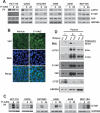
17-AAG depletes LATS1, reduces YAP Ser127 phosphorylation, and induces CTGF expression in tumor cell lines and human tumors. (A) Cell lines were treated with 0.1% DMSO vehicle (-) or 17-AAG for 6 or 24 h. Cell lysates were then immunoblotted for the indicated antigens. P-YAP, anti-phospho-Ser127-YAP. (B) Density-arrested U2OS cells were treated with vehicle (0.1% DMSO) or 1 μM 17-AAG for 3 h and fixed. Green, anti-YAP immunostain; blue, DNA stained with Hoechst 33342. Bar = 20 μm. (C) Samples from (A) were run on separate gels and immunoblotted for CTGF and HSP90. Due to low levels of CTGF in lysates, the A549 and MCF10A lysates were immunoprecipitated with anti-CTGF antibody and then immunoblotted with the same antibody; the HSP90 blot for these samples shows the starting lysates. (D) Paired patient ovarian cancer biopsies taken before (Pre) and 22-26 h after single-agent 17-AAG treatment (Post) were probed for the indicated antigens. To adjust for widely varying LATS1 levels, three exposures are shown for LATS1 immunoblots.

References

    1. Neckers L, Neckers K. Heat-shock protein 90 inhibitors as novel cancer chemotherapeutics - an update. Expert Opin Emerg Drugs. 2005;10:137–49. - PubMed
    1. Kim YS, Alarcon SV, Lee S, et al. Update on Hsp90 inhibitors in clinical trial. Curr Topics Med Chem. 2009;9:1479–92. - PMC - PubMed
    1. Saucedo LJ, Edgar BA. Filling out the Hippo pathway. Nat Rev Mol Cell Biol. 2007;8:613–21. - PubMed
    1. Zhao B, Lei QY, Guan KL. The Hippo-YAP pathway: new connections between regulation of organ size and cancer. Curr Opin Cell Biol. 2008;20:638–46. - PMC - PubMed
    1. Camargo FD, Gokhale S, Johnnidis JB, et al. YAP1 increases organ size and expands undifferentiated progenitor cells. Curr Biol. 2007;17:2054–60. - PubMed

Publication types

MeSH terms