Review
. 2010 Sep 14;3(139):re6.
doi: 10.1126/scisignal.3139re6.

ABL tyrosine kinases: evolution of function, regulation, and specificity

Affiliations
Review

ABL tyrosine kinases: evolution of function, regulation, and specificity

John Colicelli. Sci Signal. .

Erratum in

  • Sci Signal. 2011 Aug 30;4(188):er4

Abstract

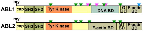
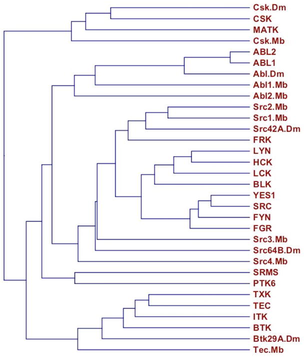
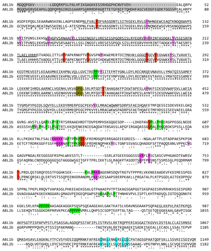
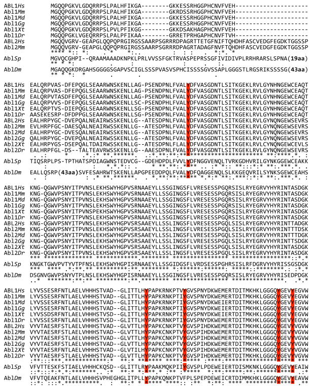
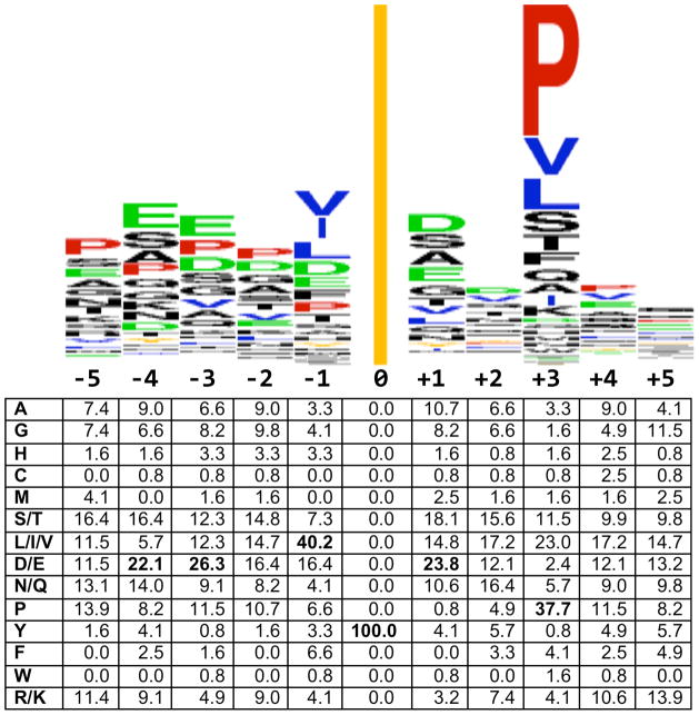
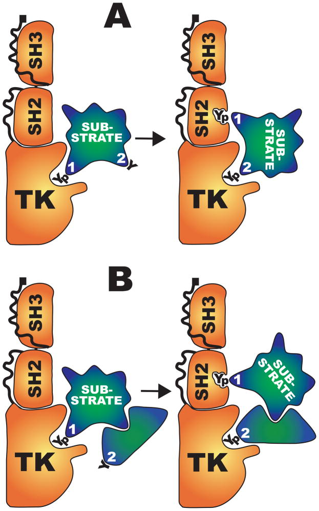
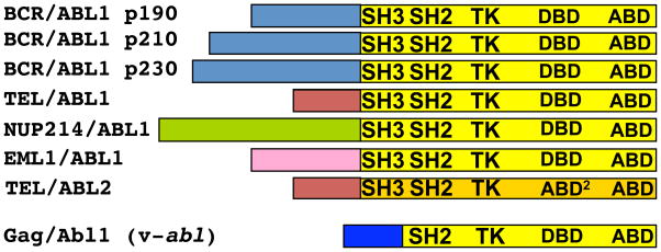
ABL-family proteins comprise one of the best conserved branches of the tyrosine kinases. Each ABL protein contains an SH3-SH2-TK (Src homology 3-Src homology 2-tyrosine kinase) domain cassette, which confers autoregulated kinase activity and is common among nonreceptor tyrosine kinases. This cassette is coupled to an actin-binding and -bundling domain, which makes ABL proteins capable of connecting phosphoregulation with actin-filament reorganization. Two vertebrate paralogs, ABL1 and ABL2, have evolved to perform specialized functions. ABL1 includes nuclear localization signals and a DNA binding domain through which it mediates DNA damage-repair functions, whereas ABL2 has additional binding capacity for actin and for microtubules to enhance its cytoskeletal remodeling functions. Several types of posttranslational modifications control ABL catalytic activity, subcellular localization, and stability, with consequences for both cytoplasmic and nuclear ABL functions. Binding partners provide additional regulation of ABL catalytic activity, substrate specificity, and downstream signaling. Information on ABL regulatory mechanisms is being mined to provide new therapeutic strategies against hematopoietic malignancies caused by BCR-ABL1 and related leukemogenic proteins.


Figures

Fig. 1
Fig. 1
ABL domain structure and motif conservation. Linear depiction of human ABL1 and ABL2 (long, b isoforms), D. melanogaster Abl, and M. brevicollis Abl1 and Abl2. my, myristoylation site (*site present but modification not verified); G BD, G-actin–binding domain; and MT BD, microtubule-binding domain. Blue triangle, NES; magenta triangle, NLS; and green triangle, proline-rich motif with capacity to bind SH3 or WW domains.
Fig. 2
Fig. 2
SH3-SH2-TK–family proteins. A dendrogram of the SH3-SH2-TK sequence cassettes from H. sapiens, D. melanogaster (Dm), and the protist M. brevicollis (Mb). The dendrogram was created using CLC Sequence Viewer (www.clcbio.com).
Fig. 3
Fig. 3
Sequence alignment of human ABL1b and ABL2b. Identity (*) and strong or weak sequence conservation [(:)colon for strong conservation; (.) period for weak conservation] are indicated. Domains are indicated with the following underlines: plain, SH3; heavy, SH2; dashed, TK; or wavy, terminal F-actin binding. Shading is used to highlight the following features: sequences deleted in human leukemogenic ABL fusions (gray), phosphorylation sites with known function (red), SH3 and WW domain proline-rich ligands (yellow), K- or R-rich NLS motifs (purple), and NES (blue). Additional reported phosphorylation sites are indicated with a red over-line. The DFG kinase signature is italicized. Alignment was performed using ClustalW (248).
Fig. 4
Fig. 4
Sequence alignment of ABL proteins from representative organisms. Representative Chordates are: Primates (Homo sapiens, Hs); Rodentia (Mus musculus, Mm); Marsupialia (Monodelphis domestica, Md); Aves (Gallus gallus); Amphibia (Xenopus tropicalis, Xt); and Pisces (Danio rerio, Dr). Representative Echinoderm is sea urchin (S. purpuratus, Sp); representative Arthropod is fruit fly (Drosophila melanogaster, Dm). Sequence identity and similarity is denoted as in Fig. 3. Color shading corresponds to the description in Fig. 3. Note that the first line shows only those vertebrate ABL2 sequences for which a long (myristoylated) isoform was identified. A few large insertions (mostly in AblDm) relative to other sequences are indicated. Sequences were obtained from Ensemble and GenBank. Alignment was performed using ClustalW.
Fig. 4
Fig. 4
Sequence alignment of ABL proteins from representative organisms. Representative Chordates are: Primates (Homo sapiens, Hs); Rodentia (Mus musculus, Mm); Marsupialia (Monodelphis domestica, Md); Aves (Gallus gallus); Amphibia (Xenopus tropicalis, Xt); and Pisces (Danio rerio, Dr). Representative Echinoderm is sea urchin (S. purpuratus, Sp); representative Arthropod is fruit fly (Drosophila melanogaster, Dm). Sequence identity and similarity is denoted as in Fig. 3. Color shading corresponds to the description in Fig. 3. Note that the first line shows only those vertebrate ABL2 sequences for which a long (myristoylated) isoform was identified. A few large insertions (mostly in AblDm) relative to other sequences are indicated. Sequences were obtained from Ensemble and GenBank. Alignment was performed using ClustalW.
Fig. 5
Fig. 5
ABL target site consensus. (Top) Sequence logo was created from 119 phosphorylation sites in Table 2 using Weblogo (249) at weblogo.berkeley.edu. The line at position zero represents the phosphorylated tyrosine. (Bottom) Amino acid position weight matrix generated using Python. Positions contributing most heavily to the consensus target site, as described in the text, are shown in bold.
Fig. 6
Fig. 6
Hierarchical processivity model. (A) The catalytically active conformation of ABL1 is depicted with SH3, SH2, and TK domains labeled (carboxy-terminal domains not shown). A “primary,” consensus, tyrosine target (1) is phosphorylated, then relocated to the SH2 domain. This guides a “secondary,” nonconsensus, tyrosine (2) into the catalytic site. (B) Same steps as in (A) except that the secondary tyrosine is in a separate protein associated with the initial substrate.
Fig. 7
Fig. 7
(Top) Seven translocation-derived ABL1 and ABL2 fusion oncoproteins. The following diseases are associated with each fusion: BCR-ABL1 p190 (also called p185) (ALL), p210 (CML), p230 (CNL), ETV6-ABL1 (CML and AML), NUP214-ABL1 (TALL), EML1-ABL1 (T-ALL), and ETV6-ABL2 (AML). (Bottom) The murine v-Abl oncoprotein results from a retroviral Gag gene fusion that deletes the Abl1 amino terminus and most of the SH3 domain. v-Abl causes B cell lymphomas.

References

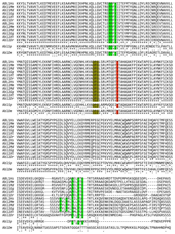
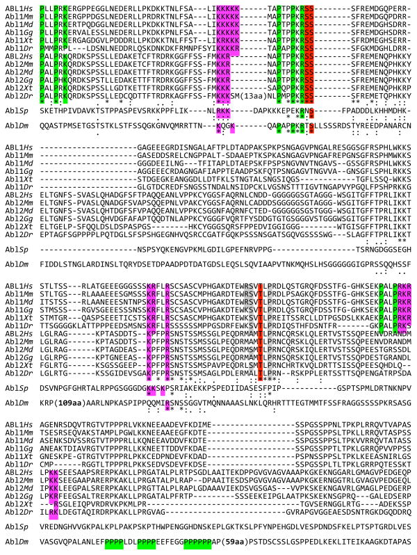
    1. Upper case is used for human genes and proteins (ABL1 and ABL2) as well as for discussion of gene and protein families (ABL). First-letter-only upper case (Abl1 and Abl2) is used when referring to genes and proteins from all other species in this review. Human Genome Organization nomenclature (www.genenames.org) is used. More common gene names are provided in the text, with additional aliases given in Table 1 and Table 2.

    1. Abelson HT, Rabstein LS. Lymphosarcoma: Virus-induced thymic-independent disease in mice. Cancer Res. 1970;30:2213–2222. - PubMed
    1. Sefton BM, Hunter T, Raschke WC. Evidence that the Abelson virus protein functions in vivo as a protein kinase that phosphorylates tyrosine. Proc Natl Acad Sci USA. 1981;78:1552–1556. - PMC - PubMed
    1. Witte ON, Dasgupta A, Baltimore D. Abelson murine leukaemia virus protein is phosphorylated in vitro to form phosphotyrosine. Nature. 1980;283:826–831. - PubMed
    1. Goff SP, Gilboa E, Witte ON, Baltimore D. Structure of the Abelson murine leukemia virus genome and the homologous cellular gene: Studies with cloned viral DNA. Cell. 1980;22:777–785. - PubMed

Publication types

MeSH terms