Review
. 2010 Oct;11(10):682-96.
doi: 10.1038/nrn2911. Epub 2010 Sep 15.

Synaptic versus extrasynaptic NMDA receptor signalling: implications for neurodegenerative disorders

Affiliations
Review

Synaptic versus extrasynaptic NMDA receptor signalling: implications for neurodegenerative disorders

Giles E Hardingham et al. Nat Rev Neurosci. 2010 Oct.

Abstract

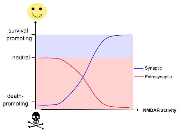
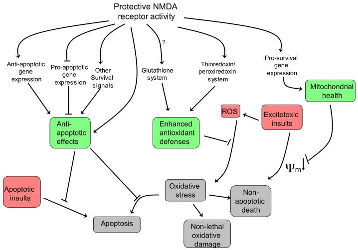
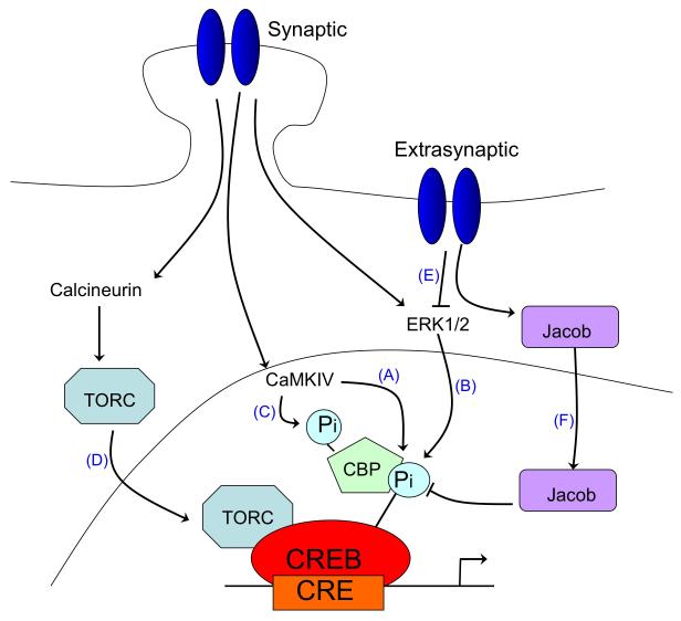
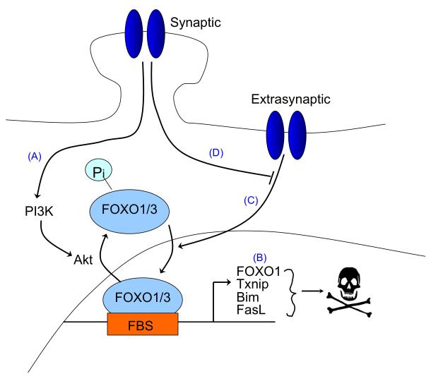
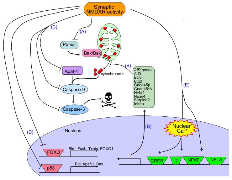
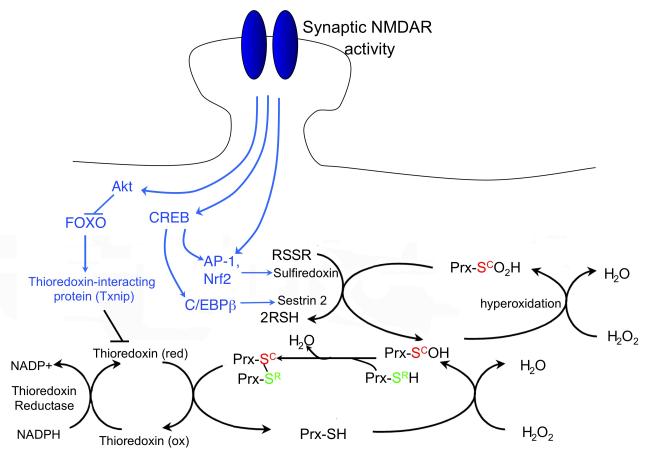
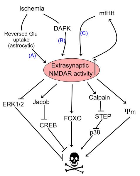
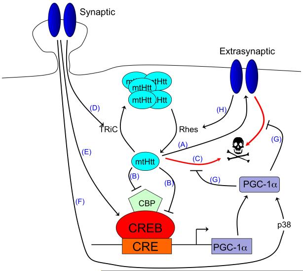
There is a long-standing paradox that NMDA (N-methyl-D-aspartate) receptors (NMDARs) can both promote neuronal health and kill neurons. Recent studies show that NMDAR-induced responses depend on the receptor location: stimulation of synaptic NMDARs, acting primarily through nuclear Ca(2+) signalling, leads to the build-up of a neuroprotective 'shield', whereas stimulation of extrasynaptic NMDARs promotes cell death. These differences result from the activation of distinct genomic programmes and from opposing actions on intracellular signalling pathways. Perturbations in the balance between synaptic and extrasynaptic NMDAR activity contribute to neuronal dysfunction in acute ischaemia and Huntington's disease, and could be a common theme in the aetiology of neurodegenerative diseases. Neuroprotective therapies should aim to both enhance the effect of synaptic activity and disrupt extrasynaptic NMDAR-dependent death signalling.


Figures

Figure 1
Figure 1. χ-shaped model of NMDAR-dependent excitotoxicity
The schematic illustrates the opposing effects of increasing synaptic and extrasynaptic N-methyl-D-aspartate receptor (NMDAR) activity on neuronal survival and resistance to trauma. Hypoactivity of synaptic NMDARs is harmful to neurons. Enhancing synaptic NMDAR activity triggers multiple neuroprotective pathways and this promotes neuronal survival. Low levels of activation of extrasynaptic NMDARs have no effects on neuronal survival but increasing the level of extrasynaptic NMDAR activity activates cell death pathways and exacerbates certain neurodegenerative processes, thus reducing neuronal survival.
Figure 2
Figure 2. Neuroprotective pathways activated by synaptic NMDAR activity
Changes in gene expression underlie the long-lasting neuroprotection exerted by activation of synaptic N-methyl-D-aspartate receptors (NMDARs). Changes include both the up-regulation of protective genes — including anti-apoptotic genes, pro-survival genes and genes involved in antioxidant responses — and the transcriptional suppression of pro-death genes. This restricts the apoptotic potential of the neuron, reduces vulnerability of mitochondria to insult-induced depolarization, and boosts intrinsic antioxidant defences (these three effects are shaded in green). These changes boost neuronal resistance to a range of traumata, including apoptotic, excitotoxic and oxidative insults (shaded in red), and thereby prevent harmful outcomes (shaded in grey) such as apoptotic and non-apoptotic cell death and cell dysfunction.ψm refers to mitochondrial membrane potential. ROS, reactive oxygen species.
Figure 3
Figure 3. Opposing effects of synaptic vs. extrasynaptic NMDAR signalling on gene expression
A. Opposing effects of synaptic vs. extrasynaptic NMDAR signalling on CREB-dependent gene expression. The activation of cyclic-AMP response element binding protein (CREB)-dependent gene expression by synaptic N-methyl-D-aspartate receptor (NMDAR) activity is a multi-step process. CREB must be phosphorylated at serine-133 in order to recruit its coactivator CREB binding protein (CBP) , ; this phosphorylation is mediated by the fast-acting nuclear CaM kinase pathway (A) and the slower acting, longer lasting Ras-ERK1/2 pathway (B) , , , both of which are promoted by activation of synaptic NMDARs CBP is subject to Ca2+-mediated transactivation by nuclear Ca2+ dependent CaM kinase IV , , which phosphorylates CBP at serine-301 (C) . In addition, nuclear translocation of transducer of regulated CREB activity (TORC) is a key step in CREB activation. Synaptic NMDAR-induced Ca2+ signals promote TORC import into the nucleus via calcineurin-dependent dephosphorylation (D) , . TORC acts at least in part by assisting in the recruitment of CBP to CREB (not shown). In contrast to these CREB-activating signals of synaptic NMDARs, extrasynaptic NMDARs suppress CREB activity , through inactivation of the Ras-ERK1/2 pathway (E) and the nuclear translocation of Jacob, which promotes CREB dephosphorylation (F) . B. Opposing effects of synaptic vs. extrasynaptic NMDAR signalling on FOXO-dependent gene expression. Synaptic NMDAR activity suppresses FOXO activity by promoting the Akt-mediated phosphorylation and nuclear export of FOXOs (A) , of which FOXO1 and FOXO3 are the predominant neuronal subtypes. FOXO1 is also regulated transcriptionally by FOXOs and thus signals that cause FOXO export also result in FOXO1 transcription being suppressed. By contrast, bath activation of NMDARs, which also triggers extrasynaptic NMDAR activity, stimulates FOXO nuclear import (B), an event that contributes, through promoting transcription of pro-death genes, to excitotoxic cell death. Synaptic NMDAR activity can exert a long-lasting block on this import signal (C) , the mechanistic basis of which remains unclear.
Figure 4
Figure 4. Synaptic NMDAR-dependent transcriptional changes have both anti-apoptotic and anti-oxidant effects
A) Synaptic NMDAR signalling suppresses the intrinsic apoptosis cascade at multiple levels. Synaptic N-methyl-D-aspartate receptor (NMDAR) activity protects neurons against apoptosis in a manner that is independent of the type of cellular insult because it suppresses the core apoptotic pathway. Transcriptional suppression of the BH3-only domain gene Puma by activation of synaptic NMDARs is a key event upstream of cytochrome c release (A) , . Up-regulation of a battery of nuclear Ca2+-regulated genes (green box) is also strongly protective , and probably also acts upstream of cytochrome c release, as it inhibits insult-induced mitochondrial membrane depolarization (B). Downstream of cytochrome c release, transcriptional suppression of components of the caspase cascade, such as Apaf-1, Caspase-9 and Caspase-3, also slows the apoptotic process (C) or raises the amount of mitochondrial cytochrome c release that is required to initiate apoptosis (as cytochrome c’s cytoplasmic target is Apaf-1) . Synaptic NMDAR activity also induces inactivation of the pro-death transcription factors FOXO and p53 (D) , , , and the subsequent suppression of the genes whose expression they regulate. Finally, transcription factors that have been shown to be important targets for NMDAR-dependent pro-survival signals are shown, namely CREB, NFAT and NFI-A (E) , , . B) Synaptic NMDAR signalling boosts intrinsic antioxidant defences. A schematic showing the effect of synaptic activity-induced changes in transcription on the thioredoxin (Trx)-peroxiredoxin (Prx) system. The dotted arrows indicate activity-dependent signalling events, which are collectively shaded in green. Briefly, reduction of peroxide levels is executed via the transfer of electrons from NADPH to peroxides through the redox-active sulfhydryl (-SH) groups of thioredoxin reductase, Trx and Prxs (the major two-cysteine Prx sub-type is shown). The catalytic redox-active cysteine residue of Prx (Sc, red) reduces peroxide, and is oxidized to cysteine sulfenic acid (SCOH). The resolving cysteine (SR, green) of a different Prx molecule then forms a disulfide bond with Prx-SCOH, thereby eliminating H2O. This intermolecular disulfide bond is then reduced by Trx, whose two-cysteine active site is reduced by Trx reductase. However, if levels of peroxide are too high, the – SCOH group becomes hyperoxidized to –SO2H (cysteine sulfinic acid). –SO2H is not a substrate for the resolving cysteine –SCH or for Trx. Instead, sulfiredoxin (and possibly sestrin 2) catalyses the reduction of hyperoxidized Prx-SO2H, returning it to the Trx cycle. Synaptic activity triggers the events shown to both enhance Trx activity and boost the reduction of hyperoxidized Prxs. These include the transcriptional activation of sulfiredoxin and sestrin 2, and the transcriptional suppression of Txnip, the gene encoding a the Trx inhibitor thioredoxin-interacting protein (Txnip). Bim (bcl2-interacting mediator of cell death); Fasl (fas antigen ligand); Apaf1 (apoptotic peptidase activating factor 1); CREB (cAMP responsive element binding protein); NFAT (nuclear factor of activated T-cells); NFI-A (nuclear factor I/A); Atf3 (activating transcription factor 3); Bcl6 (B-cell leukemia/lymphoma 6); Btg2 (B-cell translocation gene 2); Gadd45beta, GADD45gamma (growth arrest and DNA damage induced gene 45 gamma and beta, respectively); Nr4a1 (nuclear receptor subfamily 4, group A, member 1); Npas4 (neuronal PAS domain protein 4); Inhbb (Inhibin beta-A).
Figure 5
Figure 5. Activators and effectors of extrasynaptic NMDAR activity
Ischaemia results in activation of extrasynaptic N-methyl-D-aspartate receptor (NMDAR) activation through reversed glutamate uptake from astrocytes (A) . Extrasynaptic currents are also preferentially enhanced by ischaemia-induced death-associated protein kinase (DAPK) activation (B) , as well as by mutant Huntingtin (mtHtt) in a mouse model of Huntington’s disease (C) . Increased extrasynaptic (but not synaptic) NMDAR activity in turn preferentially activates a number of pro-death pathways. ψm refers to mitochondrial membrane potential, which is disrupted by extrasynaptic NMDAR activity.
Figure 6
Figure 6. Clinical relevance of the balance between synaptic and extrasynaptic NMDAR activity
A. The balance of synaptic vs. extrasynaptic NMDAR signalling influences HD pathology. Mutant Huntingtin (mtHtt) promotes neuronal dysfunction and death by enhancing the expression and activity of extrasynaptic N-methyl-D-aspartate receptors (NMDARs) (A) and by directly blocking CREB/CBP-dependent PGC-1α transcription (B) , , , as well as through other toxic mechanisms (C). Synaptic NMDAR activity opposes mtHtt toxicity by promoting TRiC-dependent aggregation of mtHtt into non-toxic inclusions (D) . Synaptic NMDAR activity also activates CREB-dependent PGC-1α transcription (E) , , and enhances PGC-1α activity post-translationally via p38 (F). PGC-1α itself protects neurons against mtHtt toxicity and extrasynaptic-NMDAR-dependent excitotoxicity (G) , , . Extrasynaptic NMDAR activity reduces the formation of non-toxic mtHtt inclusions by enhancing the expression of Rhes, a GTPase known to sumoylate and disaggregate mtHtt (H) . In addition, extrasynaptic NMDAR signals suppress CREB-dependent gene expression (not shown). B. Effect of memantine on NMDARs. Low doses of memantine preferentially block the chronic activation of extrasynaptic NMDARs (red line) by elevated levels of glutamate, which may occur as a result of ischaemia or impaired glutamate homeostasis associated with other disease processes. Low levels of memantine spare the trans-synaptic activation of synaptic NMDARs (blue line). This specificity of the effect of memantine has been demonstrated electrophysiologically , as well as by studying downstream pro-death and pro-survival signalling , , , .

References

    1. Aamodt SM, Constantine-Paton M. The role of neural activity in synaptic development and its implications for adult brain function. Adv Neurol. 1999;79:133–44. - PubMed
    1. Bliss TV, Collingridge GL. A synaptic model of memory: long-term potentiation in the hippocampus. Nature. 1993;361:31–9. - PubMed
    1. Tovar KR, Westbrook GL. The incorporation of NMDA receptors with a distinct subunit composition at nascent hippocampal synapses in vitro. The Journal of Neuroscience. 1999;19:4180–4188. - PMC - PubMed
    1. Rosenmund C, Feltz A, Westbrook GL. Synaptic NMDA receptor channels have a low open probability. J Neurosci. 1995;15:2788–95. - PMC - PubMed
    1. Cottrell JR, Dube GR, Egles C, Liu G. Distribution, density, and clustering of functional glutamate receptors before and after synaptogenesis in hippocampal neurons. J Neurophysiol. 2000;84:1573–87. - PubMed

Publication types

Substances