

Survival of chondrocytes in rabbit septal cartilage after electromechanical reshaping
- PMID: 20842431
- PMCID: PMC3010201
- DOI: 10.1007/s10439-010-0139-7
Survival of chondrocytes in rabbit septal cartilage after electromechanical reshaping
Abstract
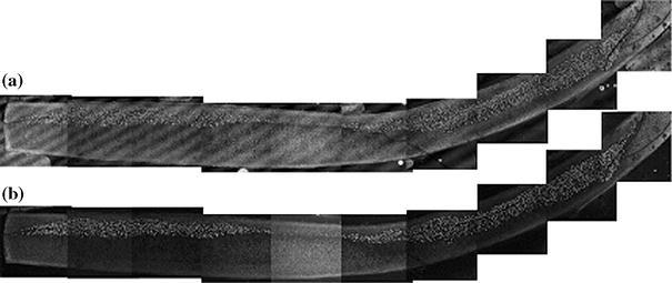
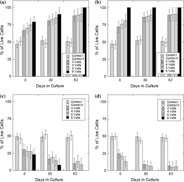
Electromechanical reshaping (EMR) has been recently described as an alternative method for reshaping facial cartilage without the need for incisions or sutures. This study focuses on determining the short- and long-term viability of chondrocytes following EMR in cartilage grafts maintained in tissue culture. Flat rabbit nasal septal cartilage specimens were bent into semi-cylindrical shapes by an aluminum jig while a constant electric voltage was applied across the concave and convex surfaces. After EMR, specimens were maintained in culture media for 64 days. Over this time period, specimens were serially biopsied and then stained with a fluorescent live-dead assay system and imaged using laser scanning confocal microscopy. In addition, the fraction of viable chondrocytes was measured, correlated with voltage, voltage application time, electric field configuration, and examined serially. The fraction of viable chondrocytes decreased with voltage and application time. High local electric field intensity and proximity to the positive electrode also focally reduced chondrocyte viability. The density of viable chondrocytes decreased over time and reached a steady state after 2-4 weeks. Viable cells were concentrated within the central region of the specimen. Approximately 20% of original chondrocytes remained viable after reshaping with optimal voltage and application time parameters and compared favorably with conventional surgical shape change techniques such as morselization.
Figures


Similar articles
-
Stress relaxation in porcine septal cartilage during electromechanical reshaping: mechanical and electrical responses.Ann Biomed Eng. 2006 Mar;34(3):455-64. doi: 10.1007/s10439-005-9051-y. Epub 2006 Feb 1. Ann Biomed Eng. 2006. PMID: 16450186
-
Needle electrode-based electromechanical reshaping of cartilage.Ann Biomed Eng. 2010 Nov;38(11):3389-97. doi: 10.1007/s10439-010-0088-1. Epub 2010 Jul 8. Ann Biomed Eng. 2010. PMID: 20614240 Free PMC article.
-
Quantitative assessment of chondrocyte viability after laser mediated reshaping: a novel application of flow cytometry.Lasers Surg Med. 2003;32(1):3-9. doi: 10.1002/lsm.10142. Lasers Surg Med. 2003. PMID: 12516064
-
Mechanobiology, chondrocyte and cartilage.Biomed Mater Eng. 2008;18(4-5):213-20. Biomed Mater Eng. 2008. PMID: 19065024 Review. No abstract available.
-
[Research progress of bioreactor biophysical factors in cartilage tissue engineering].Zhongguo Xiu Fu Chong Jian Wai Ke Za Zhi. 2013 Jul;27(7):810-3. Zhongguo Xiu Fu Chong Jian Wai Ke Za Zhi. 2013. PMID: 24063168 Review. Chinese.
Cited by
-
Electromechanical Reshaping of Face, Neck and Auricular Cartilages: A Scoping Review.Indian J Otolaryngol Head Neck Surg. 2025 Mar;77(3):1702-1721. doi: 10.1007/s12070-024-05207-4. Epub 2025 Jan 20. Indian J Otolaryngol Head Neck Surg. 2025. PMID: 40093489
-
Electromechanical reshaping of costal cartilage grafts: a new surgical treatment modality.Laryngoscope. 2011 Sep;121(9):1839-42. doi: 10.1002/lary.21892. Epub 2011 Aug 16. Laryngoscope. 2011. PMID: 22024834 Free PMC article.
-
Changes in the tangent modulus of rabbit septal and auricular cartilage following electromechanical reshaping.J Biomech Eng. 2011 Sep;133(9):094502. doi: 10.1115/1.4004916. J Biomech Eng. 2011. PMID: 22010748 Free PMC article.
-
Modular Component Assembly Approach to Microtia Reconstruction.JAMA Facial Plast Surg. 2016 Mar-Apr;18(2):120-7. doi: 10.1001/jamafacial.2015.1838. JAMA Facial Plast Surg. 2016. PMID: 26720326 Free PMC article.
-
Anatomy and Surgical Approaches to the Rabbit Nasal Septum.JAMA Facial Plast Surg. 2017 Sep 1;19(5):386-391. doi: 10.1001/jamafacial.2017.0116. JAMA Facial Plast Surg. 2017. PMID: 28472203 Free PMC article.
References
-
- Bard AJ, Faulkner LR. Electrochemical Methods: Fundamentals and Applications. New York: Wiley; 2001.
-
- Berendson, J., and D. Simonsson, Electrochemical aspects of treatment of tissue with direct current. Eur. J. Surg. Supp. 574:111–115, 1994. - PubMed
MeSH terms
Grants and funding
LinkOut - more resources
Full Text Sources
Other Literature Sources
