. 2011 Feb;24(2):114-29.
doi: 10.1002/nbm.1570. Epub 2010 Sep 15.

Fluorine (19F) MRS and MRI in biomedicine

Affiliations

Fluorine (19F) MRS and MRI in biomedicine

Jesús Ruiz-Cabello et al. NMR Biomed. 2011 Feb.

Abstract

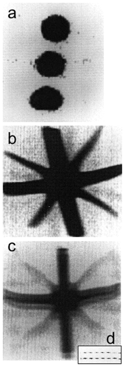
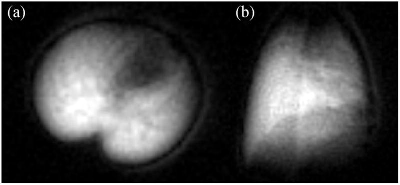
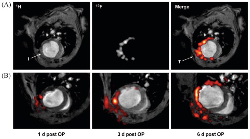
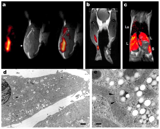
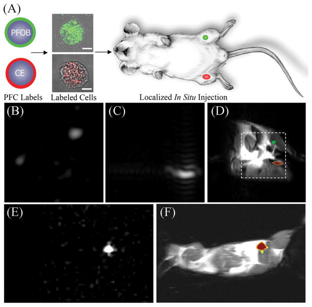
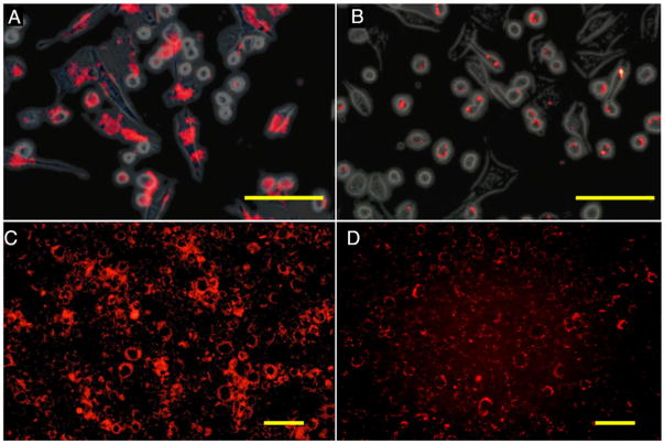
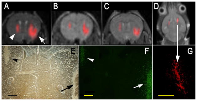
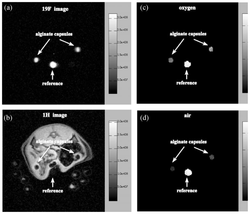
Shortly after the introduction of (1)H MRI, fluorinated molecules were tested as MR-detectable tracers or contrast agents. Many fluorinated compounds, which are nontoxic and chemically inert, are now being used in a broad range of biomedical applications, including anesthetics, chemotherapeutic agents, and molecules with high oxygen solubility for respiration and blood substitution. These compounds can be monitored by fluorine ((19)F) MRI and/or MRS, providing a noninvasive means to interrogate associated functions in biological systems. As a result of the lack of endogenous fluorine in living organisms, (19)F MRI of 'hotspots' of targeted fluorinated contrast agents has recently opened up new research avenues in molecular and cellular imaging. This includes the specific targeting and imaging of cellular surface epitopes, as well as MRI cell tracking of endogenous macrophages, injected immune cells and stem cell transplants.


Figures

Figure 1
Figure 1
The first 19F images from 1977 (1). (a) 19F MRI of three tubes of 1.4 M NaF with resolution of about 0.13 T0.13 T3 mm3. (b, c) 19F MRI of a perfluorocarbon (PFC), perfluorotributylamine, in a star-shaped ‘Union Jack’ phantom made from 2-mm (diagonal) and 6-mm tubes. The resolution is halved to about 0.6 T0.6 mm2. All images were acquired in about 400 s at 0.7 T using a steady-state free-precession (SSFP) MRI sequence (TR of several milliseconds). In (c), the image bandwidth per point is reduced, resulting in signal-to-noise ratio (SNR) loss and a ‘ghost’ image or ‘chemical shift artifact’ from the two chemically shifted moieties of this PFC, which are about 6 ppm apart. The two peaks are seen directly when the gradient in the vertical direction is switched off in the inset (d). Adapted, with permission, from Holland et al. (1).
Figure 2
Figure 2
Post-mortem 19F MRI of lung gas density using C2F6. Axial images (a) were acquired within 2 min and coronal images (b) were acquired within 3 min using a 4.7-T magnet. These images show excellent quality similar to that obtained with hyperpolarized gases, although with much more accumulation.
Figure 3
Figure 3
Infiltration of perfluorocarbons (PFCs) after myocardial infarction as detected by in vivo 19F MRI. (a) Anatomically corresponding 1H and 19F images from the mouse thorax recorded 4 days after ligation of the left anterior descending coronary artery, showing accumulation of 19F signal near the infarcted region (I) and at the location of surgery where the thorax was opened (T). PFCs were injected at day 0 (2 h after infarction) via the tail vein. (b) Sections of 1H images superimposed with the matching 19F images (red) acquired 1, 3 and 6 days after surgery (post OP) indicate a time-dependent infiltration of PFCs into injured areas of the heart and the adjacent region of the chest affected by thoracotomy. At day 4, an additional bolus of PFCs was injected to compensate for clearance of the particles from the bloodstream after 3 days. Subsequent histology demonstrated that the uptake of PFCs had occurred in cells of the monocyte/macrophage lineage. Reproduced, with permission, from Flogel et al. (81).
Figure 4
Figure 4
In vivo 19F MRI of perfluoro-crown ether (PFCE)-labeled dendritic cells in a mouse. (a) Mouse quadriceps after intramuscular injection of PFCE-labeled cells. From left to right are 19F, 1H and a composite 19F/1H image. (b) Composite image of dendritic cell migration into the popliteal lymph node following a hind foot pad injection. (c) Composite image through the torso following intravenous inoculation with perfluoropolyether (PFPE)-labeled cells. Cells are apparent in the liver (L), spleen (S) and, sporadically, lungs (Lu). Electron micrograph of a labeled fetal skin-derived dendritic cell line at a low magnification (d) and a higher magnification (e). Particles (100–200 nm) appear as smooth spheroids. Arrows show a typical multiple-membrane compartment enclosing these particles. Adapted, with permission, from Ahrens et al. (22).
Figure 5
Figure 5
Localization of labeled cells after in situ injection. (a) To determine the utility for cell tracking stem/progenitor cells labeled with either perfluoro-octyl bromide (PFOB) (green) or perfluoro-crown ether (PFCE) (red), nanoparticles were locally injected into mouse thigh skeletal muscle. (b–d) At 11.7 T, spectral discrimination permits the imaging of the fluorine signal attributable to ~ 1 × 10⁶ PFOB-loaded (b) or PFCE-loaded (c) cells individually which, when overlaid onto a conventional 1H image of the site (d), reveals PFOB- and PFCE-labeled cells localized to the left and right leg, respectively (broken line indicates 3 × 3-cm2 field of view for 19F images). (e, f) Similarly, at 1.5 T, 19F image of ~ 4 × 10⁶ PFCE-loaded cells (e) locates to the mouse thigh in a 1H image of the mouse cross-section (f). The absence of background signal in 19F images (b, c, e) enables unambiguous localization of PFCE-containing cells at both 11.7 and 1.5 T. Reproduced, with permission, from Partlow et al. (67). PFC, perfluorocarbon.
Figure 6
Figure 6
Fluorescence microscopy of cationic (a, c) and anionic (b, d) perfluoro-crown ether (PFCE)-labeled C17.2 mouse neural stem cells after 4 h of incubation with 2.4 mM PFCE. (a, b) Rhodamine fluorescent (red) and phase contrast overlay image of cells immediately after 4 h of incubation. (c,d) Rhodamine fluorescent images of cells cultured for an additional 18 h after removal of PFCE at 4 h of incubation. Note the transport and intracellular redistribution of label between the two time points. Scale bars: 100 μm in (a, b) and 50 μm in (c, d). Reproduced, with permission, from Ruiz-Cabello et al. (66).
Figure 7
Figure 7
In vivo MRI of transplanted C17.2 neural stem cells, with the 19F signal superimposed on the 1H MR images. (a–c) MR images at 1 h (a), 3 days (b) and 7 days after injection of 4 × 10⁴ (left hemisphere, arrowhead in a) or 3 × 10⁵ (right hemisphere, arrow in a) cationic perfluoro-crown ether (PFCE)-labeled cells. (e, f) Corresponding histopathology at day 7 with phase contrast (e) and anti-β-gal immunohistochemistry (f) demonstrates that implanted cells remain viable and continue to produce the marker enzyme. In (f), the right arrow indicates cells migrating from the injection site into the brain parenchyma. (d) MR image of a different animal at 14 days after injection of equal amounts of 4 × 10⁵ C17.2 cells in both hemispheres, demonstrating the persistence of the 19F signal for 2 weeks. (g) Corresponding histopathology showing rhodamine fluorescence from PFCE-labeled cells co-localizing with the 19F signal. Scale bar, 500 μm. Reproduced, with permission, from Ruiz-Cabello et al. (66).
Figure 8
Figure 8
19F (a) and 1H (b) images of a 2-mm transaxial slice through the peritoneal cavity of a rat showing two alginate capsules loaded with pure trans-1,2-bis(perfluorobutyl)-ethylene (F-44E), and the calculated 19F pO2 maps of the same slice whilst the animal is breathing oxygen (c) and air (d). The average pO2 values in the alginate capsules are 9.0 ± 1.4% (left capsule) and 12.8 ± 2.0% (right capsule) in (c) and 3.2 ± 1.0% (left capsule) and 5.0 ± 1.4% (right capsule) in (d). Images were acquired on day 1 after implantation. The scale on the right of (a) and (b) gives the signal intensity in arbitrary units; the scale in (c) and (d) gives the pO2 values in per cent. 100% corresponds to 760 mmHg. Reproduced, with permission, from Nöth et al. (89).

References

    1. Holland GN, Bottomley PA, Hinshaw WS. 19F magnetic resonance imaging. J Magn Reson. 1977;28:133–136.
    1. Bulte JWM. Hot spot MRI emerges from the background. Nat Biotech. 2005;23:945–946. - PubMed
    1. Clark LC, Gollan F. Survival of mammals breathing organic liquids equilibrated with oxygen at atmospheric pressure. Science. 1966;152:1755–1756. - PubMed
    1. Clark LC, Ackerman JL, Thomas SR, Millard RW, Hoffman RE, Pratt RG, Ragle-Cole H, Kinsey RA, Janakiraman R. Perfluorinated organic liquids and emulsions as biocompatible NMR imaging agents for 19F and dissolved oxygen. Adv Exp Med Biol. 1984;180:835–845. - PubMed
    1. Thomas SR. The biomedical application of fluorine-19 NMR. In: Partain CI, Patton JA, Kulkarni MV, James AEJ, editors. Magnetic Resonance Imaging. Vol. 2. Saunders Co; London: 1988. pp. 1536–1552.

Publication types