

Effects of 17 β-estradiol on lipopolysacharride-induced intracellular adhesion molecule-1 mRNA expression and Ca²+ homeostasis alteration in human endothelial cells
- PMID: 20843480
- PMCID: PMC2981630
- DOI: 10.1016/j.vph.2010.09.001
Effects of 17 β-estradiol on lipopolysacharride-induced intracellular adhesion molecule-1 mRNA expression and Ca²+ homeostasis alteration in human endothelial cells
Abstract
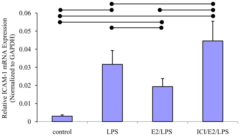
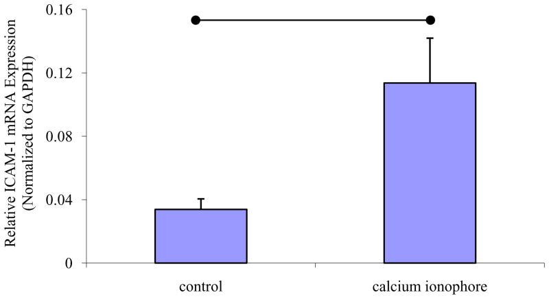
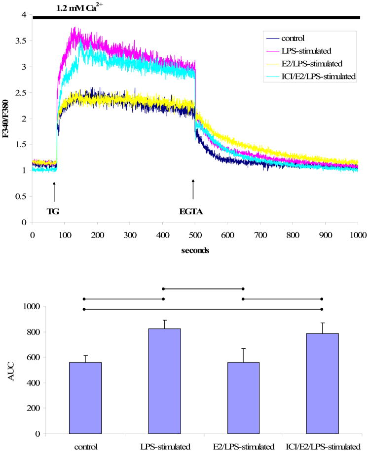
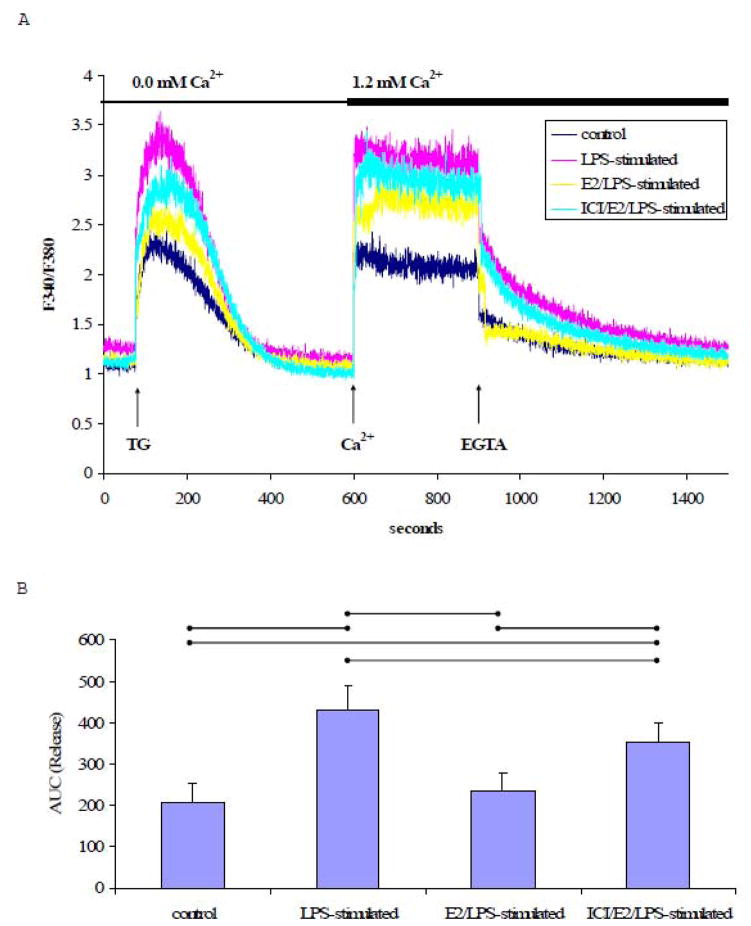
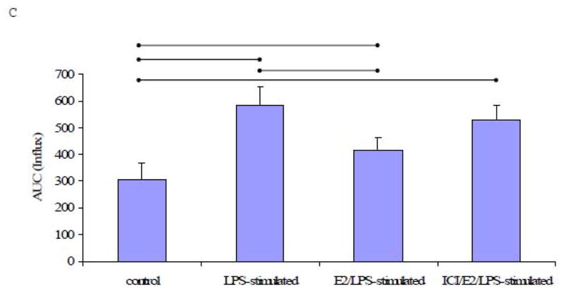
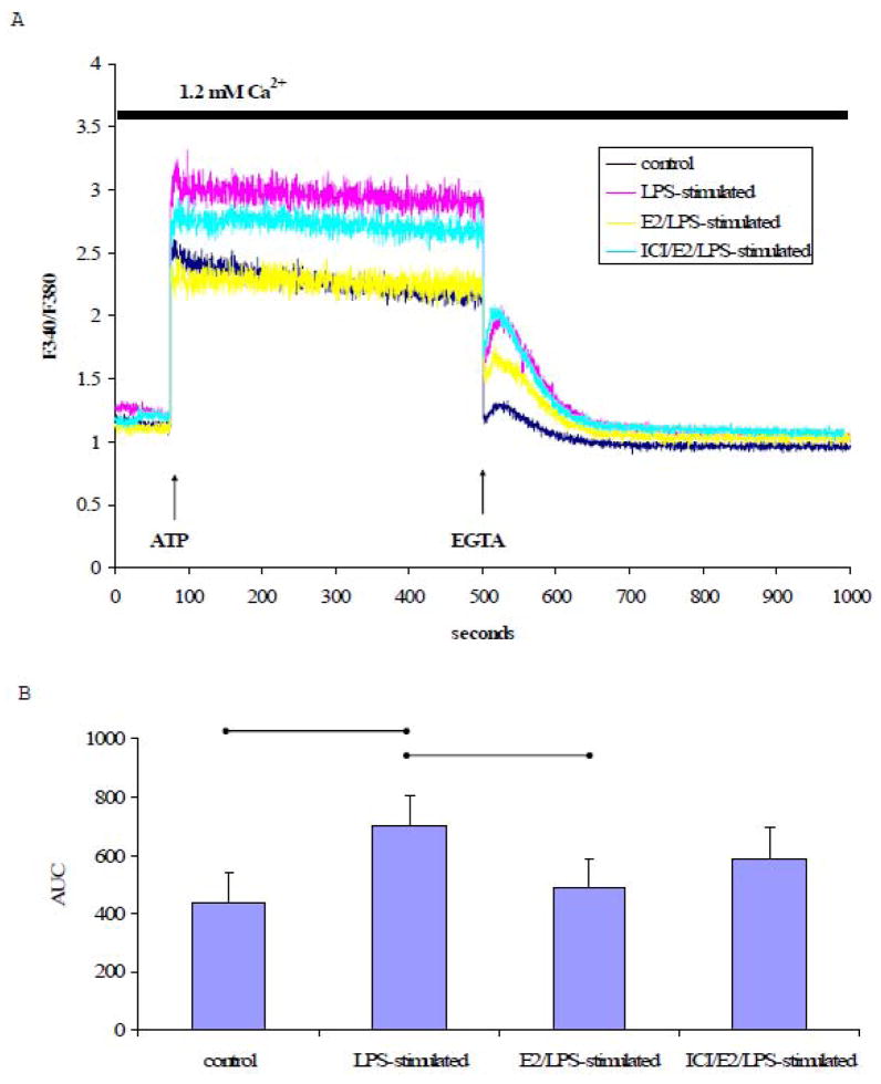
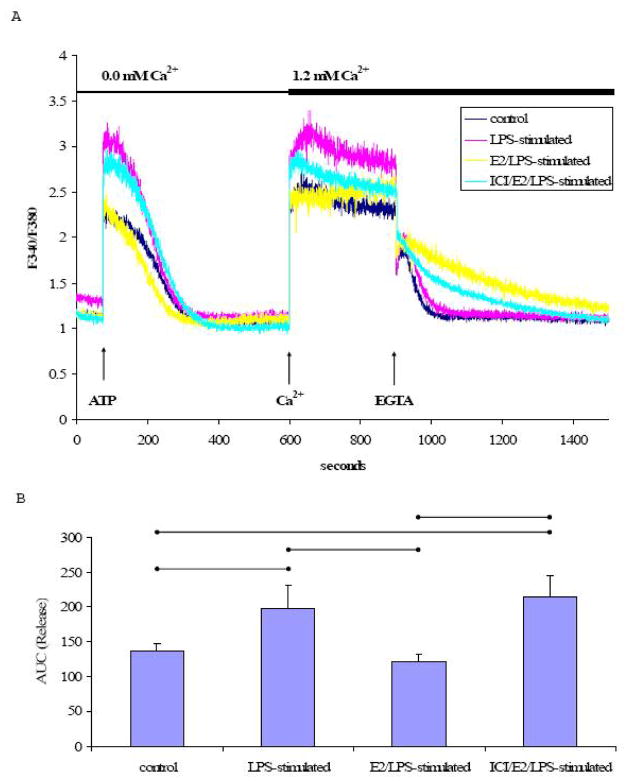
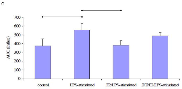
Recent evidence showed that 17 β-estradiol (E₂) decreased cytokine-induced expression of cell adhesion molecules (CAM). Changes in intracellular Ca²+ concentration ([Ca²+](i)) has been shown to be associated with CAM expression in endothelial cells. Here, the effects of E₂ (1 μM, 24 h) on the expression of intracellular adhesion molecule-1 (ICAM-1) and [Ca²+](i) were investigated in a lipopolysaccharide (LPS) (100 ng/mL, 18 h)-stimulated human endothelial cell line, EA.hy926, using real-time PCR and spectrofluorometry, respectively. PCR analysis revealed a significant increase in ICAM-1 expression in calcium ionophore A23187 (1 nM)- or LPS-stimulated cells. Pretreatment of cells with E(2) significantly inhibited LPS-induced ICAM-1 mRNA expression. [Ca²+](i) was monitored in Fura-2AM-loaded cells in the presence and absence of extracellular Ca²+ with thapsigargin (TG, 1 μM), a sarco/endoplasmic reticulum ATPase inhibitor or ATP (100 μM). The extent of TG- or ATP-induced [Ca²+](i) increase was significantly higher in LPS-stimulated cells than in control cells. Pre-treatment of LPS-stimulated cells with E₂ limited the Ca²+ response to the same level as in control cells. Furthermore, ICI 182,780, an estrogen receptor antagonist, attenuated the inhibitory actions of E₂ on ICAM-1 mRNA expression and Ca²+ responses, suggesting that estrogen receptors mediate, at least in part, the effects of estrogen. These data suggest a potential underlying mechanism for the protective effect of E₂ against atherosclerosis.
Copyright © 2010 Elsevier Inc. All rights reserved.
Figures
Similar articles
-
The effect of 17 beta-estradiol on intracellular calcium homeostasis in human endothelial cells.Eur J Pharmacol. 2010 Mar 25;630(1-3):92-9. doi: 10.1016/j.ejphar.2009.12.030. Epub 2010 Jan 4. Eur J Pharmacol. 2010. PMID: 20044991 Free PMC article.
-
Estradiol inhibits atp-induced intracellular calcium concentration increase in dorsal root ganglia neurons.Neuroscience. 2003;118(4):941-8. doi: 10.1016/s0306-4522(02)00915-6. Neuroscience. 2003. PMID: 12732239
-
Tibolone inhibits leukocyte adhesion molecule expression in human endothelial cells.Mol Cell Endocrinol. 2000 Apr 25;162(1-2):87-94. doi: 10.1016/s0303-7207(00)00206-9. Mol Cell Endocrinol. 2000. PMID: 10854701
-
Estradiol effects on intracellular Ca(2+) homeostasis in bovine brain-derived endothelial cells.Cell Tissue Res. 2012 Oct;350(1):109-18. doi: 10.1007/s00441-012-1460-2. Epub 2012 Jul 20. Cell Tissue Res. 2012. PMID: 22814863
-
Estrogen modulates TNF-alpha-induced inflammatory responses in rat aortic smooth muscle cells through estrogen receptor-beta activation.Am J Physiol Heart Circ Physiol. 2007 Jun;292(6):H2607-12. doi: 10.1152/ajpheart.01107.2006. Epub 2007 Jan 19. Am J Physiol Heart Circ Physiol. 2007. PMID: 17237237
Cited by
-
Regional Diversities in Fibrogenesis Weighed as a Key Determinant for Atrial Arrhythmogenesis.Biomedicines. 2021 Dec 14;9(12):1900. doi: 10.3390/biomedicines9121900. Biomedicines. 2021. PMID: 34944715 Free PMC article. Review.
-
Lipopolysaccharide induces endoplasmic store Ca2+-dependent inflammatory responses in lung microvessels.PLoS One. 2013 May 10;8(5):e63465. doi: 10.1371/journal.pone.0063465. Print 2013. PLoS One. 2013. PMID: 23675486 Free PMC article.
-
Sex as a Biological Variable in Atherosclerosis.Circ Res. 2020 Apr 24;126(9):1297-1319. doi: 10.1161/CIRCRESAHA.120.315930. Epub 2020 Apr 23. Circ Res. 2020. PMID: 32324497 Free PMC article. Review.
-
Sex differences in vascular physiology and pathophysiology: estrogen and androgen signaling in health and disease.Am J Physiol Heart Circ Physiol. 2017 Sep 1;313(3):H524-H545. doi: 10.1152/ajpheart.00217.2016. Epub 2017 Jun 16. Am J Physiol Heart Circ Physiol. 2017. PMID: 28626075 Free PMC article. Review.
-
Associations Between Follicular Fluid Biomarkers and IVF/ICSI Outcomes in Normo-Ovulatory Women-A Systematic Review.Biomolecules. 2025 Mar 20;15(3):443. doi: 10.3390/biom15030443. Biomolecules. 2025. PMID: 40149979 Free PMC article.
References
-
- Amrani Y, Lazaar AL, Hoffman R, Amin K, Ousmer S, Panettieri RA., Jr Activation of p55 tumor necrosis factor-alpha receptor-1 coupled to tumor necrosis factor receptor-associated factor 2 stimulates intercellular adhesion molecule-1 expression by modulating a thapsigargin-sensitive pathway in human tracheal smooth muscle cells. Mol Pharmacol. 2000;58:237–245. - PubMed
-
- Biewenga E, Cabell L, Audesirk T. Estradiol and raloxifene protect cultured SN4741 neurons against oxidative stress. Neurosci Lett. 2005;373:179–183. - PubMed
-
- Castillo C, Ceballos G, Rodriguez D, Villanueva C, Medina R, Lopez J, Mendez E, Castillo EF. Effects of estradiol on phenylephrine contractility associated with intracellular calcium release in rat aorta. Am J Physiol Cell Physiol. 2006;291:C1388–1394. - PubMed
Publication types
MeSH terms
Substances
Grants and funding
LinkOut - more resources
Full Text Sources
Miscellaneous
