. 2010;265(2):139-45.
doi: 10.1016/j.cellimm.2010.08.002. Epub 2010 Aug 14.

Chronic morphine treatment inhibits LPS-induced angiogenesis: implications in wound healing

Affiliations

Chronic morphine treatment inhibits LPS-induced angiogenesis: implications in wound healing

Josephine L Martin et al. Cell Immunol. 2010.

Abstract

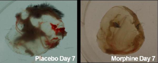
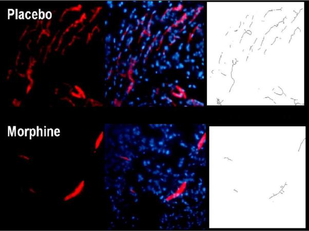
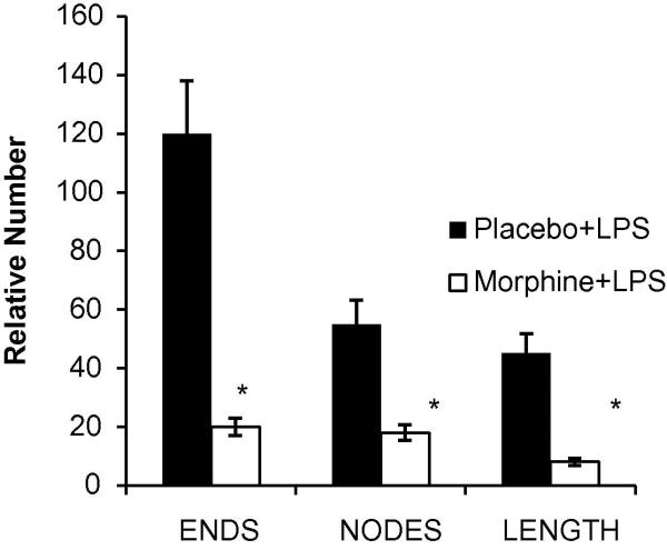
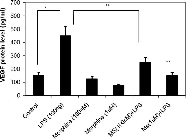
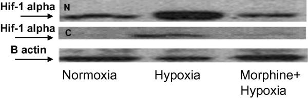
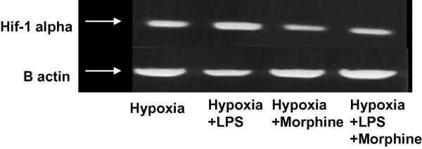
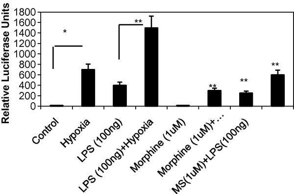
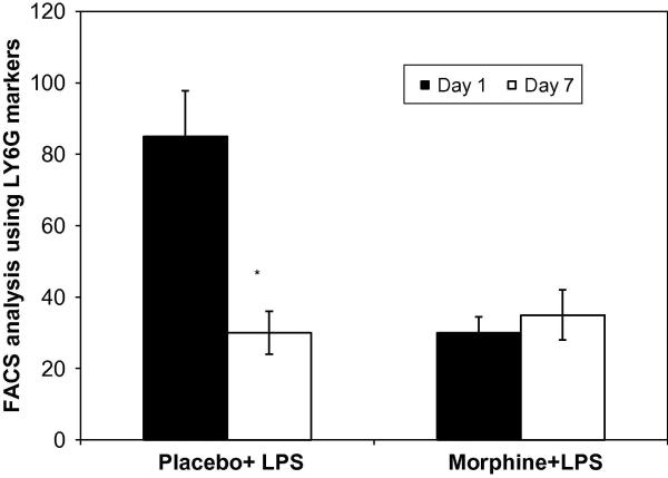
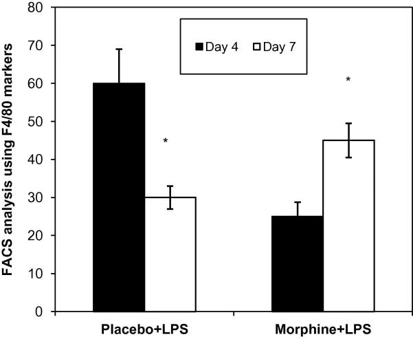
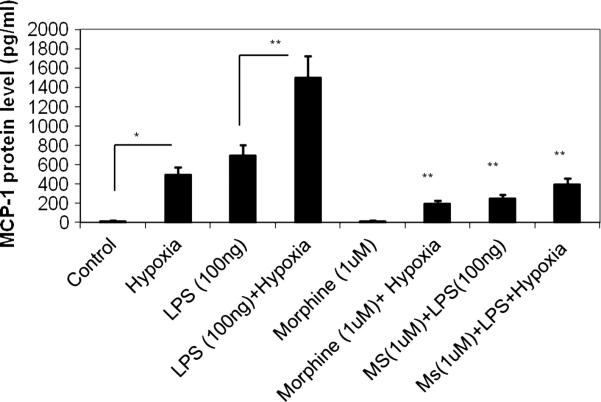
Delayed wound healing is a chronic problem in opioid drug abusers. We investigated the role chronic morphine plays on later stages of wound healing events using an angiogenesis model. Our results show that morphine treatment resulted in a significant decrease in inflammation induced angiogenesis. To delineate the mechanisms involved we investigate the role of hypoxia inducible factor 1 alpha (HIF-1 alpha), a potent inducer of angiogenic growth factor. Morphine treatment resulted in a significant decrease in the expression and nuclear translocation of HIF-1 alpha with a concurrent suppression in vascular endothelial growth factor (VEGF) synthesis. Cells of the innate immune system play a dominant role in the angiogenic process. Morphine treatment inhibited early recruitment of both neutrophils and monocytes towards an inflammatory signal with a significant decrease in the monocyte chemoattractant MCP-1. Taken together, our studies show that morphine regulates the wound repair process on multiple levels. Morphine acts both directly and indirectly in suppressing angiogenesis.


Figures

Figure 1A
Figure 1A. Morphine attenuated LPS-induced new blood vessels formation in matrigel plugs
LPS and heparin containing matrigels were injected into the hind limb of placebo or morphine pelleted mice. The non-invasive matrigel plug was removed following euthanasia. Gross morphology of matrigel plugs showed a marked presence of new blood vessels forming throughout the plugs of placebo treated mice. However, angiogenesis was attenuated following chronic morphine treatment seven days post injection.
Figure 1B
Figure 1B. LPS-induced formation of new blood vessels is suppressed in the presence of morphine
Cryostat sections from nitrogen snap frozen matrigel samples were stained using nuclear (DAPI) and PECAM (CD31) markers. In placebo treated groups, LPS-induced angiogenesis was apparent throughout matrigel plugs by Day 7. Chronic morphine treatment significantly decreased the formation of blood vessels into LPS containing plugs. Skeletonized images of newly formed blood vessels were assessed to measure vessel node, length, and end points.
Figure 1C
Figure 1C. Morphometric analysis of angiogenic events
A quantification of the skeletonized images shown in Figure 1B was evaluated for angiogenic events (node, length and end counts of new vessels). Plugs extracted from mice treated with morphine slow release pellets showed a significant reduction in blood vessel ends, nodes, and lengths when compared to placebo treated groups (*p<0.05, n=3).
Figure 2
Figure 2. Morphine suppresses LPS-induced macrophage secretion of VEGF proteins in a concentration-dependant manner
RAW-264 mouse macrophage cells were pretreated with varying concentrations of morphine 16h prior to LPS stimulation. Media-containing supernatants underwent ELISA analysis. VEGF expression was induced in the presence of LPS (lane 2). At 100 nM and 1uM morphine concentrations, LPS-stimulated VEGF protein expression was reduced to sub-basal levels (lanes 3 and 4). Morphine treatment, in the absence of LPS (lanes 5 and 6), significantly reduced VEGF expression as compared to its control treated group.
Figure 3A
Figure 3A. Morphine suppresses hypoxia-stabilized protein levels of HIF-1α in cytosolic and nuclear fractions
Peritoneal mouse macrophages were harvested and pretreated with 1μM morphine 16h prior to hypoxia stimulation. Cytosolic (lower gel) and nuclear (upper gel) extracts were analyzed using anti-HIF-1α monoclonal Ab. In the cytoplasmic fractions, hypoxia stabilization of HIF-1α protein was attenuated in the presence of morphine. Nuclear fractions of HIF-1α protein showed elevated levels in response to hypoxia. However, less HIF-1α was detected in the nuclear compartment following morphine treatment.
Figure 3B
Figure 3B. Morphine Suppresses O2-dependent HIF-1α RNA Expression
Mouse macrophage RAW-264 cells were pretreated with 1 μM morphine 16 hours prior to administration of 1 μM LPS. Cells were subjected to a hypoxic state (95% N2, 5%CO2) for 6 hours. RNA preparations, using the RNeasy Qiagen Mini kit, were reverse transcribed and PCR amplified using HIF-1α primers. Inductions of HIF-1α RNA levels were present under hypoxia (control) and LPS stimuli. Morphine significantly suppressed both hypoxia- and LPS-induced HIF-1α expression.
Figure 3C
Figure 3C. Effect of morphine on hypoxia-induced promoter activation in RAW cells
A full-length promoter (−1176 to +336) of the human VEGF gene was used to transfect Mouse macrophage RAW-264. Promoter induction was measured by firefly luciferase activity normalized to that of renilla luciferase. Fold induction of each group over respective control was expressed as relative luciferase activity.
Figure 4
Figure 4. Recruitment patterns of neutrophils into PVA sponged loaded with LPS
Following removal of PVA sponges, cells extracted were counted and labeled with the neutrophil markers LY6G/GR1 (n=4). FACs analysis revealed a significant decrease in neutrophil migration at day 1 in morphine treated animals but no differences in neutrophil migration amongst placebo+LPS and morphine+LPS treated groups seven days post injury.
Figure 5
Figure 5. Recruitment patterns of monocytes into PVA sponged loaded with LPS
Following removal of PVA sponges, cells extracted were counted and labeled with the macrophage markers F4/80(n=4). FACs analysis revealed a significant decrease in monocyte migration in the morphine treated group at day 4 but at day 7 a sustained recruitment was observed.
Figure 6
Figure 6. MCP-1 expression levels in response to morphine
The potent macrophage chemoattractants MCP-1 were analyzed using ELISA. Both LPS and hypoxia treatment alone resulted in a significant increase in MCP-1 levels. The expression was significantly greater when both insults were presented together. Morphine treatment resulted in a significant inhibition of both Hypoxia and LPS induced MCP-1 synthesis.

Similar articles

Cited by

References

    1. Lee JW, Bae SH, Jeong JW, Kim SH, Kim KW. Hypoxia-inducible factor (HIF-1) alpha: Its protein stability and biological functions. Experimental & Molecular Medicine. 2004;36(1):1–12. - PubMed
    1. Lynch SE, Colvin RB, Antoniades HN. Growth factors in wound healing. single and synergistic effects on partial thickness porcine skin wounds. The Journal of Clinical Investigation. 1989;84(2):640–646. - PMC - PubMed
    1. Bates DO, Jones RO. The role of vascular endothelial growth factor in wound healing. The International Journal of Lower Extremity Wounds. 2003;2(2):107–120. - PubMed
    1. Hoeben A, Landuyt B, Highley MS, Wildiers H, Van Oosterom AT, De Bruijn EA. Vascular endothelial growth factor and angiogenesis. Pharmacological Reviews. 2004;56(4):549–580. - PubMed
    1. Motani A, Forster L, Tull S, Anggard EE, Ferns GA. Insulin-like growth factor-I modulates monocyte adhesion to EAhy 926 endothelial cells. International Journal of Experimental Pathology. 1996;77(1):31–35. - PMC - PubMed

Publication types

MeSH terms

LinkOut - more resources