Comparative Study
. 2010 Nov 19;285(47):36909-21.
doi: 10.1074/jbc.M110.161745. Epub 2010 Sep 15.

Against the rules: human keratin K80: two functional alternative splice variants, K80 and K80.1, with special cellular localization in a wide range of epithelia

Affiliations
Comparative Study

Against the rules: human keratin K80: two functional alternative splice variants, K80 and K80.1, with special cellular localization in a wide range of epithelia

Lutz Langbein et al. J Biol Chem. .

Abstract

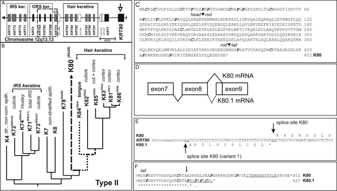
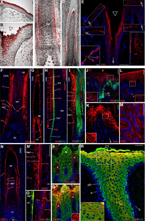
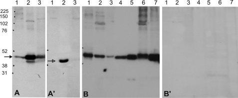
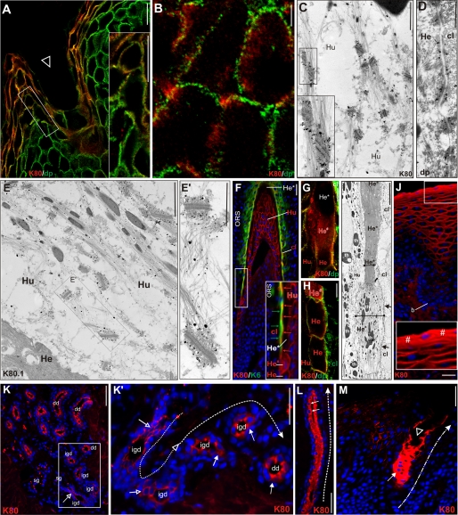
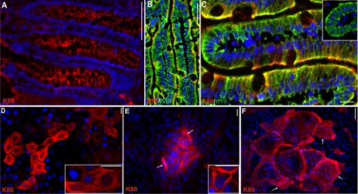
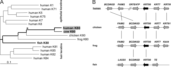
Of the 54 human keratins, five members have, at present, only been characterized at the gene level. In this study we have investigated the expression patterns of keratin K80, whose gene is located at the centromeric end of the type II keratin gene domain. K80 possesses a number of highly unusual properties. Structurally, it is distinctly closer to type II hair keratins than to type II epithelial keratins. Nonetheless, it is found in virtually all types of epithelia (stratified keratinizing/non-keratinizing, hard-keratinizing, as well as non-stratified tissues, and cell cultures thereof). This conspicuously broad expression range implies an unprecedented in vivo promiscuity of K80, which involves more than 20 different type I partners for intermediate filament (IF) formation. Throughout, K80 expression is related to advanced tissue or cell differentiation. However, instead of being part of the cytoplasmic IF network, K80 containing IFs are located at the cell margins close to the desmosomal plaques, where they are tightly interlaced with the cytoplasmic IF bundles abutting there. In contrast, in cells entering terminal differentiation, K80 adopts the "conventional" cytoplasmic distribution. In evolutionary terms, K80 is one of the oldest keratins, demonstrable down to fish. In addition, KRT80 mRNA is subject to alternative splicing. Besides K80, we describe a smaller but fully functional splice variant K80.1, which arose only during mammalian evolution. Remarkably, unlike the widely expressed K80, the expression of K80.1 is restricted to soft and hard keratinizing epithelial structures of the hair follicle and the filiform tongue papilla.


Figures

FIGURE 1.
FIGURE 1.
Analysis of the human K80 gene. A, localization of at the centromeric end of the type II keratin gene domain on chromosome 12q13.13. B, partial phylogenetic tree analysis of the α-helical rod domains of human type II keratins. C, amino acid sequence of K80. Arrowheads mark the beginning and end of the α-helical rod domain. Cysteine and proline residues in the head and tail domains are given in bold. D, schematic presentation of alternative splicing of exons 8 and 9. E, nucleotide sequence of the 5′-end of exon 9. The acceptor splice sites are underlined. An asterisk denotes an in-frame stop codon of the alternative variant. F, amino acid sequence alignment of K80 and K80.1 C-terminal ends. Underlined, peptide sequences used for specific antibody production; arrow, splice site-generated unique C-terminal peptide sequence of K80.1. Accession numbers of sequences used are given in bold under supplemental Table S1.
FIGURE 2.
FIGURE 2.
Expression pattern of K80 and K80.1. A–D, radioactive in situ hybridization. Note the cRNA probe used detects both splice variants of the mRNA (ISH signal in red). A, interfollicular epidermis. B, transition of the interfollicular epidermis with increasing labeling toward the infundibulum (triangle). C and D, follicle of plucked beard (C) and scalp (D) hair. Note the complex labeling pattern in the companion layer (cl), the three IRS compartments, Henle (He), and Huxley layer (Hu), and IRS-cuticle (icu), as well as the hair cuticle (cu). Red arrows, termination of RNA synthesis in the respective compartments. E-M, IIF staining of the larger K80 variant. K80 synthesis is strongest in upper infundibular cells (E, triangle) and decreases toward and along the interfollicular epidermis (E, upper inserts, open arrows), better visible after digital contrast enhancement (middle inserts, open arrows). K80 is also present in luminal cells of the eccrine sweat gland ducts (lower left insert, dd) and in sebaceous glands (lower right insert, seb). F, in the hair follicle, K80 is expressed in the companion layer (cl), the three IRS layers (He, Hu, and icu), and also in the hair cuticle (cu). G, note K80 expression in the cl persists considerably longer than in the IRS layers. H, double staining of K80 (red) and the hair cuticle keratin K32 (green) confirms coexpression in the upper hair cuticle (merged yellow). Note, K80 staining in the upper hair medulla (med). I, double staining of K80 (red) and the cortex keratin K31 (green; K31m, mouse monoclonal antibody against K31) confirms the absence of K80 in the hair cortex, but presence in the upper medulla (med). mc, medullary cortex cells (see Langbein and Schweizer (16)). J, double staining of K80 (red) and K6 (green) of foot sole epidermis. K80 staining occurs along the contours of upper suprabasal cells (see also inset) and is strongest (closed triangle) above the areas of K6 staining in the lower suprabasal (sb) cells. b, unstained basal cells. Prominent K80 staining of the cell margins of upper suprabasal layers of the epithelia of the tongue (K) and vagina (L) (and insets). M, cells of non-stratified, simple colon epithelium exhibit an apical cytoplasmic staining of K80. N–Q, IIF staining of the smaller K80 variant, K80.1. N and higher magnification, N′, the expression of K80.1 is restricted to the hair follicle and occurs in the middle to upper region of the cl, the IRS (Hu, icu) and strongly, in the hair cuticle (cu). In this region, the Henle layer is terminally differentiated (He*) and not labeled (N′). O, double staining of K80.1 (red) and K6 (green) shows that in cl cells, K80.1 is strictly concentrated at the side facing the Henle cells (merged yellow). P and Q, the hard-keratinizing upper cells of the filiform papillae of the tongue epithelium express both K80.1 (P, red, *) and K80 (Q, red, *). In contrast to K80, which is expressed together with K6 (green) also at the cell margins in the upper layers of the interpapillary tongue epithelium (Q and inset; serial section of P), K80.1 is absent from the interpapillary tongue epithelium (P). R, much better than in E, double staining of K80 (red) and K10 (green) of scalp emphasizes the cell margin staining of K80 in the infundibulum (merged yellow, see also inset), whereas except for basal (b) and parabasal cells (pb) K10 is cytoplasmic throughout. Blue label, DAPI staining. Bars represent 25 (A, B, E, J–M, and P–R) and 100 μm (C, D, F–I, N, and O).
FIGURE 3.
FIGURE 3.
Western blot analysis of K80 and K80.1 in epithelial tissues and cell lines. A, K80 is found most strongly at ∼50 kDa (arrow) in protein extracts of plucked beard hairs (lane 2) and to a lower extend in scalp (lane 3) and arm skin (lane 1) (A′) K80.1 is only detectable at ∼47 kDa (open arrow) in plucked hair extracts (lane 2). In A, lane 2 contains a weak band below that of K80, which is exclusively seen in extracts of plucked hairs and runs at a height comparable with that of the K80.1 variant, also present in plucked hair extracts (A′, lane 2). (Most probably the staining of this putative K80.1 band is due to a contamination with residual K80.) B, K80 is also demonstrable in extracts of the epithelia of the foot sole (lane 1), tongue (lane 2), vagina (lane 3), duodenum (lane 4), as well as in the liver carcinoma cell line PLC (lane 5), keratinocytes of the HaCaT line (lane 6) and in the mamma carcinoma cell line MCF-7 (line 7). B′, throughout, keratin extracts of the same tissues and cell lines were negative for K80.1. Dots on left sides, rainbow size markers in kDa (Amersham Biosciences).
FIGURE 4.
FIGURE 4.
Cellular localization of K80 and K80.1. A and B, confocal laser scan microscopy of K80 (red) and desmoplakin (dp, green) double labeling. A, uppermost cells of the infundibulum exhibit K80 staining at the cell borders. Higher magnification (A, inset) and particularly, the larger cells in the deeper infundibulum (B) clearly reveal an internal K80 location (red) close to but not at desmosomes (green), as seen by the paucity of merged yellow areas. Immunogold labeling of K80 confirms that it is specifically concentrated close to desmosomes. K80 is labeled at the broadening ends of cytoplasmic keratin bundles streaming toward the desmosomes of upper cells of the Huxley layer (C, and inset, Hu). As a control, immunogold staining of desmoplakin clearly labels the desmosomal plaques between cells of the companion layer and Henle layer (D, cl/He) leaving filaments unstained. Immunogold labeling of K80.1 shows also that this keratin is preferentially located in close vicinity to desmosomes, but also occurs, albeit weaker, in the cytoplasm of Huxley layer cells (E, Hu/Hu, higher magnification E′). F, IIF double staining of K80 (red) and K6 (green) on a strongly oblique hair follicle section reveals cell margin staining in Huxley cells (Hu, red) and the concentration of K80 in the inner portion of cl cells facing differentiated Henle cells (He*, merged yellow and red arrows in inset). Note, the localization of K80 (red) at cell margins of lower Henle cells (He), but a strong cytoplasmic staining of the last living cell (He#) undergoing abrupt terminal differentiation (He*) and excluding further staining of the Henle layer (He*) well detectable by double label IIF with desmoplakin (dp, in green) and confocal microscopy (G and H). This process is accompanied by dramatic morphological changes from the large and broad pre-terminal Henle cells (I, He#) to elongated and very slender terminally differentiated cells (I, He*, horizontal bars). Note that the loose keratin filaments bundles are transformed into an amorphous mass in terminally differentiated He* cells. Arrows, compact keratin belt in cl cells adjacent to Henle cells. J, IIF staining of K80 in the non-cornified stratified vaginal epithelial also shows a shift during epithelial differentiation from a marginal staining in the upper suprabasal strata to a clear cytoplasmic staining in the superficial differentiated layer (J, #). K–M, a comparable shift is observed in the eccrine sweat gland. K and K′, idg, open arrows, staining of the margins of luminal cells of the intraglandular duct; and K and K′, dd, closed arrows, subsequent cytoplasmic staining of the cells in the dermal duct. In the latter, the pronounced cytoplasmic label occurs toward the lumen (H, arrows) and is maintained along the entire dermal duct (L, dashed arrow, dd). K80 can be followed up (M, dashed-dotted arrow) to the acrosyringium (M, open triangle). Bars represent 20 (A), 10 (B, G, and H), 25 (J–M), 50 (F), and 1 μm (C, E, and I).
FIGURE 5.
FIGURE 5.
Localization of K80 in non-stratified, simple epithelia and cultured epithelial cells. A, IIF detects K80 in the apical cytoplasm of duodenum epithelium. Using double labeling, this apically concentrated K80 pattern (B and C, red) is well seen relative to the cytoplasmic staining of K19 (B and C, green). Note, less differentiated duodenum crypt cells are widely K80-negative (C), inset. In monolayer cultures of polarized epithelia, such as the mamma carcinoma line MCF-7 (D and inset), filamentous cytoplasmic K80 labeling is seen in most cells. In immortalized keratinocytes of line HaCaT (E and inset) and normal human epidermal keratinocytes (F), grown under conditions that induce local differentiation and stratification, differentiated suprabasal cells in the “domes” show cell margin staining of K80, whereas the single uppermost cells exhibit a cytoplasmic label. Note, monolayered and basal keratinocytes of the domes are K80-negative. Bars represent 25 (A, B, and D–F) and 10 μm (C).
FIGURE 6.
FIGURE 6.
Evolution of the KRT80 gene. A, phylogenetic analysis of K80 proteins of various species and representative human type II epithelial and hair keratins. Amino acid sequences of the rod domains of the keratins were aligned and a phylogenetic tree was built using the neighbor joining method. The branch lengths in the tree are proportional to the number of substitutions per site (scale: 0.1 substitution per site). B, comparison of the gene locus in vertebrates. Genes are represented by arrows pointing away from the 5′-end to the 3′-end of the respective gene. Homologous genes are connected by vertical lines. KRT, keratin; FAIM2, Fas apoptotic inhibitory molecule 2; BCDIN3D, bicoid-interacting 3 domain containing; OR7E47P, olfactory receptor family 7 subfamily E member 47 pseudogene; Kb54; Xenopus basic keratin 54; TE, transposable element derived 4, similar to piggyBac; LASS5, LAG1 homolog, ceramide synthetase 5.

References

    1. Rogers M. A., Winter H., Langbein L., Bleiler R., Schweizer J. (2004) Differentiation 72, 527–540 - PubMed
    1. Rogers M. A., Edler L., Winter H., Langbein L., Beckmann I., Schweizer J. (2005) J. Invest. Dermatol. 124, 536–544 - PubMed
    1. Schweizer J., Bowden P. E., Coulombe P. A., Langbein L., Lane E. B., Magin T. M., Maltais L., Omary M. B., Parry D. A., Rogers M. A., Wright M. W. (2006) J. Cell Biol. 174, 169–174 - PMC - PubMed
    1. Hesse M., Zimek A., Weber K., Magin T. M. (2004) Eur. J. Cell Biol. 83, 19–26 - PubMed
    1. Lu H., Zimek A., Chen J., Hesse M., Büssow H., Weber K., Magin T. M. (2006) Eur. J. Cell Biol. 85, 803–811 - PubMed

Publication types

MeSH terms