. 2010 Nov 12;285(46):35783-91.
doi: 10.1074/jbc.M110.155663. Epub 2010 Sep 15.

Structure of Escherichia coli AlkA in complex with undamaged DNA

Affiliations

Structure of Escherichia coli AlkA in complex with undamaged DNA

Brian R Bowman et al. J Biol Chem. .

Abstract

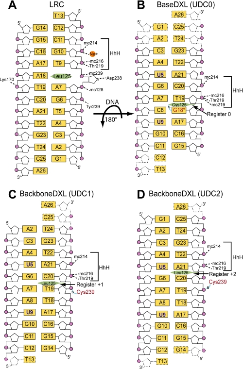
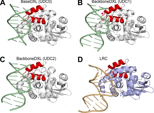
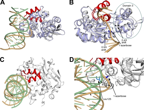
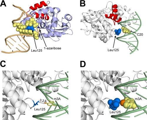
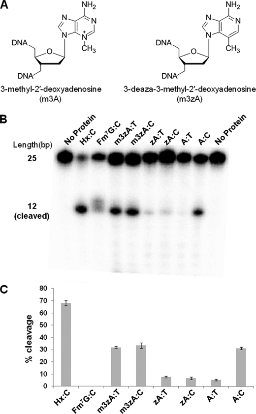
Because DNA damage is so rare, DNA glycosylases interact for the most part with undamaged DNA. Whereas the structural basis for recognition of DNA lesions by glycosylases has been studied extensively, less is known about the nature of the interaction between these proteins and undamaged DNA. Here we report the crystal structures of the DNA glycosylase AlkA in complex with undamaged DNA. The structures revealed a recognition mode in which the DNA is nearly straight, with no amino acid side chains inserted into the duplex, and the target base pair is fully intrahelical. A comparison of the present structures with that of AlkA recognizing an extrahelical lesion revealed conformational changes in both the DNA and protein as the glycosylase transitions from the interrogation of undamaged DNA to catalysis of nucleobase excision. Modeling studies with the cytotoxic lesion 3-methyladenine and accompanying biochemical experiments suggested that AlkA actively interrogates the minor groove of the DNA while probing for the presence of lesions.


Figures

FIGURE 1.
FIGURE 1.
Schematic representation of the AlkA-DNA interactions. The orientation of the DNA in the UDCs are rotated 180° with respect to the LRC. In the UDC structures, residues in the HhH motif are solely responsible for hydrogen bonding with the phosphate (purple circles) backbone with main chain atoms, designated by prefix mc. A, in the LRC structure, the lesion 1-azaribose is depicted as an extrahelical sugar interacting with Asp-238. In B, the 5-bromo-2′-deoxyuridine nucleobase is designated as a blue U, and the modified guanine, designated G18*, is shown in red. In B–D, the two-carbon thiol tether is shown as an S with a blue line through it, the interrogating residue of AlkA is a green hexagon, and the disordered nucleotides are shown as dotted lines.
FIGURE 2.
FIGURE 2.
Structure of the AlkA-DNA undamaged DNA and lesion recognition complexes. A–C, ribbon representation of the UDC structures with AlkA colored white, the DNA colored green, and the HhH colored red. In B, the ordered water molecule is pink. D, ribbon representation of the LRC with the AlkA protein colored light blue, the DNA colored brown, and the HhH colored red.
FIGURE 3.
FIGURE 3.
Cα superposition of the AlkA undamaged DNA complex with the lesion recognition complex. A, structural comparison between the AlkA undamaged DNA and lesion recognition complexes. The color scheme is the same as described for Fig. 2. B, differences in protein conformation between the UDC and LRC structures. The dotted black lines illustrate the important loop and domain movements. The 1-azaribose lesion (blue)-containing DNA strand is shown for reference. Domain 3, which contains the catalytic residue and many key active site residues, is circled by a green dotted line. C, view down the axis of the DNA duplex illustrating the overlap of the phosphate backbone between the DNA of the UDC and LRC structures within the region of the HhH motif. The AlkA monomer from the LRC has been omitted for clarity. D, a zoomed-in view (from A) into the active site of AlkA from the UDC and LRC structures. The dotted black line denotes the 8.7 Å shift in the path of the DNA between the two structures.
FIGURE 4.
FIGURE 4.
Role of Leu-125 in the undamaged DNA and lesion recognition complexes. A, the LRC of AlkA with a space-filling model of Leu-125 (blue) and the nucleobases (yellow) that are in van der Waals contact. B, view of the minor groove of the UDC with Leu-125 and the interrogated base C20 shown as a space-filling model. C, same view as in B, with the base of m3A modeled in place of C20. The N3-methyl group is shown in orange. D, same view as in C, with Leu-125 and the base of m3A shown as a space-filling model, illustrating the van der Waals contact between the Leu-125 side chain and the N3-methyl adduct (orange).
FIGURE 5.
FIGURE 5.
Biochemistry of AlkA lesion cleavage. A, chemical structures of 3-methyl-2′-deoxyadenosine and 3-deaza-3-methyl-2′-deoxyadenosine. B, cleavage assay for AlkA illustrating cleavage of the m3A lesion when paired with thymine but no cleavage of adenine when paired with thymine. There is a faint delocalized band in the Fm7G lane, which represents a breakdown of the lesion base that occurs under basic conditions rather than specific cleavage of the lesion. C, quantification of the cleavage product for each lesion tested in B. Hx, hypoxanthine.

References

    1. Friedberg E. C., Walker G. C., Siede W. (1995) DNA Repair and Mutagenesis, American Society for Microbiology, Washington D. C
    1. Lindahl T. (2004) Nature 427, 598. - PubMed
    1. Engelward B. P., Allan J. M., Dreslin A. J., Kelly J. D., Wu M. M., Gold B., Samson L. D. (1998) J. Biol. Chem. 273, 5412–5418 - PubMed
    1. Jelinsky S. A., Samson L. D. (1999) Proc. Natl. Acad. Sci. U.S.A. 96, 1486–1491 - PMC - PubMed
    1. McCullough A. K., Dodson M. L., Lloyd R. S. (1999) Annu. Rev. Biochem. 68, 255–285 - PubMed

Publication types

MeSH terms

LinkOut - more resources