. 2010 Oct;137(20):3469-76.
doi: 10.1242/dev.055160. Epub 2010 Sep 15.

A prominent requirement for single-minded and the ventral midline in patterning the dorsoventral axis of the crustacean Parhyale hawaiensis

Affiliations

A prominent requirement for single-minded and the ventral midline in patterning the dorsoventral axis of the crustacean Parhyale hawaiensis

Mario A Vargas-Vila et al. Development. 2010 Oct.

Abstract

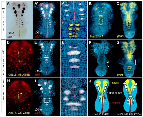
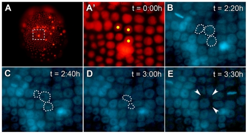
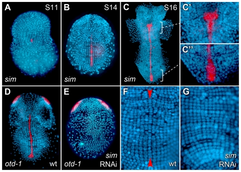
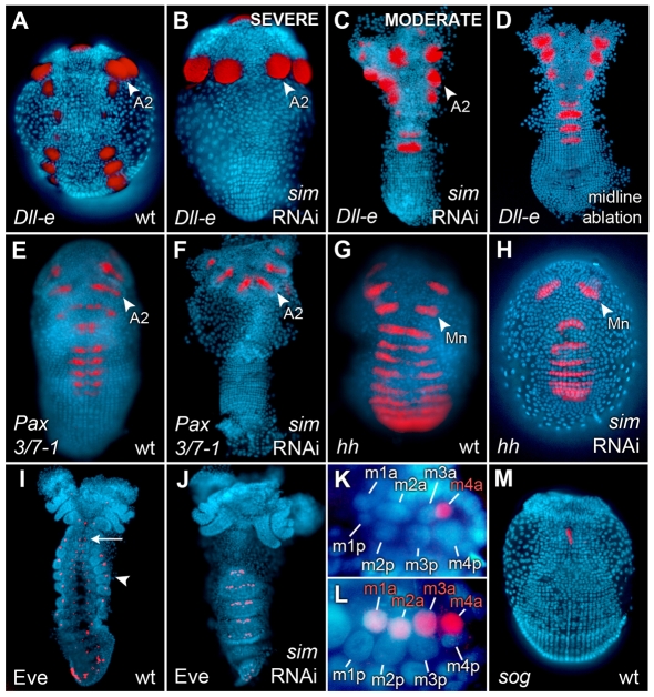
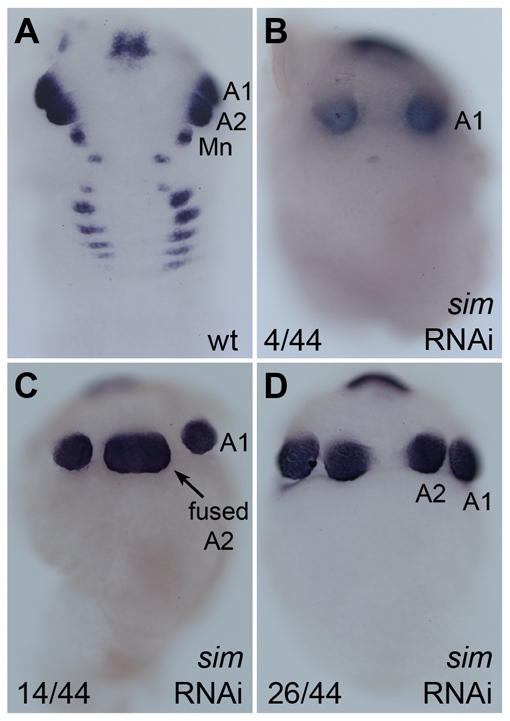
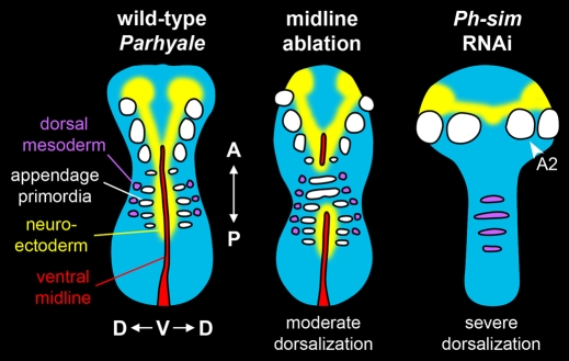
In bilaterians, establishing the correct spatial positioning of structures along the dorsoventral (DV) axis is essential for proper embryonic development. Insects such as Drosophila rely on the Dorsal activity gradient and Bone morphogenetic protein (BMP) signaling to establish cell fates along the DV axis, leading to the distinction between tissues such as mesoderm, neurogenic ectoderm and dorsal ectoderm in the developing embryo. Subsequently, the ventral midline plays a more restricted role in DV patterning by establishing differential cell fates in adjacent regions of the neurogenic ectoderm. In this study, we examine the function of the ventral midline and the midline-associated gene single-minded (Ph-sim) in the amphipod crustacean Parhyale hawaiensis. Remarkably, we found that Ph-sim and the ventral midline play a central role in establishing proper fates along the entire DV axis in this animal; laser ablation of midline cells causes a failure to form neurogenic ectoderm and Ph-sim RNAi results in severely dorsalized embryos lacking both neurogenic ectoderm and the appendage-bearing lateral ectoderm. Furthermore, we hypothesize that this role of midline cells was present in the last common ancestor of crustaceans and insects. We predict that the transition to a Dorsal-dependent DV patterning system in the phylogenetically derived insect lineage leading to Drosophila has led to a more restricted role of the ventral midline in patterning the DV axis of these insects.


Figures

Fig. 1.
Fig. 1.
Laser ablation of midline cells results in DV mispatterning. All images are ventral views with the anterior at the top. Nuclei were counterstained with DAPI (blue) and a false-color overlay of gene expression patterns was generated. All embryos shown (as well as in Figs 3,4) were processed in a similar manner. (A-A′) Wild-type expression of Ph-Dll-e (black or white, as labeled) in appendage primordia and Ph-otd-1 (red) in midline cells at stage 17. (B,B′) Wild-type expression of Ph-Pax3/7-1 (yellow) in a subset of column 0-2-derived cells. (C) Wild-type expression of Ph-pros (yellow) in neuroblasts and ganglion mother cells at stage 18. (D) Living stage 13 embryo visualized by DsRed-NLS fluorescence. Selected midline cells were ablated using a focused laser (yellow dots). (E,E′) At stage 17, the embryo shown in D shows ventrally fused Ph-Dll-e (white) domains in segments lacking midline. Ph-otd-1 is shown in red. (F,G) Two embryos in which midline cells in three consecutive parasegments were ablated at stage 13. By stage 19 (F), ventrally fused limb buds (arrowheads) are visible in segments lacking midline. At stage 18 (G), segments lacking midline show a lack of Ph-pros staining (yellow) in affected segments (bracket, T3-T4). (H) Trunk midline cells were ablated (yellow dots) at stage 13 except for one midline cell in parasegment 4 (arrowhead). (I,I′) The embryo from H is shown at stage 17. Ph-Dll-e (white) is expressed at a reproducible distance from midline cell clone (large red cell cluster). Note that some scattered red signal is visible; this is associated with debris and does not represent Ph-otd-1 hybridization signal. (J) Illustration of DV fates in wild-type and midline-ablated embryos including midline (red), presumptive neurogenic ectoderm (yellow) and presumptive appendages (white).
Fig. 2.
Fig. 2.
Visualization of cell death following laser ablation. (A) Living stage 13 embryo visualized by DsRed-NLS fluorescence. (A′) Magnification of the region indicated in A. Cells marked with yellow dots were targeted for laser ablation. (B-E) Two hours after ablation, the embryo was treated with Hoechst dye and recorded by fluorescent time-lapse videography. Times shown represent total time elapsed (t) since ablation. Ablated cells have dotted outlines. (E) By 3:30 hours after ablation, all three targeted cells were no longer visible by Hoechst fluorescence. The space previously occupied by these cells (arrowheads) was minimized as surrounding cells moved in to fill the gaps.
Fig. 3.
Fig. 3.
Ph-sim is expressed in midline cells and is required for midline differentiation. Nuclei were counterstained with DAPI (blue) and a false-color overlay of gene expression patterns was generated. (A-C) Wild-type expression of Ph-sim. (A) Stage 11. Ph-sim is first visible in midline cells during initial condensation of germ band. (B) Stage 14. Ph-sim is expressed throughout the ventral midline. (C) Ph-sim expression at stage 16. (C′,C′) Magnification of regions indicated by brackets in C. (C′) Ph-sim expression is now visible in the second antennal segment in the expanded medial domain. (C′) Magnification showing Ph-sim expression in posterior midline precursor cells (not confined to a single column of cells). (D) Wild-type expression of Ph-otd-1 in midline cells as well as in two domains in the head. (E) Ph-otd-1 expression in Ph-sim RNAi embryo. Midline staining is absent (in 23 out of 24 embryos), whereas staining in the head is unaffected (24 out of 24). (F) Ectodermal grid in wild-type embryo. The ventral midline (red arrowheads) bisects the embryo and is discernible based on morphology. (G) Ph-sim RNAi embryo. Midline cells and midline precursor cells are no longer visible.
Fig. 4.
Fig. 4.
Ph-sim RNAi causes dorsalization of embryonic trunk. Nuclei were counterstained with DAPI (blue) and a false-color overlay of gene expression patterns was generated. Where appropriate, the second antennal (A2) or mandibular (Mn) segments are labeled. (A) Ph-Dll-e expression in wild-type embryo. (B) Most Ph-sim RNAi embryos (44 out of 49) lack Ph-Dll-e expression posterior to A2. (C) Some Ph-sim RNAi embryos (4 out of 49) show a more moderate phenotype in which one or more trunk segments contain ventrally fused Ph-Dll-e spots. (D) Midline ablated embryo (from Fig. 1E) showing ventrally fused Ph-Dll-e spots, similar to the phenotype shown in C. (E) Wild-type Ph-Pax3/7-1 expression in a subset of presumptive neuroectodermal cells. (F) Ph-sim RNAi embryos lack Ph-Pax3/7-1 expression posterior to A2 (18 out of 18). (G) Wild-type Ph-hh expression in segmentally reiterated stripes. (H) Ph-sim RNAi embryos show relatively normal Ph-hh expression (16 out of 16). Decreased width of Ph-hh stripes is attributed to the dorsalized germ band of these embryos. (I) Wild-type expression of Ph-Eve in a stage 19 embryo. Protein is detected in dorsal mesoderm (arrowhead), developing neurogenic ectoderm (arrow) and posterior ectoderm. (J) In Ph-sim RNAi embryos, mesodermal Ph-Eve is expressed ectopically in ventral mesoderm suggesting embryonic dorsalization. Neurogenic staining is no longer visible as a result of deletion of this tissue. (K) Magnification of the eight mesodermal cells comprising one hemisegment in stage 18 wild-type embryo. Ventral midline is oriented to the left. Ph-Eve is expressed in the m4a cell. (L) Magnification of one hemisegment in Ph-sim RNAi embryo. Ventral midline is oriented to the left. Ph-Eve is now detected in all four anterior mesodermal cells (m1a-m4a). (M) Wild-type Ph-sog expression in a stage 15 embryo. The highest levels of expression are seen in anterior midline cells in the Mn segment. No Ph-sog expression is detected in the majority of trunk midline cells.
Fig. 5.
Fig. 5.
Head morphology variations in sim RNAi embryos. Where appropriate, the first and second antennal (A1, A2) and mandibular (Mn) segments are labeled (A) Ph-Dll-e expression in wild-type embryo. (B) In 4 out of 44 embryos showing a severe DV phenotype, the A2 appendages are completely missing. (C) In 14 out of 44 embryos, DV patterning in A2 is moderately affected leading to a single, ventrally fused appendage bud (arrow). (D) In 26 out of 44 embryos, DV patterning in the A2 segment is minimally affected and appendage buds appear distinctly separate (same embryo as shown in Fig. 4B).
Fig. 6.
Fig. 6.
Dorsalization phenotypes. Wild-type, midline-ablated, and Ph-sim RNAi embryos are depicted. In midline-ablated embryos, segments lacking midline failed to generate neuroectoderm and ventral cells were mis-specified as lateral ectoderm (bearing appendage primordia). The location of dorsal mesoderm in midline-ablated embryos was not examined, but is inferred from ectodermal markers. In Ph-sim RNAi embryos, the ventral midline, neuroectoderm and appendage primordia were absent posterior to the second antennal segment (A2). Dorsalization was confirmed by the presence of dorsal mesoderm in ventral regions of embryos.

References

    1. Akiyama-Oda Y., Oda H. (2006). Axis specification in the spider embryo: dpp is required for radial-to-axial symmetry transformation and sog for ventral patterning. Development 133, 2347-2357 - PubMed
    1. Browne W. E., Price A. L., Gerberding M., Patel N. H. (2005). Stages of embryonic development in the amphipod crustacean, Parhyale hawaiensis. Genesis 42, 124-149 - PubMed
    1. Browne W. E., Schmid B. G., Wimmer E. A., Martindale M. Q. (2006). Expression of otd orthologs in the amphipod crustacean, Parhyale hawaiensis. Dev. Genes Evol. 216, 581-595 - PubMed
    1. Brusca R. C., Brusca G. J. (2003). Invertebrates. Sunderland, MA: Sinauer Associates;
    1. Chang J., Kim I. O., Ahn J. S., Kwon J. S., Jeon S. H., Kim S. H. (2000). The CNS midline cells coordinate proper cell cycle progression and identity determination of the Drosophila ventral neuroectoderm. Dev. Biol. 227, 307-323 - PubMed

Publication types

Substances