. 2010 Nov;151(11):5537-49.
doi: 10.1210/en.2010-0424. Epub 2010 Sep 15.

Neutral antibodies to the TSH receptor are present in Graves' disease and regulate selective signaling cascades

Affiliations

Neutral antibodies to the TSH receptor are present in Graves' disease and regulate selective signaling cascades

Syed A Morshed et al. Endocrinology. 2010 Nov.

Abstract

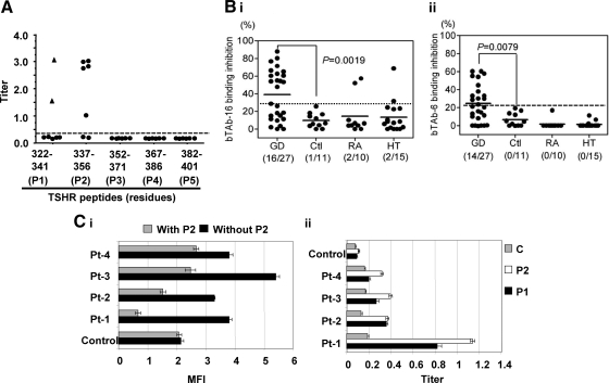
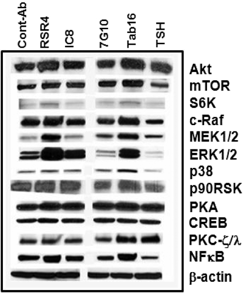
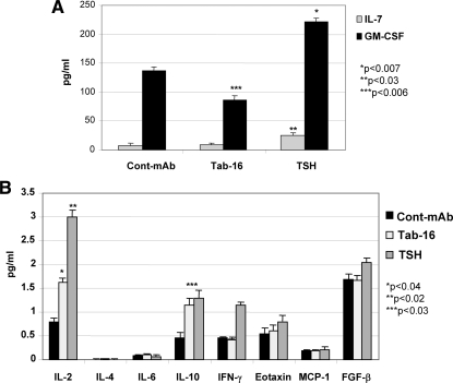
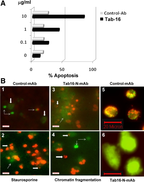
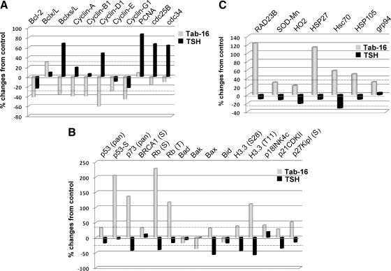
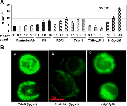
TSH receptor (TSHR) antibodies (Abs) may be stimulating, blocking, or neutral in their functional influences and are found in patients with autoimmune thyroid disease, especially Graves' disease (GD). Stimulators are known to activate the thyroid epithelial cells via both Gs- and Gq-coupled signaling pathways, whereas blockers inhibit the action of TSH and may act as weak agonists. However, TSHR neutral Abs do not block TSH binding and are unable to induce cAMP via Gsα. The importance of such neutral Abs in GD remains unclear because their functional consequence has been assumed to be zero. We hypothesized that: 1) neutral TSHR Abs are more common to GD than generally recognized; 2) they may induce distinct signaling imprints at the TSHR not seen with TSH itself; and 3) these signaling events may alter cellular function. To evaluate these hypotheses, we first confirmed the presence of neutral TSHR Abs in sera from patients with GD and then, using mouse and hamster neutral TSHR monoclonal Abs (N-mAbs) performed detailed signaling studies, including a proteomic Ab array, with rat thyrocytes (FRTL-5) as targets. This allowed us to examine a battery of signaling cascades and their downstream effectors. Neutral TSHR Abs were indeed frequently present in sera from patients with GD. Sixteen of 27 patients (59%) had detectable neutral TSHR Abs by competition assay with N-mAbs. On examining signaling cascades, we found that N-mAbs induced signal transduction, primarily via the protein kinase A II cascade. In addition to the activation of phosphatidylinositol 3K/Akt, N-mAbs, unlike TSH, had the ability to exclusively activate the mammalian target of rapamycin/p70 S6K, nuclear factor-κB, and MAPK-ERK1/2/p38α signaling cascades and their downstream effectors p90 ribosomal kinase/MAPK-interacting kinase-1/mitogen and stress-activated kinase-1 and N-mAbs activated all forms of protein kinase C isozymes. To define the downstream effector mechanisms produced by these signaling cascades, cytokine production, proliferation, and apoptosis in thyrocytes were investigated. Although N-mAbs produced less cytokines and proliferation compared with TSH, they had the distinction of inducing thyroid cell apoptosis under the experimental conditions used. When dissecting out possible mechanisms of apoptosis, we found that activation of multiple oxidative stress markers was the primary mechanism orchestrating the death signals. Therefore, using oxidative stress-induced apoptosis, N-mAbs may be capable of exacerbating the autoimmune response in GD via apoptotic cells inducing antigen-driven mechanisms. This may help explain the inflammatory nature of this common disorder.


Figures

Figure 1
Figure 1
Hamster mAbs recognized TSHR linear epitopes. A, Titers of mAbs indicated that the major epitopes were directed against the peptide sequence of 322–341 (P1) and 337–356 (P2) amino acid residues by a competitive inhibition ELISA with specific mAbs against the epitopes (Tab-16 and Tab-6, respectively). B, These two epitopes were also recognized by human IgGs from patients with GD. The frequency of these Abs was significantly higher in Grave’s patients compared with IgG from healthy individuals (P < 0.008). Ctl, Control; RA, rheumatoid arthritis; HT: Hashimoto’s thyroiditis. C, Flow cytometry recognized Abs against the human TSHR in CHO cells (i) and the indirect peptide ELISA (ii) also confirmed the presence of serum IgGs against peptides P1 and P2. A TSHR unrelated control peptide (C) failed to produce any significant binding (ii). Flow cytometry indicated that the binding of Abs was apparently inhibited by peptide (P2) as deduced by the reduction in binding of individual patient’s IgGs when incubated with the specific peptide (i). Inhibition ELISA and indirect ELISA were performed in duplicate and repeated twice. Similarly flow cytometry was repeated twice with different IgG preparations. MFI, Mean fluorescent intensity.
Figure 2
Figure 2
Representative immunoblots of signaling molecules. Quiescent, starved cells were stimulated with a fixed concentration (1.0 μg/ml) of different N-mAbs and TSH (1.0 mU/ml) over 60 min of incubation. They showed variable changes in signaling molecules as shown in these immunoblots. RSR4, with an epitope similar to Tab-16 (P1), induced a robust effect on most of the signaling molecules with increased activities of Akt/mTOR/S6K, c-Raf/MEK1/2/p38/p90RSK, PKA/CREB, PKC-ζ/λ, and NF-κB. 7G10, which recognized a different epitope (P2), did not induce such effects, whereas TSH ligand activated Akt but not mTOR/S6K. TSH also activated PKC-ζ/λ and most importantly it suppressed c-Raf/MEK1/2/p38/p90RSK signaling cascade. By contrast, isotype control mAb repeatedly did not produce any significant activity. The experiments were repeated twice and confirmed the illustrated findings.
Figure 3
Figure 3
Multiplex cytokines in the cell lysates. A, Tab-16 neutral mAb attenuated GM-CSF induction significantly compared with control Ab, whereas TSH induced both IL-7 and GM-CSF significantly higher than control mAb (Cont-mAb). B, Both Tab-16 and TSH induced IL-2 and IL-10 significantly higher than control. There was no other significant induction of cytokines either by Tab-16 or TSH. The assay was performed twice in duplicate. IFN, Interferon; MCP, monocyte chemotactic protein; FGF, fibroblast growth factor.
Figure 4
Figure 4
Annexin V binding to apoptotic cells by flow cytometry and microscopy. A, Increasing concentrations of Tab-16, but not the control Ab, induced apoptosis in thyrocytes. Cells were exposed to Tab-16 (1 μg/ml) and induced apoptosis that was stained on d 5 for flow cytometry. Four mAbs to similar epitope were used twice to ensure reproducibility. B, Apoptosis was also confirmed by microscopic observation of annexin V-positive cells (thin arrow) and propium iodide (thick arrow) staining of nucleus in the treated cells. Staurosporine, a known PKC inhibitor that induces apoptosis at 10 μm, was a positive control. Both the Tab-16 and staurosporine induced nuclear DNA fragmentation (thick arrow) as shown in B-2, B-3, and B-4. By contrast, control Ab induced significantly less effect (B-1). JC-1 staining (a mitochondrial dye or mitotracker) in live cells showed green fluorescence, as another indicator of apoptosis (B-6), whereas untreated cells exhibited both green and red fluorescence (B-5), indicating preserved mitochondrial function.
Figure 5
Figure 5
Antiapoptotic and stress-induced proapoptotic signaling molecules by protein array. A, TSH but not N-mAb activated multiple antiapoptotic proteins including Bclx/L, Bclxs/L, cyclins (A, B1, and D1), PCNA, and cell cycle-related proteins. Both TSH and Tab-16 failed to activate Bcl-2. B, N-mAb activated stress-induced DNA damage proteins including p53, p73, breast cancer type 1 susceptibility protein (BRCA1), and retinoblastoma (Rb). Tab-16 also activated many proapoptotic proteins, importantly Bcl-2-associated X protein (Bax) and cyclin-dependent kinase inhibitors. TSH did the opposite. C, N-mAb activated DNA stress (Rad23B), mitochondrial stress (SOD-Mn), hemeoxygenase (HO2), heat-shock p70-related protein (Hsc70), and stress-induced endoplasmic reticulum protein (glucose regulated protein 94). The TSH effect was minor.
Figure 6
Figure 6
Oxidative stress or ROS in cells detected by spectrofluorometry. A, N-mAbs and H2O2 activated ROS, whereas control Ab and medium alone did not. ROS was observed by relative fluorescent units (RFU) stained with H2DCF-DA. In contrast, TSH reduced ROS activity dose dependently. The highest activity of ROS was observed at the highest concentration of the N-mAbs (10 μg/ml) and H2O2 (40 μm). B, FRTL-5 cells were also stained with H2R123 (a ROS indicator) and observed under the microscope. Tab-16 (a) and H2O2 (c) activated ROS as determined by a clear increase in green fluorescent intensity, whereas such change was not seen with control mAb (b). Both spectrofluorometry and microscopy were repeated twice in duplicate.

References

    1. Kimura T, Van Keymeulen A, Golstein J, Fusco A, Dumont JE, Roger PP 2001 Regulation of thyroid cell proliferation by TSH and other factors: a critical evaluation of in vitro models. Endocr Rev 22:631–656 - PubMed
    1. Medina DL, Santisteban P 2000 Thyrotropin-dependent proliferation of in vitro rat thyroid cell systems. Eur J Endocrinol 143:161–178 - PubMed
    1. Ando T, Latif R, Pritsker A, Moran T, Nagayama Y, Davies TF 2002 A monoclonal thyroid-stimulating antibody. J Clin Invest 110: 1667–1674 - PMC - PubMed
    1. Ando T, Latif R, Daniel S, Eguchi K, Davies TF 2004 Dissecting linear and conformational epitopes on the native thyrotropin receptor. Endocrinology 145:5185–5193 - PubMed
    1. Ando T, Latif R, Davies TF 2004 Concentration-dependent regulation of thyrotropin receptor function by thyroid-stimulating antibody. J Clin Invest 113:1589–1595 - PMC - PubMed

Publication types

MeSH terms