Review
. 2010 Sep 15;24(18):2001-12.
doi: 10.1101/gad.1951710.

Interactions between Hedgehog proteins and their binding partners come into view

Affiliations
Review

Interactions between Hedgehog proteins and their binding partners come into view

Philip A Beachy et al. Genes Dev. .

Abstract

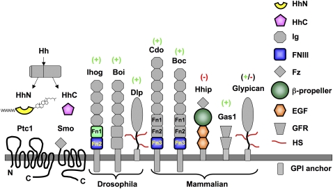
Hedgehog (Hh) proteins are secreted signaling molecules that mediate essential tissue-patterning events during embryonic development and function in tissue homeostasis and regeneration throughout life. Hh signaling is regulated by multiple mechanisms, including covalent lipid modification of the Hh protein and interactions with multiple protein and glycan partners. Unraveling the nature and effects of these interactions has proven challenging, but recent structural and biophysical studies of Hh proteins and active fragments of heparin, Ihog, Cdo, Boc, Hedgehog-interacting protein (Hhip), Patched (Ptc), and the monoclonal antibody 5E1 have added a new level of molecular detail to our understanding of how Hh signal response and distribution are regulated within tissues. We review these results and discuss their implications for understanding Hh signaling in normal and disease states.


Figures

Figure 1.
Figure 1.
Domain organization of proteins in the Hh signaling pathway. Domains for which high-resolution structures have been determined are shown in color, and those that are positive or negative regulators of Hh pathway activity are indicated by (+) or (−), respectively. (Ig) Immunoglobulin; (Fz) Frizzled-like cysteine-rich domain; (EGF) epidermal growth factor; (GFR) glial-derived neurotrophic factor family receptor; (HS) heparan sulfate; (GPI) glycosylphosphatidylinositol.
Figure 2.
Figure 2.
ShhN and HhC. (A) Ribbon diagrams of the N-terminal signaling domain of Sonic hedgehog is shown with zinc-coordinating residues colored in light blue, calcium-coordinating residues colored red, calcium ions colored green, and the zinc ion colored cyan. The view in the right panel is rotated ∼80° about a vertical axis and ∼30° about a horizontal axis relative to the view shown in the left panel. The side chain of the zinc-coordinating aspartate (D148) has been removed in the right panel to show the zinc-binding cleft more clearly. (B) Near-orthogonal views of a superposition of α carbon traces of the core structural elements of ShhN (yellow) and D-Ala-D-Ala-carboxypeptidase (green) are shown. The side chains of zinc-coordinating residues are shown as sticks, and the zinc ions are shown as cyan spheres. (C) Ribbon diagrams of the HhC (left) and Mtu intein (right) (Hiraga et al. 2009) structures are shown. The ribbons are colored in a rainbow color gradient from blue (N terminus) to red (C terminus). All structure figures were made using PyMol (http://www.pymol.org).
Figure 3.
Figure 3.
HhN:IhogFn12 complex. (A) A ribbon diagram of the 2:2 HhN:IhogFn12 heterotetramer is shown. HhN is colored yellow, the first FNIII domain of IhogFn12 is colored green, and the second FNIII domain of IhogFn2 is colored blue. (B, top) A single HhN:IhogFn12 complex is shown with HhN oriented similarly to the orientation of ShhN in the left panel of Figures 2A and 8. (Bottom) The electrostatic surface potential of the HhN:IhogFn12 complex in the same orientation as in the top panel is shown. A basic region that spans the HhN:IhogFn12 interface is outlined in yellow (portion contributed by HhN) and green (portion contributed by IhogFn12). Blue represents regions of positive charge, and red represents regions of negative charge. The scale is ±10 kT/e.
Figure 4.
Figure 4.
ShhN:CdoFn3. (A) Ribbon diagram of the ShhN:CdoFn3 complex. ShhN is shown in yellow, and CdoFn3 is shown in blue. Calcium ions are shown as green spheres, and the zinc ion is shown as a cyan sphere. (B) A closeup view of the calcium-coordinating region of murine ShhN is shown. The side chain of D88, which is homologous to D89 in human ShhN and the site of a holoprosencephaly-causing mutation in human ShhN, is colored purple. The side chains of E95, D100, and E131, which are homologous to sites of BDA1-causing mutations in human IhhN, are colored cyan, and the calcium ions are shown as green spheres.
Figure 5.
Figure 5.
Different HhN-binding modes of Ihog and Cdo. Ribbon diagrams of the ShhN:CdoFn3 and HhN:IhogFn12 complexes are shown following superposition of ShhN and HhN. ShhN is colored yellow, HhN is red, CdoFn3 is blue, and IhogFn12 is green/light blue. The ShhN-bound calcium and zinc ions are shown as green and cyan spheres, respectively.
Figure 6.
Figure 6.
ShhN:Hhip. (A) Ribbon diagrams of ShhN (yellow) and HhipΔN (the β-propeller region is colored dark green, the first EGF repeat is shown in orange, and the second EGF repeat is colored brown) are shown in orientations parallel and perpendicular to the plane of the β-propeller. The side chain of D383 is colored red, calcium ions are colored green, and the zinc ion is colored cyan. In the bottom panel, the EGF repeats have been removed for clarity. (B) Ribbon diagrams of ShhN (yellow) in orientations similar to those shown in Figure 2A are shown with the blade 3 loop of Hhip (dark green). Zinc-coordinating side chains are colored blue, the Hhip D383 side chain is colored red, the zinc ion is shown as a cyan sphere, and the calcium ions are shown as green spheres. The calcium ions have been removed from the right panel for clarity.
Figure 7.
Figure 7.
ShhN:5E1 Fab. A ribbon diagram of ShhN (yellow) complexed with the Fab fragment of the 5E1 antibody (red) is shown. Calcium and zinc ions are colored green and cyan, respectively.
Figure 8.
Figure 8.
HhN-binding surfaces. Molecular surfaces of ShhN or dHhN are shown with atoms within 4 Å of the indicated binding partner colored blue (CdoFn3), light green (IhogFn12), dark green (Hhip), and red (5E1 Fab). The orientation of each surface is the same as that of the ribbon diagram of ShhN in the top left, in which calcium and zinc ions are shown as green and cyan spheres, respectively, with the exception of the side view of the IhogFn12-binding surface (top right), which is rotated 90° about a vertical axis relative to the ShhN ribbon. When present, zinc ions are shown as cyan spheres. The calcium ions bound to ShhN are buried and are not visible in surface representations, but their position can be inferred by reference to the ribbon diagram of ShhN (which is at top left).

Similar articles

Cited by

References

    1. Alcedo J, Ayzenzon M, Von Ohlen T, Noll M, Hooper JE 1996. The Drosophila smoothened gene encodes a seven-pass membrane protein, a putative receptor for the hedgehog signal. Cell 86: 221–232 - PubMed
    1. Allen BL, Tenzen T, McMahon AP 2007. The Hedgehog-binding proteins Gas1 and Cdo cooperate to positively regulate Shh signaling during mouse development. Genes Dev 21: 1244–1257 - PMC - PubMed
    1. Amanai K, Jiang J 2001. Distinct roles of Central missing and Dispatched in sending the Hedgehog signal. Development 128: 5119–5127 - PubMed
    1. Beckett K, Franch-Marro X, Vincent JP 2008. Glypican-mediated endocytosis of Hedgehog has opposite effects in flies and mice. Trends Cell Biol 18: 360–363 - PubMed
    1. Bishop B, Aricescu AR, Harlos K, O'Callaghan CA, Jones EY, Siebold C 2009. Structural insights into hedgehog ligand sequestration by the human hedgehog-interacting protein HHIP. Nat Struct Mol Biol 16: 698–703 - PMC - PubMed

Publication types

LinkOut - more resources