. 2010 Sep 15;24(18):2068-80.
doi: 10.1101/gad.1963210.

Ephrin-B1 forward signaling regulates craniofacial morphogenesis by controlling cell proliferation across Eph-ephrin boundaries

Affiliations

Ephrin-B1 forward signaling regulates craniofacial morphogenesis by controlling cell proliferation across Eph-ephrin boundaries

Jeffrey O Bush et al. Genes Dev. .

Abstract

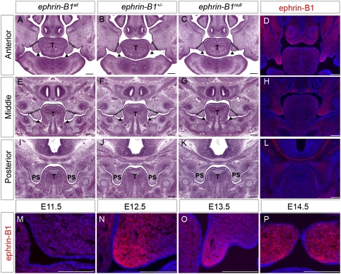
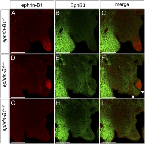
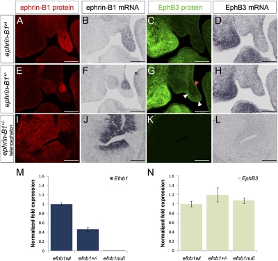
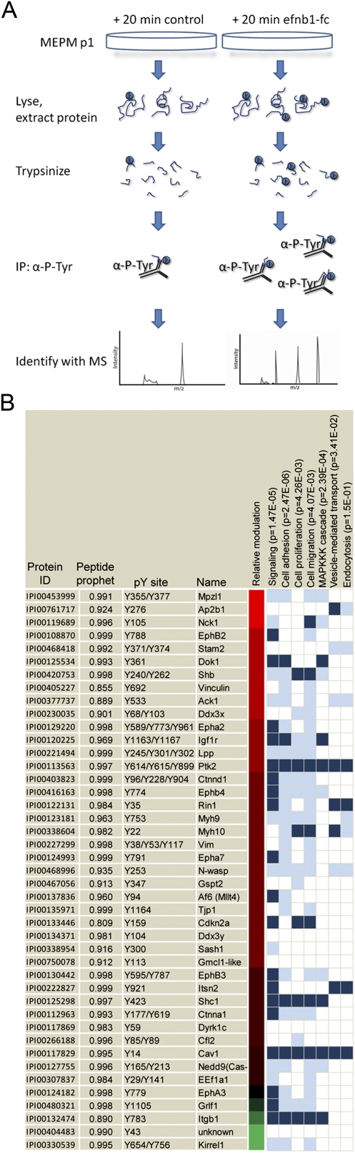
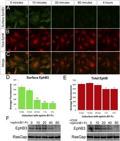
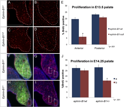
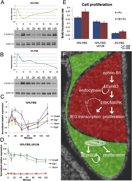
Mutations in the X-linked human EPHRIN-B1 gene result in cleft palate and other craniofacial anomalies as part of craniofrontonasal syndrome (CFNS), but the molecular and developmental mechanisms by which ephrin-B1 controls the underlying developmental processes are not clear. Here we demonstrate that ephrin-B1 plays an intrinsic role in palatal shelf outgrowth in the mouse by regulating cell proliferation in the anterior palatal shelf mesenchyme. In ephrin-B1 heterozygous mutants, X inactivation generates ephrin-B1-expressing and -nonexpressing cells that sort out, resulting in mosaic ephrin-B1 expression. We now show that this process leads to mosaic disruption of cell proliferation and post-transcriptional up-regulation of EphB receptor expression through relief of endocytosis and degradation. The alteration in proliferation rates resulting from ectopic Eph-ephrin expression boundaries correlates with the more severe dysmorphogenesis of ephrin-B1(+/-) heterozygotes that is a hallmark of CFNS. Finally, by integrating phosphoproteomic and transcriptomic approaches, we show that ephrin-B1 controls proliferation in the palate by regulating the extracellular signal-regulated kinase/mitogen-activated protein kinase (ERK/MAPK) signal transduction pathway.


Figures

Figure 1.
Figure 1.
Ephrin-B1 is required for anterior palatal shelf morphogenesis. (A–C,E–G,I–K) Histological frontal sections of E13.5 embryonic heads reveal reduced anterior palatal shelf outgrowth in ephrin-B1+/− (arrowheads in B) and ephrin-B1null (arrowheads in C) compared with the finger-like projection in ephrin-B1wt (arrowheads in A). The palatal shelves appear slightly affected in middle positions of ephrin-B1+/− (arrowheads in F) and ephrin-B1null (arrowheads in G) mutant embryos compared with ephrin-B1wt at the same position (arrowheads in E). The posterior palate is unaffected in ephrin-B1+/− (J) and ephrin-B1null (K) mutant embryos. (D,H,L,M–P) Immunofluorescence detection of ephrin-B1 expression in ephrin-B1wt embryos. At E13.5, Ephrin-B1 expression is restricted to the anterior palatal shelf mesenchyme (D), is more weakly expressed in the middle (H), and is not detected in the posterior palate (L). Ephrin-B1 is not significantly detectable in the palatal shelf primordia at E11.5 (M), but becomes strongly detectable within the palatal mesenchyme by E12.5 (N). Ephrin-B1 expression remains highly restricted to the mesenchyme during outgrowth (E13.5) (O) and after elevation (E14.5) (P) of the palatal shelves. Bar, 200 μm. (T) Tongue; (PS) palatal shelves.
Figure 2.
Figure 2.
EphB3 protein is up-regulated in ephrin-B1 mutant domains in the palate. (A–I) Immunofluorescence detection of ephrin-B1 (red) and EphB3 (green) in frontal sections of E13.5 anterior palatal shelves. (B,C) EphB3 protein expression is not detectable in the palatal shelves of ephrin-B1wt E13.5 embryos. In ephrin-B1+/− mutant embryos (D–F), the expression of ephrin-B1 is mosaic (D,F) and EphB3 protein is up-regulated in domains lacking ephrin-B1 expression within the palatal shelf mesenchyme (E,F). (F) In addition, a boundary one to two cells in width separates the ephrin-B1-expressing and EphB3-expressing domains (arrowheads). (H,I) EphB3 protein is up-regulated throughout the anterior palatal shelf mesenchyme of ephrin-B1null mutant embryos. Bar, 200 μm.
Figure 3.
Figure 3.
EphB3 is post-transcriptionally up-regulated in ephrin-B1 mutant palates. Adjacent frontal sections of E13.5 anterior palatal shelves analyzed by antibody staining for ephrin-B1 (red) (A,E,I), in situ hybridization for ephrin-B1 (B,F,J), antibody staining for EphB3 (green) (C,G,K), and in situ hybridization for EphB3 (D,H,L). In ephrin-B1wt embryos (A–D), ephrin-B1 protein (A) and mRNA (B) are detected throughout the palatal shelf mesenchyme. Although EphB3 protein is not detectable (C), EphB3 mRNA is expressed in the ephrin-B1wt palatal shelf (D). In ephrin-B1+/− embryos (E–H), ephrin-B1 protein (E) and mRNA (F) are expressed in an identical mosaic pattern. Whereas EphB3 protein is up-regulated specifically in ephrin-B1 mutant domains (white arrowheads in G), EphB3 mRNA does not display this complementary pattern of regulation, and instead maintains the wild-type expression pattern (H). In E13.5 ephrin-B1+/− telencephalon (I–L), mosaic domains of ephrin-B1 protein (I) and mRNA (J) but no complementary up-regulation of EphB3 protein are observed (K). (L) EphB3 mRNA is not detectable in the telencephalon at this stage. (M) QRT–PCR analysis of dissected palates indicates that ephrin-B1 is down-regulated in ephrin-B1+/− and ephrin-B1null palatal shelves relative to ephrin-B1wt (relative mean normalized to GAPDH ± SD). (N) EphB3 mRNA expression levels are not significantly different between ephrin-B1wt, ephrin-B1+/−, and ephrin-B1null mutant palatal shelves. Bar, 200 μm.
Figure 4.
Figure 4.
Phosphoproteomic identification of phosphorylation targets of ephrin-B1 forward signaling in palate cells. (A) Schematic representation of strategy for the identification of ephrin-B1 forward signaling targets. Induction of forward signaling was performed for 20 min with 2 μg/mL preclustered ephrin-B1-Fc. Briefly, cells were lysed and protein was extracted, followed by digestion into tryptic peptides. Immunoprecipitation with an antibody against phosphorylated tyrosine was followed by elution of peptides and LC-MS/MS to identify differentially phosphorylated targets. (B) Proteins identified as phosphorylated are listed by name and protein ID. The site(s) of phosphorylation and peptide prophet score (see the Materials and Methods) are listed. Chromatic representation of the ratio of the relative number of spectral counts (induced to uninduced) was estimated based on the log10 ratio of 1 + (induced peptides to uninduced); complete spectral counts are found in Supplemental Table 1. GO annotations are listed in vertical columns, along with associated P-values. Dark blue represents official assignment to a GO category, and light blue represents manual assignments based on literature searching.
Figure 5.
Figure 5.
EphB3 is internalized and degraded in response to activation of forward signaling in palatal cell cultures. (A,C) Immunostaining of MEP cells with an antibody recognizing the extracellular domain of EphB3 (green), without cell permeabilization to detect cell surface EphB3. (D) Confocal quantification indicates EphB3 is removed from the cell surface over a time course of ephrin-B1 activation of forward signaling (shown in B,C,E) (mean ± SEM; [**] P < 0.0001, Student's t-test). Total EphB staining (red) diminished only slightly (mean ± SEM; [**] P < 0.02, Student's t-test). (F) Immunoprecipitation of EphB3 from MEP cell lysate followed by Western blotting indicates that total EphB3 protein is reduced over a time course of forward signaling activation. Induction of forward signaling in the presence of 10 μg/mL cycloheximide indicates that EphB3 protein reduction is not a consequence of an effect on translation rate.
Figure 6.
Figure 6.
Cell proliferation rate is disrupted in ephrin-B1 mutant palates. (A–D) Frontal sections of E13.5 palatal shelves showing BrdU incorporation (red). A significant reduction in BrdU incorporation was detected in ephrin-B1null (C,E) anterior palatal shelf mesenchyme compared with ephrin-B1wt (A,E) (mean ± SEM; [*] P < 0.001, Student's t-test). No significant reduction was observed in the posterior palatal shelf mesenchyme of ephrin-B1null embryos (D,E) compared with ephrin-B1wt at this position (B,E). (F–J) Frontal sections of E14.25 palatal shelves showing double staining for BrdU incorporation (red), ephrin-B1 (green), and nuclei (DAPI, blue). Ephrin-B1-expressing (box a) and Ephrin-B1-negative (box b) regions were chosen in frontal sections of ephrin-B1+/− palates. (F,G) These regions were matched in ephrin-B1wt for control. (H,I,J) The proliferation rate was significantly reduced in ephrin-B1-negative domains compared with neighboring positive domains (n = 46 regions, 30 sections, mean ± SEM; [*] P < 0.001, Student's t-test). (F,G,J) The proliferation rate in comparable areas of ephrin-B1wt palatal shelves indicated that this was not a consequence of regional differences in the proliferation rate (n = 22 regions, 12 sections, mean ± SEM).
Figure 7.
Figure 7.
Ephrin-B1 forward signaling activates ERK/MAPK signaling in palatal cells. (A,B) Phosphorylated ERK1/ERK2 were detected by Western blot and quantified using ImageJ. (A) Time course of induction with 2 μg/mL preclustered ephrin-B1-Fc in media containing 10% FBS resulted in two phases of ERK1/ERK2 activation. (B) In starvation conditions (media containing 0.2% FBS), an activation of ERK1/ERK2 was observed at 2 min, followed by decay. (C,D) Real-time QRT–PCR quantification of immediate early transcriptional targets of ephrin-B1 forward signaling in MEP cells. (C) Regulation of expression of immediate early targets shadows the activation of the ERK/MAPK signaling. QRT–PCR over a time course of forward signaling induction shows initial up-regulation, down-regulation by 1 h, and increase in expression by 2 h (relative mean normalized to GAPDH ± SD) (D) Inhibition of Ras–MAPK activation eliminates the induction of IEG transcriptional targets. Real-time QRT–PCR over a time course of forward signaling induction in the presence of 10 μM U0126 (relative mean normalized to GAPDH ± SD). (E) BrdU incorporation after 3 h of treatment with 2 μg/mL preclustered ephrin-B1-Fc indicates that cell proliferation is induced by ephrin-B1 forward signaling in media containing 10% FBS (n = 9, mean ± SEM; P < 0.008) or 0.2% FBS (n = 9, mean ± SEM; P < 0.006); this effect is eliminated when Ras–MAPK signaling is blocked by treatment with 10 μM U0126 (n = 9, mean ± SEM). (F) Model of ephrin-B1 regulation of palatal morphogenesis. Ephrin-B1+/− heterozygous females constitute mosaic expression of ephrin-B1 (red). Ephrin-B1 activates forward signaling, resulting in endocytosis and degradation of the EphB receptor. In ephrin-B1 mutant domains, relief of this feedback allows receptor up-regulation (green); several candidate mediators of this feedback were identified as phosphorylation targets of forward signaling (Rin1, Stam2, and Ap2β1). Activation of this forward signal also leads to the phosphorylation of several signaling adaptors (Shc1, Shb, and Dok1) that are capable of regulating ERK/MAPK. The transcriptional regulation of IEGs by activation of the ERK/MAPK may also play a role in feedback regulation of the pathway. Cell proliferation, which is critical for the outgrowth of the palatal shelves, is also dependent on signaling through ERK/MAPK.

References

    1. Bentires-Alj M, Kontaridis MI, Neel BG 2006. Stops along the RAS pathway in human genetic disease. Nat Med 12: 283–285 - PubMed
    1. Braybrook C, Lisgo S, Doudney K, Henderson D, Marcano AC, Strachan T, Patton MA, Villard L, Moore GE, Stanier P, et al. 2002. Craniofacial expression of human and murine TBX22 correlates with the cleft palate and ankyloglossia phenotype observed in CPX patients. Hum Mol Genet 11: 2793–2804 - PubMed
    1. Bush JO, Soriano P 2009. Ephrin-B1 regulates axon guidance by reverse signaling through a PDZ-dependent mechanism. Genes Dev 23: 1586–1599 - PMC - PubMed
    1. Bush JO, Lan Y, Maltby KM, Jiang R 2002. Isolation and developmental expression analysis of Tbx22, the mouse homolog of the human X-linked cleft palate gene. Dev Dyn 225: 322–326 - PubMed
    1. Carbon S, Ireland A, Mungall CJ, Shu S, Marshall B, Lewis S 2009. AmiGO: Online access to ontology and annotation data. Bioinformatics 25: 288–289 - PMC - PubMed

Publication types

MeSH terms

Associated data