. 2010 Dec;84(23):12274-84.
doi: 10.1128/JVI.00260-10. Epub 2010 Sep 15.

Respiratory syncytial virus induces host RNA stress granules to facilitate viral replication

Affiliations

Respiratory syncytial virus induces host RNA stress granules to facilitate viral replication

Michael E Lindquist et al. J Virol. 2010 Dec.

Abstract

Mammalian cell cytoplasmic RNA stress granules are induced during various conditions of stress and are strongly associated with regulation of host mRNA translation. Several viruses induce stress granules during the course of infection, but the exact function of these structures during virus replication is not well understood. In this study, we showed that respiratory syncytial virus (RSV) induced host stress granules in epithelial cells during the course of infection. We also showed that stress granules are distinct from cytoplasmic viral inclusion bodies and that the RNA binding protein HuR, normally found in stress granules, also localized to viral inclusion bodies during infection. Interestingly, we demonstrated that infected cells containing stress granules also contained more RSV protein than infected cells that did not form inclusion bodies. To address the role of stress granule formation in RSV infection, we generated a stable epithelial cell line with reduced expression of the Ras-GAP SH3 domain-binding protein (G3BP) that displayed an inhibited stress granule response. Surprisingly, RSV replication was impaired in these cells compared to its replication in cells with intact G3BP expression. In contrast, knockdown of HuR by RNA interference did not affect stress granule formation or RSV replication. Finally, using RNA probes specific for RSV genomic RNA, we found that viral RNA predominantly localized to viral inclusion bodies but a small percentage also interacted with stress granules during infection. These results suggest that RSV induces a host stress granule response and preferentially replicates in host cells that have committed to a stress response.


Figures
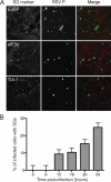
FIG. 1.
FIG. 1.
Stress granules (SGs) are induced during RSV infection. (A) HEp-2 cells were infected with RSV (MOI = 1.0) for 24 h, fixed, and processed for immunofluorescence. Anti-G3BP, anti-eIF3η, and anti-TIA-1 were used as stress granule markers and appear red in the merged image. Anti-RSV P was used as the viral inclusion body marker and appears green in the merged image. The collapsed z-sections are shown for each image. (B) HEp-2 cells were infected with RSV (MOI = 1.0) for the indicated times. The percentages of infected cells and cells containing stress granules per HPF were quantified as described in Materials and Methods. Error bars show standard deviations.
FIG. 2.
FIG. 2.
RSV-induced stress granule formation is dependent on virus replication. HEp-2 cells mock infected (top row), inoculated with replication-competent RSV (MOI = 1.0), or inoculated with UV-inactivated RSV for 48 h were fixed and processed for immunofluorescence. Anti-G3BP was used as a marker for stress granules and appears red in the merged image. Anti-RSV F was used as a marker for viral infection and appears green in the merged image. The collapsed z-sections are shown for each image.
FIG. 3.
FIG. 3.
RSV protein levels are higher in cells with stress granules. HEp-2 cells were infected with RSV (MOI = 1.0) for 24 h. At least 10 infected cells containing or not containing stress granules were analyzed. (A) The total average number of inclusion bodies (IBs) per cell was determined as described in Materials and Methods for cells containing stress granules (+SGs) or not containing stress granules (No SGs). (B) The average volumes (μm3) of individual inclusion bodies were determined for cells containing or not containing stress granules. (C) The volumes of all individual inclusion bodies in single cells containing or not containing stress granules were totaled on a per cell basis to determine average total inclusion body volume per cell. Error bars show standard deviations.
FIG. 4.
FIG. 4.
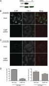
Decreased levels of G3BP inhibit stress granule formation. (A) Wild-type or representative G3BP-deficient HEp-2 cells were analyzed for G3BP expression via Western blotting. (B) Wild-type or G3BP-deficient HEp-2 cells not treated with arsenite were analyzed for G3BP (green in merged image) and TIA-1 (red in merged image) expression using indirect immunofluorescence. (C) Wild-type or G3BP-deficient HEp-2 cells were treated with arsenite, fixed, and processed for immunofluorescence. Stress granule proteins were stained using anti-G3BP (green in merged image) and TIA-1 (red in merged image) antibodies. (D) Each cell type was examined for the percentage of cells containing stress granules per HPF and the size of stress granules per cell using TIA-1 as a marker for stress granules, as described in Materials and Methods. Error bars show standard deviations. KD, knockdown.
FIG. 5.
FIG. 5.
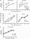
RSV replication is inhibited in G3BP-deficient cells. (A) Wild-type (WT) or representative G3BP-deficient cells were infected with RSV (MOI = 1.0) for indicated times. Cell-associated or supernatant virus was collected at each time point. Viral titer for each sample was determined by plaque assay. (B) Wild-type or G3BP-deficient cells were infected with RSV or rotavirus for indicated times. Viral RNA was collected and assayed for fold change of RNA during infection. (C) Wild-type or G3BP-overexpressing cells derived from U2OS cells (RGD3) were infected with RSV (MOI = 1.0) for indicated times. Cell-associated virus was collected at each time point, and viral titer for each sample was determined by plaque assay. Error bars show standard deviations.
FIG. 6.
FIG. 6.
HuR protein colocalizes with RSV inclusion bodies but is not necessary for replication. (A) HEp-2 cells were infected with RSV (MOI = 1.0) for 24 h, fixed, and prepared for immunofluorescence. Anti-G3BP was used to mark stress granules and appears green in the merged image. Anti-RSV P was used to mark RSV inclusion bodies and appears red in the merged image. HuR appears blue in the merged image. White squares are used to mark areas in which HuR is contained in stress granules or inclusion bodies, respectively. The collapsed z-sections are shown for each image. (B) Cells transduced with nontargeting (NonTgt) or HuR shRNA were compared with wild-type HEp-2 cells for expression levels of HuR. Total HuR levels were quantified for each cell type and compared to wild-type levels. (C) Wild-type HEp-2 cells, cells treated with nontargeting shRNA, or HuR-deficient cells were infected with RSV (MOI = 0.1) for the indicated times. Viral RNA was collected and assayed for the fold change in RNA levels during infection. (D) Wild-type or HuR-deficient cells were infected with RSV (MOI = 0.1) for the indicated times. Cell-associated virus and supernatant virus were collected at each time point. The viral titer for each sample was determined by plaque assay. Error bars show standard deviations.
FIG. 7.
FIG. 7.
Viral genomic RNA predominantly colocalizes with RSV inclusion bodies. HEp-2 cells were mock-infected or infected with RSV (MOI = 1) for the indicated times. RSV RNA-specific probes were added as described in Materials and Methods. Cells were fixed and processed for immunofluorescence. Anti-RSV N was used as an inclusion body marker and appears green in the merged image. Anti-G3BP was used as a stress granule marker and appears blue in the merged image. RSV RNA (vRNA) appears red in the merged image (fourth column). The main images are xy cross sections, and the images above and to the left represent xz and yz cross sections, respectively. The horizontal lines are scale bars, while the diagonal or vertical lines were used to calculate the intensity profiles. Intensity profiles at each time point demonstrate the strong correlation between the N protein and viral genomic RNA (fifth column). Images of N (green), viral genomic RNA (red), and G3BP (blue) in mock infection and at 1, 6, 12, and 24 h postinfection at an image plane near the cell surface are in the sixth column. PI, postinfection; a.u., absorbance units.

References

    1. Brown, G., H. W. Rixon, J. Steel, T. P. McDonald, A. R. Pitt, S. Graham, and R. J. Sugrue. 2005. Evidence for an association between heat shock protein 70 and the respiratory syncytial virus polymerase complex within lipid-raft membranes during virus infection. Virology 338:69-80. - PubMed
    1. Carlos, T. S., D. F. Young, M. Schneider, J. P. Simas, and R. E. Randall. 2009. Parainfluenza virus 5 genomes are located in viral cytoplasmic bodies whilst the virus dismantles the interferon-induced antiviral state of cells. J. Gen. Virol. 90:2147-2156. - PMC - PubMed
    1. Carromeu, C., F. M. Simabuco, R. E. Tamura, L. E. Farinha Arcieri, and A. M. Ventura. 2007. Intracellular localization of human respiratory syncytial virus L protein. Arch. Virol. 152:2259-2263. - PubMed
    1. Cristea, I. M., J. W. Carroll, M. P. Rout, C. M. Rice, B. T. Chait, and M. R. MacDonald. 2006. Tracking and elucidating alphavirus-host protein interactions. J. Biol. Chem. 281:30269-30278. - PubMed
    1. Dang, Y., N. Kedersha, W. K. Low, D. Romo, M. Gorospe, R. Kaufman, P. Anderson, and J. O. Liu. 2006. Eukaryotic initiation factor 2alpha-independent pathway of stress granule induction by the natural product pateamine A. J. Biol. Chem. 281:32870-32878. - PubMed

Publication types

MeSH terms