Review
. 2010 Dec;84(23):12100-9.
doi: 10.1128/JVI.01169-10. Epub 2010 Sep 15.

Herpesviruses and chromosomal integration

Affiliations
Review

Herpesviruses and chromosomal integration

Guillaume Morissette et al. J Virol. 2010 Dec.

Abstract

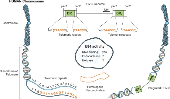
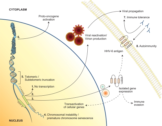
Herpesviruses are members of a diverse family of viruses that colonize all vertebrates from fish to mammals. Although more than one hundred herpesviruses exist, all are nearly identical architecturally, with a genome consisting of a linear double-stranded DNA molecule (100 to 225 kbp) protected by an icosahedral capsid made up of 162 hollow-centered capsomeres, a tegument surrounding the nucleocapsid, and a viral envelope derived from host membranes. Upon infection, the linear viral DNA is delivered to the nucleus, where it circularizes to form the viral episome. Depending on several factors, the viral cycle can proceed either to a productive infection or to a state of latency. In either case, the viral genetic information is maintained as extrachromosomal circular DNA. Interestingly, however, certain oncogenic herpesviruses such as Marek's disease virus and Epstein-Barr virus can be found integrated at low frequencies in the host's chromosomes. These findings have mostly been viewed as anecdotal and considered exceptions rather than properties of herpesviruses. In recent years, the consistent and rather frequent detection (in approximately 1% of the human population) of human herpesvirus 6 (HHV-6) viral DNA integrated into human chromosomes has spurred renewed interest in our understanding of how these viruses infect, replicate, and propagate themselves. In this review, we provide a historical perspective on chromosomal integration by herpesviruses and present the current state of knowledge on integration by HHV-6 with the possible clinical implications associated with viral integration.


Figures

FIG. 1.
FIG. 1.
Hypothetical model of the integration of the HHV-6 genome into the subtelomeric and/or telomeric region of human chromosomes, along with the possible contributions of the U94 gene product in homologous recombination events. Drawing is not to scale.
FIG. 2.
FIG. 2.
Structure, integrity, and orientation of the HHV-6 genome following integration of linear (A), episomal (B), or concatemeric (C) viral DNA. For simplicity, only one recombination event with the chromosome's telomeric region, yielding a chromosome ending with the viral genome, is presented. (A) Recombination of linear viral DNA through the (TAACCC)n repeats in DRR results in the loss of the pac2 at the right end of the genome. Recombination within (TAACCC)n of DRL (not shown) would result in the loss of the majority of the genome. Without additional recombination events, these structures would not be compatible with a replication/packaging-competent HHV-6. (B) Recombination of episomal viral DNA would result in two different viral genomic architectures, depending on whether recombination occurred within DRL or DRR. Once again, in the absence of additional recombination events, these structures would not be compatible with a replication/packaging-competent HHV-6. (C) Recombination of concatemeric viral DNA through the (TAACCC)n of one DRL is presented. Considering that the pac1-pac2 junction forms a viral genomic cleavage site provided that two such junctions are present, the possibility exists that a full-length genome could be excised once integrated. Similar conclusions could be drawn if the recombination event were to occur within the DRR (not shown). Please refer to main text for further details.
FIG. 3.
FIG. 3.
Schematic representation of the hypothetical cellular consequences associated with CIHHV-6 (see text for details), as follows: (1) no viral gene transcription; (2) viral gene expression, replication, and virion production; (3) expression of a subset of HHV-6 genes; (4 and 5) impact of HHV-6 integration on telomere function, architecture, and chromosome stability; (6) trans and/or cis activation of cellular gene expression following integration; (7) immune tolerance due to expression of HHV-6 genes during embryogenesis; (8) destruction of tissues or cells expressing HHV-6 antigens (from integrated HHV-6) by immune defense mechanisms developed in response to natural HHV-6 infection.

References

    1. Ablashi, D. V., N. Balachandran, S. F. Josephs, C. L. Hung, G. R. Krueger, B. Kramarsky, S. Z. Salahuddin, and R. C. Gallo. 1991. Genomic polymorphism, growth properties, and immunologic variations in human herpesvirus-6 isolates. Virology 184:545-552. - PubMed
    1. Achour, A., I. Malet, F. Le Gal, A. Dehee, A. Gautheret-Dejean, P. Bonnafous, and H. Agut. 2008. Variability of gB and gH genes of human herpesvirus-6 among clinical specimens. J. Med. Virol. 80:1211-1221. - PubMed
    1. Adams, A., T. Lindahl, and G. Klein. 1973. Linear association between cellular DNA and Epstein-Barr virus DNA in a human lymphoblastoid cell line. Proc. Natl. Acad. Sci. U. S. A. 70:2888-2892. - PMC - PubMed
    1. Andersson-Anvret, M., and T. Lindahl. 1978. Integrated viral DNA sequences in Epstein-Barr virus-converted human lymphoma lines. J. Virol. 25:710-718. - PMC - PubMed
    1. Anvret, M., A. Karlsson, and G. Bjursell. 1984. Evidence for integrated EBV genomes in Raji cellular DNA. Nucleic Acids Res. 12:1149-1161. - PMC - PubMed

LinkOut - more resources