. 2010 Nov;84(22):11747-60.
doi: 10.1128/JVI.01445-10. Epub 2010 Sep 15.

Nuclear export of human papillomavirus type 31 E1 is regulated by Cdk2 phosphorylation and required for viral genome maintenance

Affiliations

Nuclear export of human papillomavirus type 31 E1 is regulated by Cdk2 phosphorylation and required for viral genome maintenance

Amélie Fradet-Turcotte et al. J Virol. 2010 Nov.

Abstract

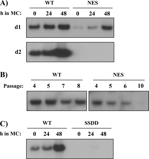
The initiator protein E1 from human papillomavirus (HPV) is a helicase essential for replication of the viral genome. E1 contains three functional domains: a C-terminal enzymatic domain that has ATPase/helicase activity, a central DNA-binding domain that recognizes specific sequences in the origin of replication, and a N-terminal region necessary for viral DNA replication in vivo but dispensable in vitro. This N-terminal portion of E1 contains a conserved nuclear export signal (NES) whose function in the viral life cycle remains unclear. In this study, we provide evidence that nuclear export of HPV31 E1 is inhibited by cyclin E/A-Cdk2 phosphorylation of two serines residues, S92 and S106, located near and within the E1 NES, respectively. Using E1 mutant proteins that are confined to the nucleus, we determined that nuclear export of E1 is not essential for transient viral DNA replication but is important for the long-term maintenance of the HPV episome in undifferentiated keratinocytes. The findings that E1 nuclear export is not required for viral DNA replication but needed for genome maintenance over multiple cell divisions raised the possibility that continuous nuclear accumulation of E1 is detrimental to cellular growth. In support of this possibility, we observed that nuclear accumulation of E1 dramatically reduces cellular proliferation by delaying cell cycle progression in S phase. On the basis of these results, we propose that nuclear export of E1 is required, at least in part, to limit accumulation of this viral helicase in the nucleus in order to prevent its detrimental effect on cellular proliferation.


Figures

FIG. 1.
FIG. 1.
Mutation of the major donor splicing site in the HPV31 E1 open reading frame increases expression of full-length EYFP-31E1. (A) Western blot analysis of C33A cells transfected with an EYFP-31E1 expression vector either containing (+) or lacking (−) the major splicing site mutation (Spl mut.), or transfected with and EYFP expression vector, as a control. E1 proteins were detected using an anti-GFP antibody. Tubulin was used as a loading control. (B) Effect of the major splicing site mutation on the intracellular localization of EYFP-31E1. The indicated EYFP-31E1 expression vectors were transfected in C33A cells. After 24 h, the cells were fixed, mounted, and visualized by fluorescence confocal microscopy. Nuclei (DNA) were stained with TO-PRO-3.
FIG. 2.
FIG. 2.
Nuclear export of HPV31 E1 is regulated by Cdk2 activity. (A) Alignment of the N-terminal regions from different papillomavirus E1 encompassing the bipartite nuclear NLS, the NES, and the CBM. (B) Intracellular localization of WT and mutant HPV31 E1. C33A cells transiently expressing the WT or indicated mutant EYFP-31E1 proteins were visualized by fluorescence confocal microscopy. Nuclei (DNA) were stained with TO-PRO-3. When indicated, cells were also transfected with Cdk2dn or p21 to inhibit Cdk2 activity or were treated with 15 μg of LMB/ml to inhibit CRM1-dependent nuclear export. Quantification of these results is presented in Table 1. (C) Interaction of HPV31 E1 with cyclin A and Cdk2 in vitro. The indicated HPV11 and HPV31 E1 fragments were purified as GST fusion proteins and used in pulldown assays with EcR-293 whole-cell extracts. Bound proteins were separated by SDS-PAGE and analyzed by Western blotting with anti-cyclin A (CycA) and anti-Cdk2 antibodies, as indicated. The bottom panel shows a Coomassie blue-stained SDS-PAGE gel of the purified GST-E1 proteins used in these pulldown assays. The sizes of molecular weight markers are indicated on the left of the gel. (D) Interaction of HPV31 E1 with cyclin E and A in vivo is mediated by the CBM motif. EYFP-31E1 WT (W) or CBM (C), NES (N), or NLS (n) E1 mutants were cotransfected with cyclin E (Cyc E) or cyclin A (Cyc A) fused to a 6xmyc epitope (myc). Coimmunoprecipitated proteins were separated by SDS-PAGE and analyzed by Western blotting with anti-GFP and anti-myc antibodies, as indicated. The lower panels show the amount of EYFP proteins expressed in the different whole-cell extracts prior to their immunoprecipitation (input). Tubulin was used as a loading control (α-Tub.). (E) Western blot analysis of total protein extracts prepared from transfected C33A cells expressing either WT or the indicated EYFP-31E1 mutant proteins. Extracts were prepared 24 h posttransfection. E1 proteins were detected by using an anti-GFP antibody (α-GFP), and tubulin was used as a loading control (α-Tub.).
FIG. 3.
FIG. 3.
Serines 92 and 106 of HPV31 E1 are essential for its nuclear accumulation. Intracellular localization of EYFP-31E1 proteins in which one, two (A) or several (B) putative Cdk2 phosphorylation sites have been substituted for alanine. AAAAA refers to a HPV31 E1 mutant in which all five putative Cdk2 phosphorylation sites have been substituted for alanine, as described in the text. Where indicated, cells were treated with 15 μg of LMB/ml to inhibit nuclear export. Quantification of these results is presented in Table 1. (C) Western blot analysis of C33A cells expressing WT or the indicated EYFP-31E1 mutant proteins as described in Fig. 2E. E1 proteins were detected by using an anti-GFP antibody (α-GFP), and tubulin was used as a loading control (α-Tub.).
FIG. 4.
FIG. 4.
Evidence that HPV31 E1 is phosphorylated on serines 92 and 106 by Cdk2. (A) Western blots of immunoprecipitated EYFP-31E1 fusion proteins, or of EYFP alone as a control, using an anti-phospho-Ser/Thr-Pro antibody (upper panel). Because these proteins were immunoprecipitated with an anti-GFP antibody, an anti-GFP Western blot was also performed to control for the efficiency of immunoprecipitation (lower panel). WT E1 (WT) or mutant derivatives in which all five putative Cdk2 sites were mutated to alanine (AAAAA) or in which only S92 (SAAAA), S106 (ASAAA), or both serines (SSAAA) were left intact were used. (B and C) Same as in panel A, but using EYFP-31E1 fusion protein immunoprecipitated from C33A cells cotransfected with expression vectors for cyclin E and Cdk2 (B, +Cyc E-Cdk2) or for p21 (C, +p21). The positions of EYFP, EYFP-31E1, and of the anti-GFP antibody heavy (H) and light (L) chains are indicated.
FIG. 5.
FIG. 5.
Cytoplasmic E1 proteins are relocalized to the nucleus by E2 and are able to support transient viral DNA replication. (A) Intracellular localization of WT or the indicated mutant EYFP-31E1 proteins coexpressed with either WT (left panel) or E39Q (right panel) HPV31 E2. Quantification of these results is presented in Table 2. (B) Transient DNA replication activities of the indicated EYFP-31E1 proteins in C33A cells. DNA replication activities were measured by determining the ratio of firefly (Fluc) to Renilla (Rluc) luciferase activity as described in Materials and Methods. Cells transfected with EYFP only (No E1) were used as a negative control. Replication activities are reported as a percentage of the Fluc/Rluc ratio obtained with the highest amount of WT E1 expression vector.
FIG. 6.
FIG. 6.
Nuclear export of HPV31 E1 is inhibited by phosphomimetic substitution of S92 and S106 and is dispensable for transient viral DNA replication. (A) Intracellular localization of EYFP-31E1 and of mutant derivatives carrying a mutation in the NES or in which S92 and/or S106 have been changed to an aspartic acid as a phosphomimetic. In the right panel, the same proteins were cotransfected with the Cdk2 inhibitor p21. Quantification of these results is presented in Table 1. (B) Western blot analysis of C33A cells expressing WT or the indicated EYFP-31E1 mutant proteins as described in Fig. 2E. E1 proteins were detected by using an anti-GFP antibody (α-GFP), and tubulin was used as a loading control (α-Tub.). (C) Transient DNA replication activities of the indicated EYFP-31E1 proteins in C33A cells. DNA replication activities were measured by determining the ratio of firefly (Fluc) to Renilla (Rluc) luciferase activity as described in Materials and Methods. Cells transfected with EYFP only (No E1) were used as a negative control. Replication activities are reported as a percentage of the Fluc/Rluc ratio obtained with the highest amount of WT E1 expression vector.
FIG. 7.
FIG. 7.
Nuclear export of HPV31 E1 is required for genome maintenance in HFKs. (A) Maintenance and amplification of HPV31 genomes expressing either the WT or NES mutant E1 protein. HFKs immortalized with the WT or the NES genome were resuspended in methylcellulose (MC) for 24 and 48 h to induce their differentiation. DNA was harvested from undifferentiated (0 h) and from differentiated cells (24 and 48 h). The figure shows the Southern blots used to detect the presence of episomal genomes. Two independent experiments are presented that were performed using HFKs from two different donors (d1 and d2). (B) Southern blots showing the maintenance of the HPV31 WT or NES genome in immortalized HFKs at the indicated number of cell passages. (C) Maintenance and amplification of an HPV31 genome expressing the S92D/S106D (SSDD) E1 mutant in HFKs from donor d2. Experiments were performed as described in panel A.
FIG. 8.
FIG. 8.
HPV31 E1 impairs cell cycle progression in S-phase. (A and B) C33A cells were transfected with the indicated EYFP-31E1 or EYFP expression vector either alone or together with an expression vector for 3F-31E2 and the ori-containing plasmid. After 48 h, cells were treated with trypsin, and their DNA was stained with Hoechst. The DNA content of EYFP-expressing cells was then analyzed by flow cytometry. For each transfection condition described on the left side of the figure, the cells were either grown asynchronously, synchronized in G1/S with mimosine (T = 0) or synchronized with mimosine for 24 h and then released in nocodazole for an additional 24 h (T = 24 h), as indicated. The cell cycle distribution of each sample was derived from the analysis of 5,000 EYFP-expressing cells and is represented by a histogram. For the analysis of untransfected cells (Mock), 5,000 Hoechst-positive cells were collected. Quantification of these results is reported in Table 3.
FIG. 9.
FIG. 9.
Nuclear export of E1 alleviates its detrimental effect on S-phase progression. (A) Cell cycle distribution. C33A cells were transfected with the indicated EYFP-31E1, prepared, and analyzed as described in Fig. 8. The histograms show that nuclear E1, but not cytoplasmic E1, delays S phase. Quantification of these results is reported in Table 3. (B) Colony formation assays (CFA). CFAs were performed with C33A cells transfected with the indicated EYFP-31E1 proteins (expressed from a neomycin-resistant plasmid). After a 3-week selection in G418, the colonies were stained with methylene blue. The results indicate that nuclear but not cytoplasmic E1 impairs colony formation. (C) Cells were transfected with an expression vector for Flag-tagged HPV31 E1 (3F-31E1) and colony formation assayed as described in panel B. An expression vector for Flag-tagged HPV31 E2 (3F-31E2) and the empty vector expressing the three-Flag epitope (3F) alone were used as controls.
FIG. 10.
FIG. 10.
Sequence alignment of the E1 N-terminal regions from different HPV types. The conserved Cdk2-phosphorylation sites are highlighted in black. The NES and CBM are boxed. The amino acid number of the first and last amino acid in each sequence is given in parentheses.

Similar articles

Cited by

References

    1. Abbate, E. A., J. M. Berger, and M. R. Botchan. 2004. The X-ray structure of the papillomavirus helicase in complex with its molecular matchmaker E2. Genes Dev. 18:1981-1996. - PMC - PubMed
    1. Amin, A. A., S. Titolo, A. Pelletier, D. Fink, M. G. Cordingley, and J. Archambault. 2000. Identification of domains of the HPV11 E1 protein required for DNA replication in vitro. Virology 272:137-150. - PubMed
    1. Androphy, E. J., D. R. Lowy, and J. T. Schiller. 1987. Bovine papillomavirus E2 transactivating gene product binds to specific sites in papillomavirus DNA. Nature 325:70-73. - PubMed
    1. Chow, L. T., and R. Broker. 1997. Small DNA tumor viruses, p. 267-301. In N. Nathanson (ed.), Viral pathogenesis. Lippincott-Raven, Philadelphia, PA.
    1. Clower, R. V., J. C. Fisk, and T. Melendy. 2006. Papillomavirus E1 protein binds to and stimulates human topoisomerase I. J. Virol. 80:1584-1587. - PMC - PubMed

Publication types

MeSH terms

Substances