. 2010 Nov 1;21(21):3654-68.
doi: 10.1091/mbc.E10-06-0523. Epub 2010 Sep 15.

Autocrine transforming growth factor-{beta}1 activation mediated by integrin {alpha}V{beta}3 regulates transcriptional expression of laminin-332 in Madin-Darby canine kidney epithelial cells

Affiliations

Autocrine transforming growth factor-{beta}1 activation mediated by integrin {alpha}V{beta}3 regulates transcriptional expression of laminin-332 in Madin-Darby canine kidney epithelial cells

Jose V Moyano et al. Mol Biol Cell. .

Abstract

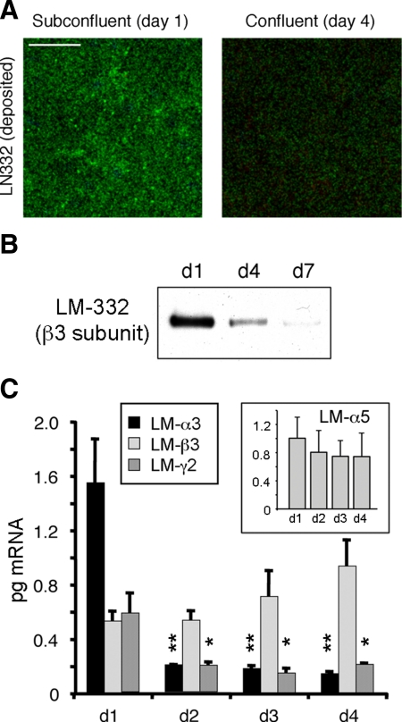
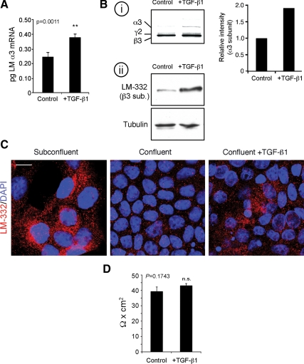
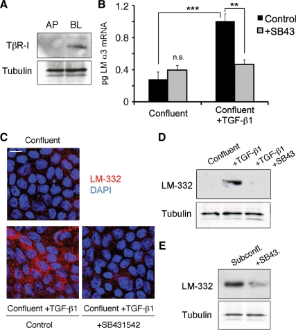
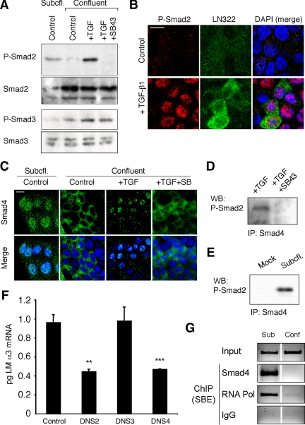
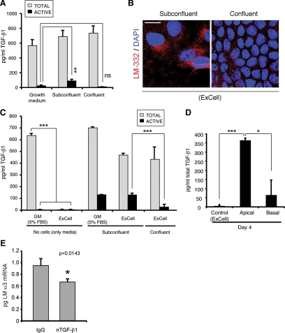
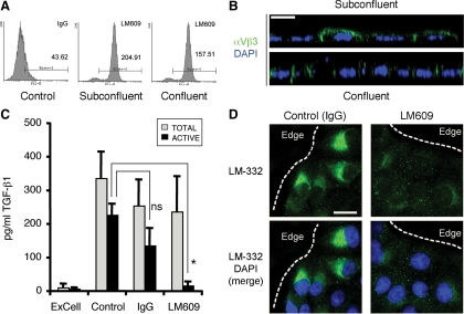
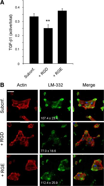
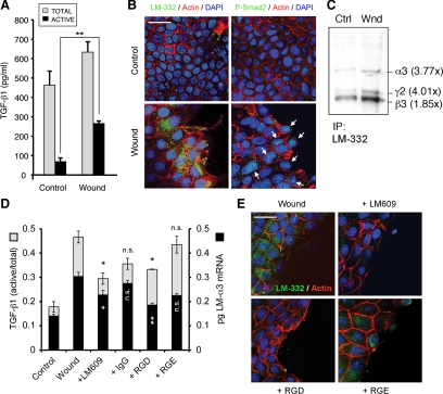
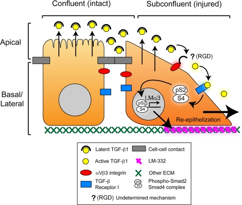
Laminin (LM)-332 is an extracellular matrix protein that plays a structural role in normal tissues and is also important in facilitating recovery of epithelia from injury. We have shown that expression of LM-332 is up-regulated during renal epithelial regeneration after ischemic injury, but the molecular signals that control expression are unknown. Here, we demonstrate that in Madin-Darby canine kidney (MDCK) epithelial cells LM-332 expression occurs only in subconfluent cultures and is turned-off after a polarized epithelium has formed. Addition of active transforming growth factor (TGF)-β1 to confluent MDCK monolayers is sufficient to induce transcription of the LM α3 gene and LM-332 protein expression via the TGF-β type I receptor (TβR-I) and the Smad2-Smad4 complex. Significantly, we show that expression of LM-332 in MDCK cells is an autocrine response to endogenous TGF-β1 secretion and activation mediated by integrin αVβ3 because neutralizing antibodies block LM-332 production in subconfluent cells. In confluent cells, latent TGF-β1 is secreted apically, whereas TβR-I and integrin αVβ3 are localized basolaterally. Disruption of the epithelial barrier by mechanical injury activates TGF-β1, leading to LM-332 expression. Together, our data suggest a novel mechanism for triggering the production of LM-332 after epithelial injury.


Figures

Figure 1.
Figure 1.
Laminin-332 (LM-332) expression is regulated as a function of confluence. (A) LM-332 deposition only occurs in subconfluent cells. Subconfluent (day 1) or confluent (day 4) MDCK cells cultured on 0.4-μm Transwell supports were immunostained for LM-332 with an anti-β3 subunit mAb (green). Confocal sections corresponding to the basal plane (deposited extracellular matrix [ECM]) are shown. Bar, 10 μm. (B) Significant amounts of LM-332 are deposited into the substratum only in subconfluent (day 1) cultures. Cells plated on Transwell supports were removed by treatment with 20 mM NH4OH at the indicated time points (days 1–7). Deposited ECM proteins were extracted, resolved by SDS-polyacrylamide gel electrophoresis and Western-blotted for LM-332 with an anti-β3 mAb. (C) Laminin α3 and γ2 subunit expression is transcriptionally regulated as a function of cell confluence. RNA from Transwell cultures was isolated at different time points (days 1–4) and analyzed by qRT-PCR using canine-specific primers. Inset, qRT-PCR for the α5 subunit of LM-511 (LM-α5). The histograms represent the average abundance of mRNAs from three independent experiments expressed in picograms ± SD. **p < 0.01 or *p < 0.05 relative to day 1 (d1) levels.
Figure 2.
Figure 2.
Exogenous active TGF-β1 is sufficient to induce LM-332 expression in confluent cells. (A) TGF-β1 induces transcription of the laminin α3 subunit gene. Confluent MDCK cells untreated (control) or treated with 5 ng/ml active TGF-β1 for 6 h in serum-free medium (+TGF-β1) were collected and analyzed for laminin α3 subunit mRNA by q-RT-PCR. **p = 0.0011 (B) TGF-β1 induces LM-332 protein synthesis. i, untreated control (control) or TGF-β1-treated (+TGF-β1) confluent cultures were metabolically labeled with [35S]Met/Cys for 20 min and extracted with RIPA buffer. The extracts were immunoprecipitated with a polyclonal antibody against LM-332, and the immunoprecipitates were resolved by SDS-polyacrylamide gel electrophoresis and detected by autoradiography. The graph represents the intensities of the α3 bands in this experiment. ii, equal amounts of total protein from RIPA lysates were analyzed by Western blot for LM-332 using a mAb against the β3 subunit with immunoblotting for tubulin as loading control. (C) Confocal immunofluorescence localization of LM-332 after TGF-β1 treatment. Subconfluent and confluent MDCK cell cultures without added exogenous TGF-β1, and TGF-β1-treated confluent MDCK cells were immunostained with a polyclonal antibody against LM-332 (red) and optical sections of a mid/basal plane visualized. Nuclei were stained with DAPI (blue). Bar, 10 μm. (D) Transepithelial resistance of MDCK cells is not affected by TGF-β1 treatment. Transepithelial electrical resistance of confluent MDCK cultures grown on Transwell supports in either the absence or presence of exogenous active TGF-β1 was measured with a Millicell-ERS device. The experiment was repeated four times, and the average is shown. n.s., not statistically significant, p = 0.1743.
Figure 3.
Figure 3.
Kinase activity of the TGF-β receptor type I (TβR-I) is required for LM-332 expression. (A) TβR-I is localized basolaterally in polarized MDCK cells. Confluent cultures of MDCK cells grown on permeable supports were biotinylated either apically (AP) or basolaterally (BL). Biotinylated proteins were captured on streptavidin-conjugated beads and immunoblotted with anti-TβR-I antibodies. The unbound protein fraction was used for immunoblotting with an mAb against tubulin as loading control. These experiments were repeated twice with similar results. (B) Inhibition of TβR-I signaling abolishes laminin α3 subunit mRNA expression. Confluent MDCK cells were pretreated with DMSO (control) or with 5 μM TβR-I kinase activity inhibitor SB431542 (+SB43) for 30 min. Then, cells were either incubated with or without exogenous TGF-β1 for 6 h and analyzed for laminin α3 subunit mRNA expression by qRT-PCR. n.s., not statistically significant; ***p < 0.001; **p < 0.01 (C) LM-332 protein expression is also diminished after blocking TβR-I signaling in TGF-β1–treated confluent MDCK cells. Confluent cultures of MDCK cells untreated or pretreated with SB431542 were then incubated with or without exogenous TGF-β1 for 6 h, and LM-332 expression was detected by immunofluorescence. Control/confluent, no TGF-β1 or SB431542; control/confluent + TGF-β1, treated with TGF-β1 but no SB431542; +SB431452/confluent+TGF-β1, treated with both TGF-β1 and SB431452. Bar, 10 μm. (D) Production of LM-332 was examined by Western blotting of MDCK cell extracts using an anti-β3 subunit monoclonal from cultures treated as described in C. SB43, treated with SB431452. (E) LM-332 expression is also dependent on TβR-I signaling in subconfluent cells. Subconfluent MDCK cell cultures without addition of exogenous TGF-β1 were incubated with or without SB431452 (SB43) for 18 h, and extracts were Western blotted for the β3 subunit of LM-332.
Figure 4.
Figure 4.
Phospho-Smad2 and Smad4 regulate LM α3 subunit gene transcription. (A) Differential phosphorylation of Smads. Subconfluent (Subcfl.) or confluent cultures either untreated (control) or treated with 5 ng/ml TGF-β1 for 6 h in the absence or presence of the TβR-I inhibitor SB431542 (+TGF-β1 or + TGF-β1+SB43, respectively) were analyzed by Western blotting for phospho-Smad2 (P-Smad2), total Smad2, P-Smad3, and total Smad3. (B) Phospho-Smad2 is localized to the nuclei after TGF-β1 treatment in confluent cells. Untreated (control) or TGF-β1–treated confluent cells (+TGF-β1) were stained with antibodies against P-Smad2 (red) and LM-332 (β3 subunit; green) and analyzed by confocal fluorescence microscopy. Nuclear staining with DAPI, blue. Bar, 10 μm. (C) Smad 4 is also localized to the nuclei in subconfluent and TGF-β1–treated confluent cells. Subconfluent MDCK cell cultures without added exogenous TGF-β1 and confluent cultures either untreated (control) or treated with of TGF-β1 in the presence or absence of SB431542 (SB) were stained with antibodies against Smad4 (green) and analyzed by confocal fluorescence microscopy. Nuclear staining with DAPI, blue. Bar, 10 μm. (D) Phosho-Smad2 and Smad4 form a complex dependent on TβR-I signaling. Confluent cultures treated with TGF-β1 to induce LM-332 expression in the absence or presence of the TβR-I inhibitor SB431542 (+TGF or +TGF+SB43, respectively) were extracted in RIPA buffer. Extracts were immunoprecipitated with an anti-Smad4 antibody (mouse) and Western blotted with anti-P-Smad2 antibody (rabbit). (E) P-Smad2 and Smad4 also form a complex in subconfluent cells. Extracts of subconfluent MDCK cell cultures without added exogenous TGF-β1 were immunoprecipitated with a control IgG (mock) or with an anti-Smad4 antibody (mouse), and Western blotted for P-Smad2 (rabbit). (F) DN Smad2 and Smad4, but not Smad3, impairs α3 subunit transcription in subconfluent cultures. Subconfluent MDCK cell cultures were transiently transfected with DN-Smad2, DN-Smad3, or DN-Smad4, and α3 subunit mRNA expression was analyzed by qRT-PCR after 24 h. **p < 0.05; ***p < 0.001 (G) Smad4 binds to an Smad binding element in the α3 subunit gene promoter. Subconfluent (6 h) or confluent (4 d) MDCK cell extracts were subjected to ChIP with anti-Smad4, RNA-polymerase II, or nonspecific antibodies (IgG). Enrichment of the SBE in the immunoprecipitated chromatin was determined by touchdown-PCR using primers specific for the α3 subunit promoter. Products were resolved by agarose gel electrophoresis (negative staining is shown) and compared with whole chromatin lysates (input).
Figure 5.
Figure 5.
MDCK cells constitutively secrete endogenous TGF-β1 but only activate it under subconfluent conditions. (A) Latent TGF-β1 is activated only in subconfluent cells. Subconfluent and confluent cultures were grown in normal growth medium (containing 5% FBS) and the conditioned media were analyzed for total and active TGF-β1 by sandwich ELISA. **p < 0.01 (B) Cells grown in ExCell also express LM-332 only when subconfluent. MDCK cells were grown in ExCell, a defined serum-free growth media, on Transwell supports for 1 d (subconfluent) or 4 d (confluent); fixed; stained; and analyzed for LM-332 expression (red) by confocal immunofluorescence microscopy; nuclei were stained with DAPI (blue). Bar, 10 μm. (C) MDCK cells constitutively secrete latent TGF-β1. Conditioned media of cells grown in ExCell and normal serum-containing growth medium (GM) were analyzed for total and active TGF-β1 using sandwich ELISA. ***p < 0.001 (D) Confluent MDCK cells secrete latent TGF-β1 apically. MDCK cells were grown in ExCell serum-free medium on Transwell supports for 4 d to achieve full apicobasal polarization. Conditioned medium from either the apical or basal compartments was analyzed for total TGF-β1 by sandwich ELISA. *p < 0.05 (E) Neutralization of endogenous active TGF-β1 reduces LM α3 subunit expression. Subconfluent MDCK cell cultures grown in ExCell in the presence of 1 μg of neutralizing antibody against active TGF-β1 (nTGF-β1) or IgG as negative control were analyzed for α3 subunit mRNA levels by qRT-PCR. *p = 0.0143.
Figure 6.
Figure 6.
αVβ3 integrins regulate TGF-β1 activation and LM-332 expression. (A) MDCK cells express integrin αVβ3 on the cell surface. Subconfluent and confluent cells were incubated with monoclonal anti-αVβ3 (LM609) or mouse IgG (control), and analyzed by flow cytometry to determine cell surface expression levels. (B) Integrin αVβ3 is localized to the apical and lateral plasma membrane in subconfluent cells but is basolaterally polarized in confluent cultures of MDCK cells. Subconfluent and confluent (polarized) MDCK cell cultures grown in ExCell on Transwell supports were stained for αVβ3 integrins with the LM609 (green) and analyzed by confocal microscopy. XZ stacks (lateral views) are shown. Nuclei, blue. Bar, 10 μm. (C) Integrin αVβ3 participates in TGF-β1 activation. MDCK cells were pretreated with a nonspecific antibody (IgG) or anti-αVβ3 function blocking antibody (LM609) for 30 min and then grown for 18 h in ExCell (subconfluent cultures). The conditioned media was analyzed by sandwich ELISA to determine TGF-β1 activation. IgG, nonspecific immunoglobulin. n.s., not statistically significant; *p < 0.05 (D) Inhibition of αVβ3 integrin prevents LM-332 expression. Subconfluent MDCK cell cultures grown on Transwell supports in ExCell were pretreated with either anti-αVβ3 function blocking antibody (LM609) or a nonspecific antibody (IgG), and after 18 h LM-332 (green) was detected by confocal immunofluorescence. The dotted line indicates the edge of an “island” of cells. Nuclei, blue. Bar, 10 μm.
Figure 7.
Figure 7.
TGF-β1 activation and LM-332 expression in subconfluent MDCK cells is regulated by RGD. (A) The RGD peptide inhibits TGF-β1 activation. Subconfluent MDCK cells were grown in ExCell for 18 h in the absence (Subcnf.) or presence of either RGD or RGE peptides (1 mM). The conditioned media were collected and analyzed for TGF-β1 activation by ELISA. Histograms represent the ratio of active versus total TGF-β1. **p < 0.01 (B) RGD inhibits LM-332 expression. Cell cultures corresponding to A were immunostained for LM-332 with a polyclonal antibody (green). Actin, red; nuclei, blue. The numbers indicate relative units of fluorescence (±SD) for the green channel (LM-332). Bar, 10 μm.
Figure 8.
Figure 8.
Wounded confluent MDCK monolayers express LM-332. (A) Wounded epithelial monolayers activate TGF-β1. Conditioned media from a confluent MDCK cell culture (control) and a culture wounded by scraping with a pipette tip were analyzed by sandwich ELISA for total and active TGF-β1. **p < 0.01 (B) LM-332 expression is localized to the cells close to the wound edge. A confluent MDCK cell monolayer (control) and a culture injured by scraping (Wound) were analyzed after 6 h for LM-332 expression (green; left) or P-Smad2 (green/arrows; right). F-actin, red; nuclei, blue. Bar, 25 μm. (C) LM-332 synthesis is up-regulated in wounded cultures. Confluent MDCK cell monolayers (Ctrl) or wounded cultures (Wnd) were metabolically labeled with [35S]Met/Cys for 20 min 6 h after wounding, and synthesized LM-332 was analyzed by immunoprecipitation. Autoradiography revealed increased levels of α3, β3, and γ2 subunit synthesis in wounded monolayers compared with the unwounded control. The numbers between parentheses indicate fold-increase in band intensity compared with the control. Note that α3 and γ2 subunits show a similar fold-increase, consistent with coregulation of the α3 and γ2 mRNAs. (D) Blocking αVβ3 integrin with LM609 or RGD prevents TGF-β1 activation and LM-α3 mRNA expression. Confluent MDCK cell monolayers were treated with either anti-αVβ3 function blocking antibody (LM609), a nonspecific antibody (IgG), RGD or RGE peptides in ExCell. After 6 h, the conditioned media were analyzed for TGF-β1 activation (gray bars), and the cells were analyzed for LM-α3 mRNA expression by qRT-PCR (black bars). n.s., not statistically significant compared with “wound”; *p < 0.05; **p < 0.01 (E) Wounded MDCK monolayers treated as described in D were analyzed by immunofluorescence for LM-332 with a polyclonal antibody (green). F-actin, red; nuclei, blue. Bar, 25 μm.
Figure 9.
Figure 9.
Proposed model for the regulation of LM-332 expression by TGF-β1 and the epithelial barrier. Confluent (polarized) epithelial cells form an intact epithelium with differentiated apical and basolateral domains. Latent TGF-β1 is secreted apically but is separated from the activation machinery (composed at least by integrin αVβ3) and the TGF-β Receptor I (TβR-I) by intact epithelial junctional complexes. When the epithelium is wounded (cell–cell contacts disrupted), latent TGF-β1 is able to interact with integrin αVβ3 which activates it by an unidentified mechanism dependent on RGD [indicated by “? (RGD)”; see Discussion]. Activated TGF-β1 then binds to TβR-I, initiating transcription and expression of LM-332 and facilitating the restoration of a continuous epithelium (reepithelialization).

Similar articles

Cited by

References

    1. Aberdam D., Virolle T., Simon-Assmann P. Transcriptional regulation of laminin gene expression. Microsc. Res. Tech. 2000;51:228–237. - PubMed
    1. Aumailley M., et al. A simplified laminin nomenclature. Matrix Biol. 2005;24:326–332. - PubMed
    1. Aumailley M., El Khal A., Knoss N., Tunggal L. Laminin 5 processing and its integration into the ECM. Matrix Biol. 2003;22:49–54. - PubMed
    1. Basile D. P., Martin D. R., Hammerman M. R. Extracellular matrix-related genes in kidney after ischemic injury: potential role for TGF-β in repair. Am. J. Physiol. 1998;275:F894–F903. - PubMed
    1. Brown K. A., Pietenpol J. A., Moses H. L. A tale of two proteins: differential roles and regulation of Smad2 and Smad3 in TGF-β signaling. J. Cell. Biochem. 2007;101:9–33. - PubMed

Publication types

MeSH terms