. 2010 Nov 1;21(21):3617-29.
doi: 10.1091/mbc.E10-03-0246. Epub 2010 Sep 15.

Centriolar association of ALMS1 and likely centrosomal functions of the ALMS motif-containing proteins C10orf90 and KIAA1731

Affiliations

Centriolar association of ALMS1 and likely centrosomal functions of the ALMS motif-containing proteins C10orf90 and KIAA1731

Victoria J Knorz et al. Mol Biol Cell. .

Abstract

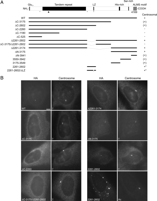
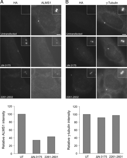
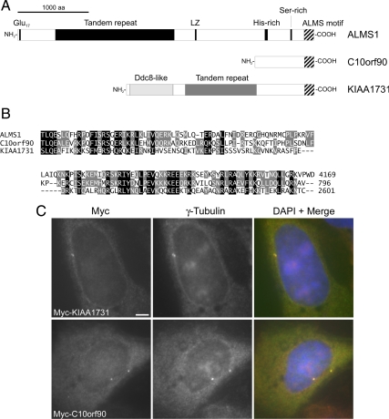
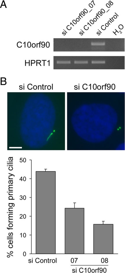
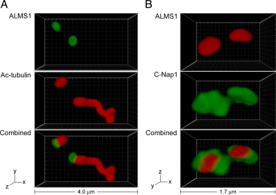
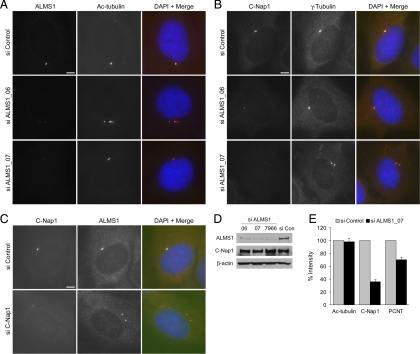
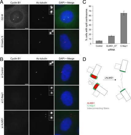
Mutations in the human gene ALMS1 cause Alström syndrome, a rare progressive condition characterized by neurosensory degeneration and metabolic defects. ALMS1 protein localizes to the centrosome and has been implicated in the assembly and/or maintenance of primary cilia; however its precise function, distribution within the centrosome, and mechanism of centrosomal recruitment are unknown. The C-terminus of ALMS1 contains a region with similarity to the uncharacterized human protein C10orf90, termed the ALMS motif. Here, we show that a third human protein, the candidate centrosomal protein KIAA1731, contains an ALMS motif and that exogenously expressed KIAA1731 and C10orf90 localize to the centrosome. However, based on deletion analysis of ALMS1, the ALMS motif appears unlikely to be critical for centrosomal targeting. RNAi analyses suggest that C10orf90 and KIAA1731 have roles in primary cilium assembly and centriole formation/stability, respectively. We also show that ALMS1 localizes specifically to the proximal ends of centrioles and basal bodies, where it colocalizes with the centrosome cohesion protein C-Nap1. RNAi analysis reveals markedly diminished centrosomal levels of C-Nap1 and compromised cohesion of parental centrioles in ALMS1-depleted cells. In summary, these data suggest centrosomal functions for C10orf90 and KIAA1731 and new centriole-related functions for ALMS1.


Figures

Figure 1.
Figure 1.
Deletion analysis identifies two potential centrosome-targeting regions in ALMS1. (A) ALMS1 primary structure and summary of HA-tagged deletion constructs. All constructs lack exon 2 (not shown), which encodes 42 residues but appears to be rarely expressed in humans (Collin et al., 2002; our unpublished observations). The ability of transiently transfected constructs to localize to the centrosome is summarized to the right: +, compact centrosomal; (+), diffuse or less compact centrosomal; −, no detectable centrosomal staining. Asterisks denote constructs that gave additional prominent punctate/aggregate-like staining throughout the cytoplasm. LZ, putative leucine zipper; Glu17, stretch of consecutive glutamic acid residues. Gray bars denote regions with >0.8 probability of forming coiled coils (COILS program, window size 21; Lupas, 1996); the arrowhead indicates the epitope of the antibody used in subsequent experiments to detect endogenous ALMS1. (B) Immunofluorescence data for selected constructs. Cells were costained with antibodies to HA and either γ-tubulin or acetylated tubulin (Ac), as indicated, to mark the centrosome. Size bar, 5 μm.
Figure 2.
Figure 2.
Transiently expressed ALMS1 deletion constructs appear to displace endogenous ALMS1 from the centrosome. U2OS cells were transfected with HA-tagged constructs ΔN-3175 or 2261–2602, representing two putative centrosome-targeting domains in ALMS1, and costained with antibodies to HA and either endogenous ALMS1 (A) or γ-tubulin (B). The epitope recognized by the ALMS1 antibody is not present in either deletion construct, allowing constructs to be distinguished from endogenous ALMS1. The mean intensity of ALMS1 and γ-tubulin immunofluorescence in cells expressing each construct, relative to that in untransfected (UT) cells, is shown; 11–20 cells were analyzed for each condition.
Figure 3.
Figure 3.
The candidate centrosomal protein KIAA1731 contains a C-terminal ALMS motif, and tagged KIAA1731 and C10orf90 localize to the centrosome. (A) Primary structures of human ALMS1, C10orf90 (GenBank accession no. BAG59968) and KIAA1731 (GenBank accession no. NP_203753); the ALMS motif is denoted by striped shading. In addition to its C-terminal ALMS motif, KIAA1731 contains a region with similarity to Ddc8 and a degenerate tandem repeat apparently unrelated to that in ALMS1. (B) Multiple sequence alignment of human ALMS motifs. The C-terminus of each protein is shown; numbers denote amino acid positions. Black and gray shading denote positions conserved in all or 2/3 sequences, respectively, with conservative substitutions permitted. (C) Immunolocalization of Myc-tagged KIAA1731 (full-length) and C10orf90 (residues 243–796). U2OS cells were transiently transfected and coimmunostained for Myc and γ-tubulin, as indicated. In this and subsequent figures, DNA was visualized by DAPI staining (blue). Size bar, 5 μm.
Figure 4.
Figure 4.
siRNA-mediated depletion of KIAA1731 leads to loss of centrosome markers in hTERT-RPE1 cells. (A) qRT-PCR analysis showing depletion of KIAA1731 mRNA by two different siRNA duplexes. Results are expressed relative to the negative control siRNA and represent the mean ± SD of triplicate assays. (B) Quantification of cells in which immunostaining of centriolar acetylated tubulin and either ALMS1 or γ-tubulin was undetectable. The mean of three independent experiments is shown; in each experiment 100–300 cells were counted for each condition. Error bars, SE. (C) Examples of siRNA-treated cells costained with antibodies to ALMS1 and acetylated tubulin. The middle and bottom panels show cells with markedly diminished and undetectable immunostaining, respectively. (D) siRNA-treated cells costained with antibodies against C-Nap1 and pericentrin, showing loss of both markers in a cell treated with KIAA1731-directed siRNA. Size bars, 5 μm.
Figure 5.
Figure 5.
siRNA-mediated depletion of KIAA1731 leads to progressive loss of centrioles and spindle pole abnormalities. (A) Representative images of siRNA-treated cells stained with an antibody to acetylated tubulin (green) to label centrioles. The number of clearly visible centrioles is indicated. The chart shows the percentage of cells containing >2, 2, 1, and 0 centrioles after 48-, 72-, and 96-h siRNA treatment. Approximately 120 cells were counted for each experimental condition. (B) Examples of mitotic siRNA-treated cells costained with antibodies to acetylated tubulin and γ-tubulin. Size bars, 5 μm.
Figure 6.
Figure 6.
Ciliation is decreased in C10orf90-depleted hTERT-RPE1 cells. (A) RT-PCR analysis showing depletion of C10orf90 by two different siRNA duplexes, using HPRT1 as control. (B) Examples of siRNA-treated cells stained with an antibody to acetylated tubulin (green) to visualize ciliary axonemes and centrioles. Cells were transfected with siRNAs for 72 h and then incubated in serum-free medium for a further 24 h to induce primary cilia formation. The mean percentage of ciliated cells is shown; data are from three independent experiments in which 150–300 cells were counted for each siRNA. Error bars, SE. Size bar, 5 μm.
Figure 7.
Figure 7.
ALMS1 localizes specifically to the proximal ends of centrioles and basal bodies and colocalizes with the centrosome cohesion protein C-Nap1. (A) 4Pi microscopy analysis of a ciliated hTERT-RPE1 cell costained with antibodies to ALMS1 (green) and acetylated tubulin (red), which marks the immature parental centriole, basal body, and ciliary axoneme. (B) 4Pi microscopy analysis of a cell costained with antibodies to ALMS1 (red) and C-Nap1 (green), a marker for the proximal ends of centrioles. The axes and total width of images are indicated.
Figure 8.
Figure 8.
Centriolar levels of C-Nap1 are diminished in ALMS1-depleted cells. (A) Immunofluorescence microscopy analysis showing depletion of ALMS1 by two different siRNA duplexes (ALMS1_06 and ALMS1_07). hTERT-RPE1 cells were transfected with the indicated siRNAs for 96 h and coimmunostained for ALMS1 and the centriole marker acetylated tubulin. (B) To assess levels of C-Nap1 at the centrosome after siRNA-mediated depletion of ALMS1, cells were costained with antibodies to C-Nap1 and γ-tubulin. (C) In the reciprocal experiment, cells were depleted of C-Nap1 by RNAi and costained with antibodies to ALMS1 and C-Nap1. Size bars, 5 μm. (D) Immunoblot analysis of total cellular levels of C-Nap1 and ALMS1 after siRNA treatment, using β-actin as loading control. ALMS1_7966 is a previously described siRNA duplex (Graser et al., 2007a). HEK293 cells were used due to more efficient extraction of C-Nap1 and ALMS1 compared with RPE1 cells; diminished centrosomal C-Nap1 immunostaining was confirmed in HEK293 cells depleted of ALMS1 (data not shown). (E) Mean intensity of centrosome marker immunostaining in siRNA-treated cells. Mitotic cells were excluded from analysis. Results are expressed relative to the mean intensity in cells treated with a negative control siRNA. Data are from three independent experiments in which 20–50 cells were analyzed for each siRNA; error bars, SE. PCNT, pericentrin.
Figure 9.
Figure 9.
Depletion of ALMS1 causes centrosome splitting. (A) Cyclin B1 immunostaining was used to determine cell cycle stage, allowing cells entering mitosis to be excluded from analysis. Examples of control cells at the G2-M transition and in G1/early S phase are shown. Centrioles were visualized by costaining with an antibody to acetylated tubulin. Note the separating centrosomes (each containing one parental and one progeny centriole) at G2-M and the close association of the two parental centrioles in G1/early S phase. (B) Immunofluorescence microscopy analysis of centrosome splitting in G1/early S phase cells. Cells were treated with the indicated siRNAs and costained with antibodies to cyclin B1 and acetylated tubulin. Interphase cells with undetectable cyclin B1 and with centrioles >2 μm apart were classed as having a split centrosome. (C) Quantification of centrosome splitting, after siRNA treatment, in interphase cells with undetectable cyclin B1. Depletion of the centrosome cohesion protein C-Nap1 was used as a positive control. The mean ± SE of three independent experiments is shown; in each experiment at least 100 cells were counted for each condition. (D) Model of ALMS1 function in centrosome cohesion, based on the model of C-Nap1/rootletin–dependent centrosome cohesion proposed by Bahe et al. (2005). Depletion of ALMS1 causes, by an unknown mechanism, reductions in the level of C-Nap1 at centrioles. This is predicted to perturb docking of interconnecting or “entangling” fibers with the proximal end of each centriole, leading to centrosome splitting. Procentrioles, which assemble orthogonally to parental centrioles during S and G2 phase, are depicted by dashed lines. Note that C-Nap1 is not thought to localize to the interface between the procentriole and centriole or be involved in maintaining their attachment (Mayor et al., 2000); it is not known if ALMS1 localizes to this interface.

References

    1. Alstrom C. H., Hallgren B., Nilsson L. B., Asander H. Retinal degeneration combined with obesity, diabetes mellitus and neurogenous deafness: a specific syndrome (not hitherto described) distinct from the Laurence-Moon-Bardet-Biedl syndrome: a clinical, endocrinological and genetic examination based on a large pedigree. Acta Psychiatr. Neurol. Scand. 1959;34:1–35. - PubMed
    1. Andersen J. S., Wilkinson C. J., Mayor T., Mortensen P., Nigg E. A., Mann M. Proteomic characterization of the human centrosome by protein correlation profiling. Nature. 2003;426:570–574. - PubMed
    1. Azimzadeh J., Bornens M. Structure and duplication of the centrosome. J. Cell Sci. 2007;120:2139–2142. - PubMed
    1. Badano J. L., Mitsuma N., Beales P. L., Katsanis N. The ciliopathies: an emerging class of human genetic disorders. Annu. Rev. Genom. Hum. Genet. 2006;7:125–148. - PubMed
    1. Bahe S., Stierhof Y. D., Wilkinson C. J., Leiss F., Nigg E. A. Rootletin forms centriole-associated filaments and functions in centrosome cohesion. J. Cell Biol. 2005;171:27–33. - PMC - PubMed

Publication types

MeSH terms

Associated data