. 2010 Sep 15;30(37):12400-13.
doi: 10.1523/JNEUROSCI.6407-09.2010.

Functional role of the interaction between polysialic acid and extracellular histone H1

Affiliations

Functional role of the interaction between polysialic acid and extracellular histone H1

Bibhudatta Mishra et al. J Neurosci. .

Abstract

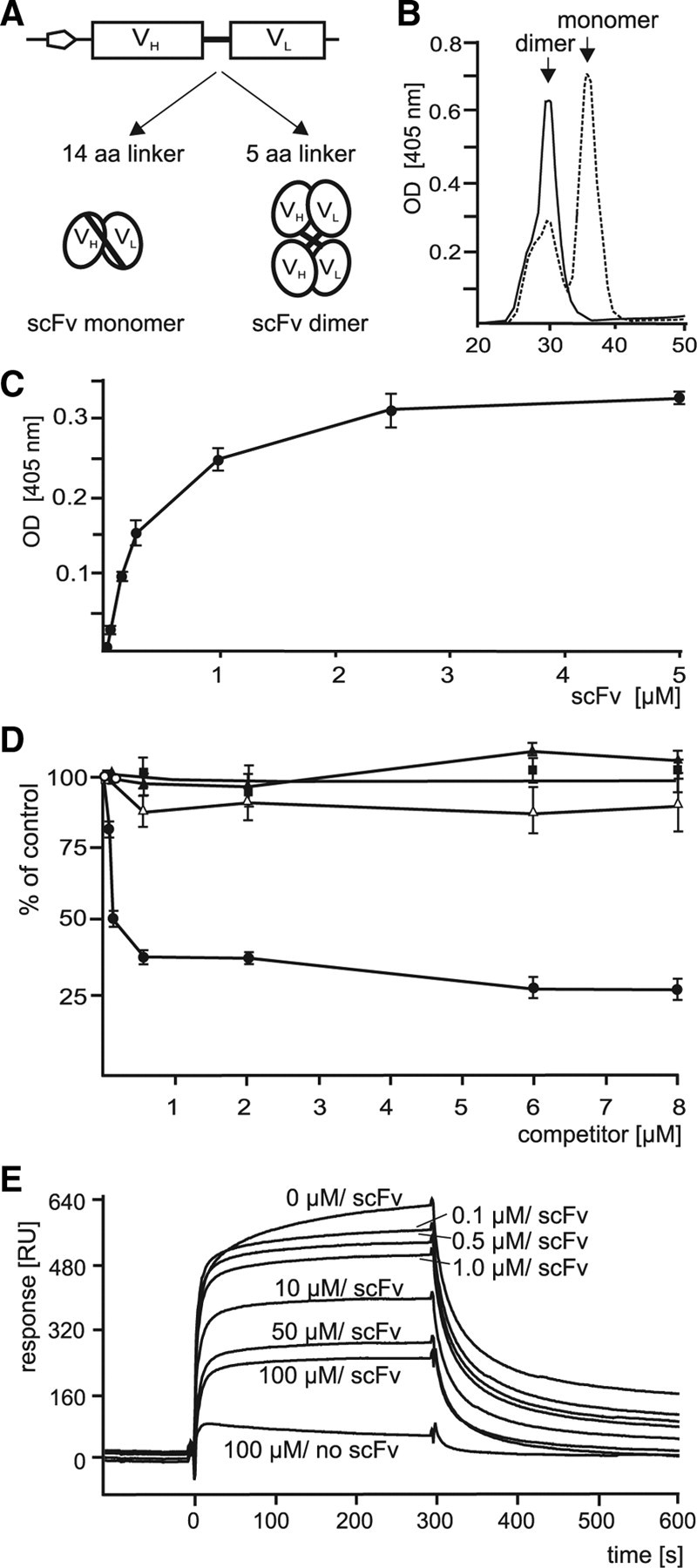
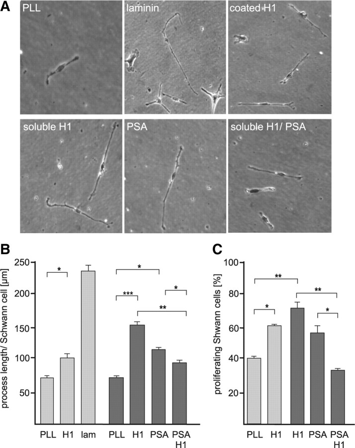
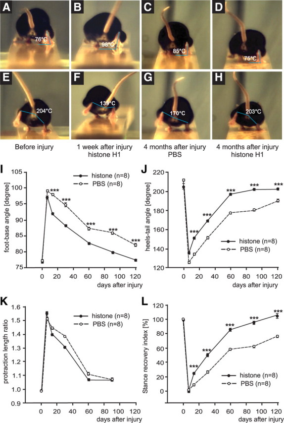
Polysialic acid (PSA) is a large and highly negatively charged glycan that plays crucial roles in nervous system development and function in the adult. It has been suggested to facilitate cell migration, neurite outgrowth, and synaptic plasticity because its hydration volume could enhance flexibility of cell interactions. Evidence for receptors of PSA has so far been elusive. We now identified histone H1 as binding partner of PSA via a single-chain variable fragment antibody using an anti-idiotypic approach. Histone H1 directly binds to PSA as shown by ELISA. Surface biotinylation of cultured cerebellar neurons indicated an extracellular localization of histone H1. Immunostaining of live cerebellar neurons and Schwann cells confirmed that an extracellular pool of histone H1 colocalizes with PSA at the cell surface. Histone H1 was also detected in detergent-insoluble synaptosomal membrane subfractions and postsynaptic densities. When applied in vitro, histone H1 stimulated neuritogenesis, process formation and proliferation of Schwann cells, and migration of neural precursor cells via a PSA-dependent mechanism, further indicating that histone H1 is active extracellularly. These in vitro observations suggested an important functional role for the interaction between histone H1 and PSA not only for nervous system development but also for regeneration in the adult. Indeed, histone H1 improved functional recovery, axon regrowth, and precision of reinnervation of the motor branch in adult mice with femoral nerve injury. Our findings encourage investigations on the therapeutic potential of histone H1 in humans.


Figures

Figure 1.
Figure 1.
Anti-idiotypic scFv antibodies directed against a PSA antibody mimic PSA. A, Substrate-coated PSA antibody was preincubated with different concentrations of colominic acid (closed circles), chondroitin sulfate A (closed triangles), chondroitin sulfate C (open triangles), or heparin (closed rectangles) and incubated with purified scFv antibody with the sequence GLPIDS. Binding of the scFv antibody to the PSA antibody was determined by ELISA and binding in the presence of competitor was related to the binding in the absence of competitor, which was set to 100%. Mean values ± SD from three independent experiments are shown. B, C, Surface plasmon resonance experiments were performed using immobilized PSA antibody and different concentrations (0.01, 0.05, 0.1, 0.5, and 1 μm) of scFv antibody with the sequence GLPIDS for injection (B) or with a constant amount of scFv antibody (5 μm) and different concentrations of colominic acid (0, 1, 10, and 100 μm) for injection or without scFv and 100 μm colominic acid (100 μm/no scFv) (C). Binding of scFv was monitored by measuring the response signal in arbitrary units (RU). A representative experiment is shown.
Figure 2.
Figure 2.
Affinity of PSA-mimicking scFv antibodies to a PSA antibody after linker truncation. A, The scFv antibody contains a linker of 14 aa (14 aa linker) between the VH and VL chains before truncation and 5 aa (5 aa linker) after truncation, which should result in increased formation of scFv antibody dimers. B, Purified scFv antibody with nontruncated (dashed line) or with truncated linker region (solid line) were separated by FPLC. The retention times of the monomeric and dimeric forms are indicated. C, Increasing concentrations of scFv antibody with truncated linker region were used in ELISA with substrate-coated PSA antibody. D, In the competition ELISA, different concentrations of colominic acid (closed circles), chondroitin sulfate A (closed triangles), chondroitin sulfate C (open triangles), or heparin (closed rectangles) and a constant amount of scFv antibody with truncated linker region were incubated with substrate-coated PSA antibody. C, D, Mean values ± SD from three independent experiments are shown for the binding of scFv antibody to the PSA antibody relative to the binding in the absence of competitor, which was set to 100%. E, Surface plasmon resonance experiments were performed using immobilized PSA antibody and different concentrations of colominic acid (0, 0.1, 0.5, 1.0, 10, 50, and 100 μm) together with a constant amount of scFv antibody (5 μm) with truncated linker region or without scFv and 100 μm colominic acid (100 μm/no scFv) for injection. Binding of scFv antibody in a representative experiment is shown. B–E, Results obtained for the scFv antibody with the sequence GLPIDS are shown.
Figure 3.
Figure 3.
Identification of histone H1 as extracellular membrane-attached PSA binding partner using a PSA-mimicking scFv antibody for affinity chromatography. A, Detergent-solubilized membrane (memb) or soluble (sol) brain proteins isolated from 7-d-old (7 d) or 2-month-old (adult) mice were used as substrate coat and incubated without (control) or with either scFv antibody with the sequence GLPIDS or LNDGPVTSA and truncated linker region. Mean values ± SD from three independent experiments are shown for the binding of the scFv antibodies to soluble or membrane proteins. B, Purified scFv antibody with the sequence GLPIDS was immobilized and used for affinity chromatography with membrane-attached brain proteins obtained by alkaline treatment of membranes. Proteins binding to the scFv antibody were eluted and subjected to silver staining together with purified scFv antibody as a control. The position of the 30 kDa protein band that was analyzed by mass spectrometry and identified as histone H1 is indicated by an arrow. C, Histone H1, H2A, H2B, H3, and H4 as well as GAPDH as negative control were substrate-coated and incubated with increasing concentrations of colominic acid. The binding of colominic acid was detected by ELISA using PSA antibody and corresponding HRP-conjugated secondary antibodies. Mean values ± SD from three independent experiments performed in triplicate are shown. D, Triton X-100-soluble (Tx sol), Triton X-100-insoluble membrane proteins (Tx insol), and PSDs were subjected to Western blot analysis using histone H1-specific polyclonal rabbit antibody. E, Cultured live cerebellar neurons were incubated with biotinylation reagent. Biotinylated surface proteins (surface) were precipitated by streptavidin beads and subjected to Western blot analysis using a monoclonal histone H1 antibody, a calreticulin antibody, or HRP-conjugated streptavidin (SA). Total cell lysate (lysate) and post-pull-down supernatant (post-PD) were used as controls. The biotinylated histone H1 form with apparent molecular weights of ∼60 kDa (black arrowhead) and ∼30 kDa (gray arrowhead) are indicated.
Figure 4.
Figure 4.
Colocalization of histone H1 and PSA at the surface of live cerebellar neurons and Schwann cells. A, Cultured live cerebellar neurons (A) or Schwann cells (B) were immunostained with rabbit polyclonal histone H1 antibody, mouse monoclonal PSA antibody, and the corresponding fluorescent dye-labeled secondary antibodies. After fixation and permeabilization of cells, nuclei were stained with DAPI. Superimpositions of histone H1 and PSA staining show partial colocalization (seen in yellow) at the surface of cell bodies and processes. Scale bars, 10 μm.
Figure 5.
Figure 5.
Histone H1 promotes neurite outgrowth of cerebellar neurons in a PSA-dependent manner. Primary cultures of cerebellar neurons were grown on substrate-coated PLL or PLL with 10 μg/ml histone H1, H2A, H2B, H3, or H4, or laminin (lam), on PLL with increasing amounts of histone H1 or on PLL in the presence of increasing amounts of soluble histone H1 or on PLL in the presence of 10 μg/ml soluble PSA and histone H1 (A, C), on PLL or laminin in the absence and presence of PSA-specific antibody (735) or endoglycosidase N (endo) and/or histone H1, H2A, H2B, H3, or H4 (D), or on substrate-coated PLL or laminin in the absence or presence of soluble PSA and/or histone H1 antibody (αH1) (E). A, Images of representative neuron on substrate-coated PLL in the absence or presence of PSA and/or soluble histone H1, on substrate-coated histone H1 or on control substrate laminin are shown. B–E, The total length of neurites per cerebellar neuron was determined. The asterisks, double asterisks, and triple asterisks denote p < 0.05, p < 0.01, and p < 0.001 obtained by the two-tailed t test, respectively, from n = ∼100 neurons in three independent experiments.
Figure 6.
Figure 6.
Histone H1 enhances process elongation and proliferation of Schwann cells in a PSA-dependent manner. Schwann cells were grown on substrate-coated PLL, histone H1 (H1), or laminin (lam) or on substrate-coated PLL in the presence of soluble PSA and/or histone H1 (A, B). A, Images of representative Schwann cells on substrate-coated PLL in the absence or presence of PSA and/or soluble histone H1, on substrate-coated histone H1 or laminin are shown. The process length per Schwann cell (B) and the number of BrdU-labeled proliferating Schwann cells (C) were determined. B, C, The asterisks, double asterisks, and triple asterisks denote p < 0.05, p < 0.01, and p < 0.001 obtained by the two-tailed t test, respectively, from n = ∼100 (B) or n = ∼1000 (C) Schwann cells in three independent experiments.
Figure 7.
Figure 7.
Histone H1 and PSA are expressed by neurospheres and affect migration of neural progenitor cells. A, Slices of neurospheres were stained with histone H1- and PSA-specific antibodies followed by DAPI staining. Superimpositions of histone H1 and PSA staining show partial colocalization (seen in yellow) at the surface of neural precursor cells. Scale bar, 10 μm. B, Neurospheres were seeded onto substrate-coated PLL (PLL), histone H1 (H1), or laminin (lam) or on substrate-coated PLL in the presence of soluble PSA and/or histone H1. Micrographs displaying neurospheres (borders are indicated by dashed lines) with cells migrating out of the neurospheres in the absence (PLL) or presence of soluble PSA (PSA), soluble histone H1 (H1), or both soluble PSA and soluble histone H1 (PSA/H1) (left panel) and the quantification of the total distance reached by all migrating cells measured from the border of the neurosphere (right panel) are shown. Mean values ± SD from three independent experiments are shown. The asterisks and double asterisks denote p < 0.05 and p < 0.01, respectively, obtained by the two-tailed t test from 20 neurospheres per group from three independent experiments.
Figure 8.
Figure 8.
Analysis of motor function after femoral nerve injury and application of histone H1. FBA (A–D) and HTA (E–H) were determined by analyzing single video frames from recordings of beam walking of mice before injury (A, E), 1 week (B, F) and 4 months (C, D, G, H) after injury and application of histone H1 (B, D, F, H) or PBS as vehicle solution (C, G) to the lesion site. I–L, Time course of motor recovery after application of histone H1 or PBS are shown as mean values ± SEM of foot–base angle (I) and heels–tail angle (J), protraction length ratio (K), and stance recovery index (L) at different time points after injury and application of histone H1 or PBS. Preinjury values are plotted for day 0 and for other time points indicated in the graphs. Numbers of animals studied per group are indicated in brackets. The asterisks indicate significant differences (p < 0.05, one-way ANOVA with Tukey's post hoc test) between the histone H1-treated group and the PBS control group at the given time points.
Figure 9.
Figure 9.
Analysis of regrowth of motoneuron axons after femoral nerve injury. Four months after injury and application of histone H1 or vehicle (PBS), animals were subjected to retrograde labeling of motoneurons. A, Mean numbers + SEM of motoneurons labeled through the motor branch representing correctly projecting neurons (correct), through the sensory branch representing incorrectly projecting neurons (incorrect), through both branches (mixed), and the sum of labeled neurons (total number) are shown. B, Morphometric analysis of soma size of regenerated motoneurons was performed. Mean values + SEM of soma area of correctly and incorrectly projecting motoneurons after application of histone H1 or PBS are shown. A, B, Significant differences between the groups of eight animals are indicated by asterisks (*p < 0.05, **p < 0.01, ***p < 0.001, one-way ANOVA with Tukey's post hoc test). C, Analysis of myelinated nerve fibers in regenerated and intact nerves. Shown are normalized frequency distributions of g ratios (axon/fiber diameter) in the motor nerve branch of noninjured mice (intact) and mice treated with histone H1 (histone) or vehicle (PBS) 4 months after injury.

Similar articles

Cited by

References

    1. Ahlborn P, Schachner M, Irintchev A. One hour electrical stimulation accelerates functional recovery after femoral nerve repair. Exp Neurol. 2007;208:137–144. - PubMed
    1. Al-Majed AA, Neumann CM, Brushart TM, Gordon T. Brief electrical stimulation promotes the speed and accuracy of motor axonal regeneration. J Neurosci. 2000;20:2602–2608. - PMC - PubMed
    1. Angata K, Huckaby V, Ranscht B, Terskikh A, Marth JD, Fukuda M. Polysialic acid-directed migration and differentiation of neural precursors are essential for mouse brain development. Mol Cell Biol. 2007;27:6659–6668. - PMC - PubMed
    1. Asahara T, Lin M, Kumazawa Y, Takeo K, Akamine T, Nishimura Y, Kayahara T, Yamamoto T. Long-term observation on the changes of somatotopy in the facial nucleus after nerve suture in the cat: morphological studies using retrograde labeling. Brain Res Bull. 1999;49:195–202. - PubMed
    1. Bolton SJ, Perry VH. Histone H1; a neuronal protein that binds bacterial lipopolysaccharide. J Neurocytol. 1997;26:823–831. - PubMed

Publication types

MeSH terms