. 2010 Sep 15;30(37):12545-56.
doi: 10.1523/JNEUROSCI.3042-10.2010.

Supporting cells eliminate dying sensory hair cells to maintain epithelial integrity in the avian inner ear

Affiliations

Supporting cells eliminate dying sensory hair cells to maintain epithelial integrity in the avian inner ear

Jonathan E Bird et al. J Neurosci. .

Abstract

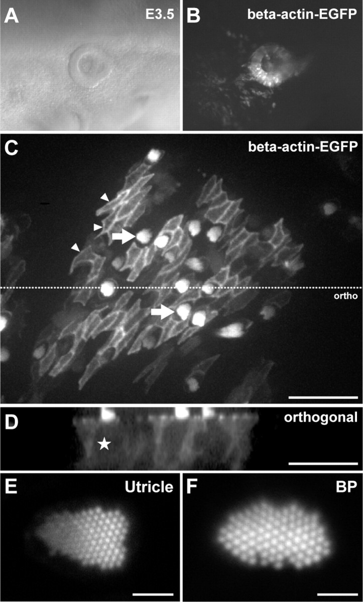
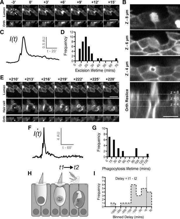
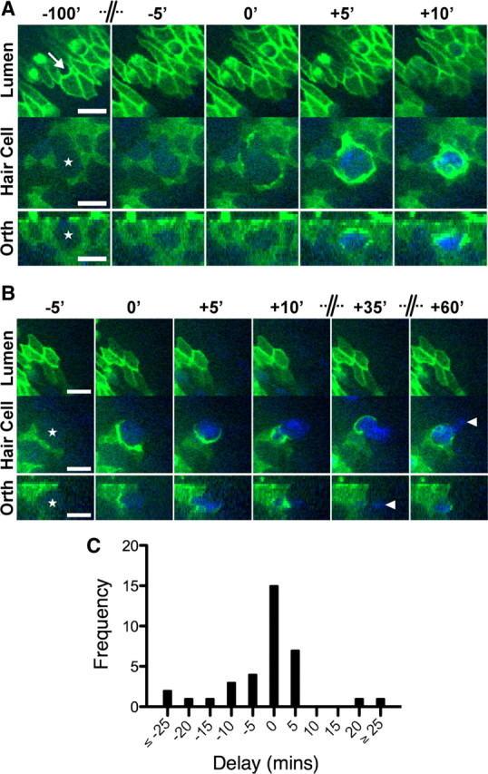
Epithelial homeostasis is essential for sensory transduction in the auditory and vestibular organs of the inner ear, but how it is maintained during trauma is poorly understood. To examine potential repair mechanisms, we expressed β-actin-enhanced green fluorescent protein (EGFP) in the chick inner ear and used live-cell imaging to study how sensory epithelia responded during aminoglycoside-induced hair cell trauma. We found that glial-like supporting cells used two independent mechanisms to rapidly eliminate dying hair cells. Supporting cells assembled an actin cable at the luminal surface that extended around the pericuticular junction and constricted to excise the stereocilia bundle and cuticular plate from the hair cell soma. Hair bundle excision could occur within 3 min of actin-cable formation. After bundle excision, typically with a delay of up to 2-3 h, supporting cells engulfed and phagocytosed the remaining bundle-less hair cell. Dual-channel recordings with β-actin-EGFP and vital dyes revealed phagocytosis was concurrent with loss of hair cell integrity. We conclude that supporting cells repaired the epithelial barrier before hair cell plasmalemmal integrity was lost and that supporting cell activity was closely linked to hair cell death. Treatment with the Rho-kinase inhibitor Y-27632 did not prevent bundle excision but prolonged phagocytic engulfment and resulted in hair cell corpses accumulating within the epithelium. Our data show that supporting cells not only maintain epithelial integrity during trauma but suggest they may also be an integral part of the hair cell death process itself.


Figures

Figure 1.
Figure 1.
Live-cell imaging of β-actin-EGFP in the avian inner ear. RCASBP(B)-β-actin-EGFP was transfected into an E2.5 otic cup by in ovo electroporation. A, B, Twenty-four hours later, β-actin-EGFP was expressed throughout the otic vesicle. C, An example of β-actin-EGFP expression in a utricle isolated from a transgenic E18 embryo. The extrastriolar region was imaged live using a spinning-disk confocal microscope. The cellular mosaic of β-actin-EGFP expression allowed the cytoskeleton of hair cells and supporting cells to be discriminated. β-actin-EGFP was particularly concentrated in the apical junctions of supporting cells (small arrowheads) and hair cell stereocilia (arrows). D, Orthogonal projection of C. Hair cell somas (e.g., star) are identified by their low cytosolic β-actin-EGFP fluorescence compared to adjacent supporting cells. E, F, Expression of β-actin-EGFP did not affect development of actin-based structures at the subcellular level. Laser-scanning confocal imaging of live stereocilia bundles in the utricle (E) and basilar papilla (F) revealed a normal architecture. Scale bars: C, D, 50 μm; E, F, 2 μm.
Figure 2.
Figure 2.
Supporting cells drive epithelial repair in the avian utricle using two distinct mechanisms. Transgenic utricles (E17–E19) expressing β-actin-EGFP were time-lapse imaged live in the presence of 1 mm streptomycin to induce hair cell death. A, E, A time-lapse sequence of hair cell removal and epithelial repair after at least 12 h of streptomycin treatment. Panels depict the exact same hair cell at different times during the repair process. In this mosaic, only supporting cells (stars) were expressing β-actin-EGFP. A nonexpressing hair cell was present in the center of the mosaic (arrow). A, Activity was initially restricted to the epithelial lumen where supporting cells formed an actin cable (0′) that invaded into the hair cell (+3′) and excised the stereocilia bundle (+6′). Junction complexes reformed after removal of the stereocilia bundle (+15′). B, Higher-resolution laser-scanning confocal image of a different hair cell shows how the actin cable constricts beneath the cuticular plate to eject the stereocilia bundle. C, Changes in fluorescence intensity of supporting cell β-actin-EGFP measured at the luminal surface during stereocilia excision. D, Distribution of supporting cell activity during stereocilia excision (n = 36 events). E, After stereocilia excision (shown in A) supporting cells engulfed the remaining hair cell soma (star) within a phagosome highlighted by β-actin-EGFP. F, Supporting cell activity during phagocytosis was heterogeneous and was best modeled as a spiking phenomenon. Frame-by-frame differential fluorescence intensity was used to detect suprathreshold activity. The initial burst of activity represents formation of the phagosome, followed by an extended period where the cell is internalized. G, Distribution of supporting cell activity during phagocytosis (n = 22 events). H, Schematic of the hair cell removal process. I, Distribution of delay (t1 − t2) between onset of stereocilia excision (t1) and phagocytosis (t2). The majority of stereocilia excision events occurred in advance of hair cell phagocytosis. Scale bars: A, E, 10 μm; B, 5 μm. Times are expressed in minutes (′).
Figure 3.
Figure 3.
Supporting cells phagocytosis is closely correlated with hair cell death. Utricles (E17–E19) expressing β-actin-EGFP were incubated with 1 mm streptomycin and 0.1 μm TOTO-3, a fluorescent nucleic acid dye excluded by the plasmalemma of viable cells. Dual-wavelength time-lapse images were captured for EGFP (green) and TOTO-3 (blue). A, B, Two examples where supporting cells expressing β-actin-EGFP remove nonexpressing hair cells (stars) from the sensory epithelium. The position of the hair cell bundle is marked in A (arrow). A, Excision of the stereocilia bundle occurs with negligible uptake of TOTO-3 into the hair cell cytosol (−100′ to −5′) (supplemental Movie 3, available at www.jneurosci.org as supplemental material). Significant uptake of TOTO-3 is observed as supporting cells engulf the hair cell soma within a phagosome (+5′). TOTO-3 fluorescence confirms that the phagosome contains chromatin and that the hair cell confined within is dead. B, In the second example, uptake of TOTO-3 occurs with formation of the supporting cell phagosome. After the initial engulfment, TOTO-3-labeled hair cell chromatin is transported into the supporting cells' cytoplasm. Not all of the hair cell nucleus is engulfed by the β-actin-EGFP-expressing supporting cell. Fragments of TOTO-3-labeled chromatin (arrowheads) remain either in the extracellular space or within supporting cells that did not express β-actin-EGFP. C, Histogram of time delays between the onset of supporting cell phagocytosis and TOTO-3/PI uptake in hair cells (n = 35 events). The modal response was for these events to occur in the same frame, i.e., a delay of 0 min. Negative delays indicate that TOTO-3/PI uptake preceded phagosome formation and vice versa. Scale bars: 10 μm. Time is expressed in minutes, and the axis has been compressed where indicated.
Figure 4.
Figure 4.
Supporting cell phagosomes are present in aminoglycoside-treated utricles, both in vitro and in vivo. Wild-type chick utricles (E21) were cultured with 1 mm streptomycin sulfate in vitro to induce hair cell toxicity. Paraformaldehyde fixed samples were labeled with anti-HCS-1, phalloidin, and DAPI, and then visualized using confocal microscopy. A, Normal organization of the sensory epithelium after 24 h in vitro. Phalloidin highlights the stereocilia bundles at the epithelial lumen (arrows) and anti-HCS-1 labels the hair cell pericuticular junction (arrowheads). Within the epithelium, hair cell somas (stars) are packed densely together with HCS-1 labeling the basolateral membranes. B, After 24 h exposure to 1 mm streptomycin, there is significant hair cell loss. Few stereocilia bundles are visible and this correlates with expansion of supporting cells at the epithelial lumen. At the luminal surface, the HCS-1-positive rings associated with the hair cell pericuticular regions are no longer visible. At the level of the hair cell nuclei, there are HCS-1-labeled cells containing pyknotic DNA. F-actin accumulates in phagosomes containing HCS-1 and pyknotic DNA (inset, overlay of boxed regions). C, Quantification of hair cell and phagosome density in utricles incubated with streptomycin for varying durations reveals an inverse relationship. A significant change is observed at 24 h (compared to sham-operated controls; n = 5 utricles per group; formula image
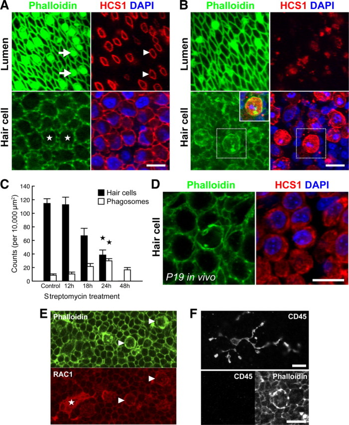
p < 0.001). D, Phagosomes were also detected in P19 utricles after systemic administration of streptomycin in vivo. E, F, Phagosomes colocalize with phagocytic, but not macrophages markers. Phagosomes (arrowheads) and a macrophage (star) both label with anti-RAC1 (CED-10) (E). However, phagosomes do not label with anti-CD45 (F, bottom), unlike macrophages (F, top). Scale bars: 10 μm. Data are expressed as mean ± SEM.
Figure 5.
Figure 5.
Rho kinase is required for efficient phagocytosis of hair cells during aminoglycoside treatment. Utricles expressing β-actin-EGFP were imaged in the continuous presence of 1 mm streptomycin and 30 μm Y-27632, a Rho-kinase inhibitor. A, B, Identical hair cell at different times during the repair process. Supporting cells and the hair cell (star) are both expressing β-actin-EGFP in this example. The hair cell stereocilia bundle is clearly visible (−5′; arrowhead). A, Inhibition of Rho kinase did not prevent stereocilia excision or repair of the epithelial junctions. After excision, the stereocilia bundle remains lying on the surface of the epithelium (+30′; arrowhead). No β-actin-EGFP activity was observed about the hair cell soma during this process. B, Rho-kinase treatment did not block formation of the supporting phagosome (+260′), but did interfere with the uptake of the hair cell remains (star). The supporting cell phagosome persisted within the epithelium for extended periods, suggesting that Rho kinase was required for ingestion of the hair cell corpse. C, D, Quantification revealed that although Y-27632 did not prevent stereocilia excision, it significantly extended the duration of supporting cell activity (n = 36 events) when compared to controls in Figure 2 (*p < 0.001; Student's t test). E, F, Treatment with Y-27632 significantly extended the duration of supporting cells' activity (n = 27 events) during phagocytosis compared with controls from Figure 2 (*p < 0.001; Mann–Whitney U test). Data are expressed as mean ± SEM. Scale bars: 10 μm. Time is expressed in minutes.
Figure 6.
Figure 6.
Inhibition of Rho kinase leads to accumulation of supporting cell phagosomes during aminoglycoside treatment. Wild-type chick utricles (E21) were incubated for 24 h in vitro with 1 mm streptomycin and 30 μm Y-27632. A, Phalloidin labeling reveals supporting cell phagosomes (arrows) within the epithelium of utricles treated with streptomycin alone. B, Simultaneous treatment with streptomycin and 30 μm Y-27632 increases the density of phagosomes within the epithelium. C, Y-27632 treatment significantly increases the number of supporting cell phagosomes observed (formula image
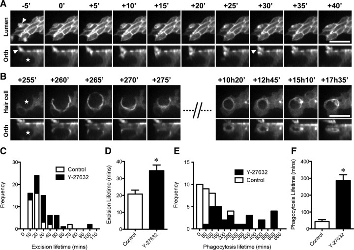
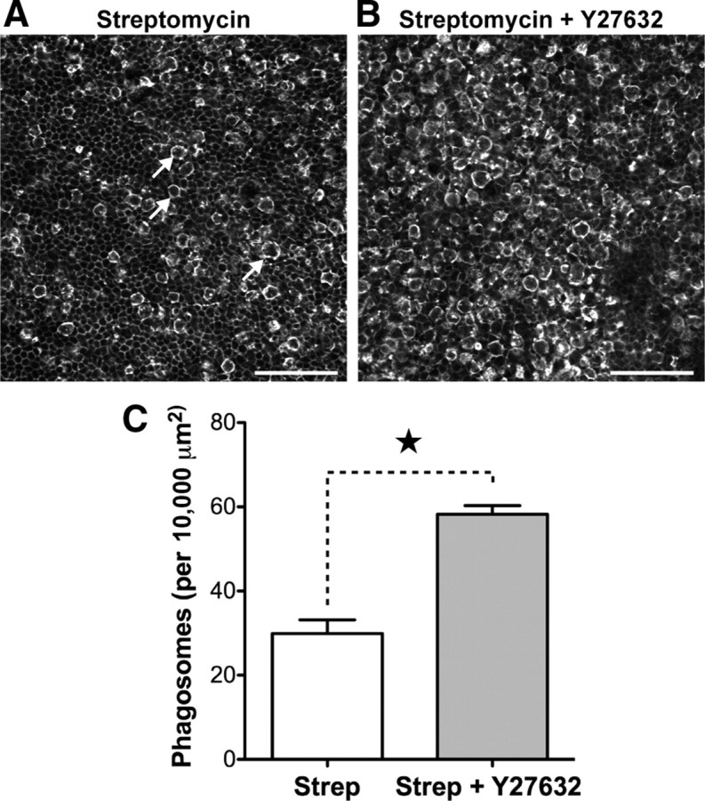
p < 0.001). Data from Figure 4 were used as controls for statistical tests in C. Data are expressed as mean ± SEM. Scale bars: 50 μm.

References

    1. Abrashkin KA, Izumikawa M, Miyazawa T, Wang CH, Crumling MA, Swiderski DL, Beyer LA, Gong TW, Raphael Y. The fate of outer hair cells after acoustic or ototoxic insults. Hear Res. 2006;218:20–29. - PubMed
    1. Bement WM, Forscher P, Mooseker MS. A novel cytoskeletal structure involved in purse string wound closure and cell polarity maintenance. J Cell Biol. 1993;121:565–578. - PMC - PubMed
    1. Ben-Yosef T, Belyantseva IA, Saunders TL, Hughes ED, Kawamoto K, Van Itallie CM, Beyer LA, Halsey K, Gardner DJ, Wilcox ER, Rasmussen J, Anderson JM, Dolan DF, Forge A, Raphael Y, Camper SA, Friedman TB. Claudin 14 knockout mice, a model for autosomal recessive deafness DFNB29, are deaf due to cochlear hair cell degeneration. Hum Mol Genet. 2003;12:2049–2061. - PubMed
    1. Bhave SA, Oesterle EC, Coltrera MD. Macrophage and microglia-like cells in the avian inner ear. J Comp Neurol. 1998;398:241–256. - PubMed
    1. Bohne BA, Rabbitt KD. Holes in the reticular lamina after noise exposure: implication for continuing damage in the organ of Corti. Hear Res. 1983;11:41–53. - PubMed

Publication types

MeSH terms

LinkOut - more resources