. 2010 Oct 15;185(8):4659-72.
doi: 10.4049/jimmunol.1000715. Epub 2010 Sep 15.

Development of a nascent galectin-1 chimeric molecule for studying the role of leukocyte galectin-1 ligands and immune disease modulation

Affiliations

Development of a nascent galectin-1 chimeric molecule for studying the role of leukocyte galectin-1 ligands and immune disease modulation

Filiberto Cedeno-Laurent et al. J Immunol. .

Abstract

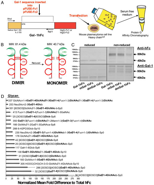
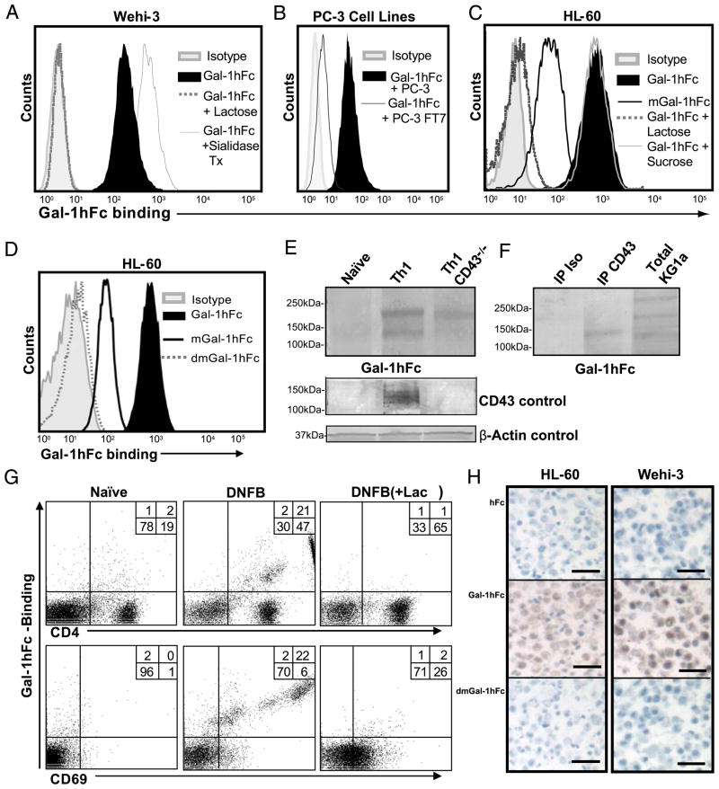
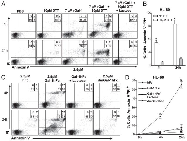
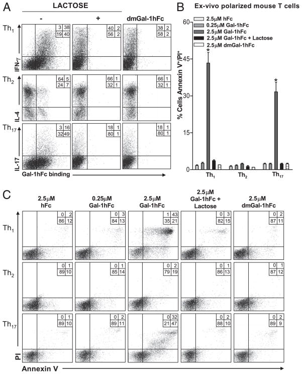
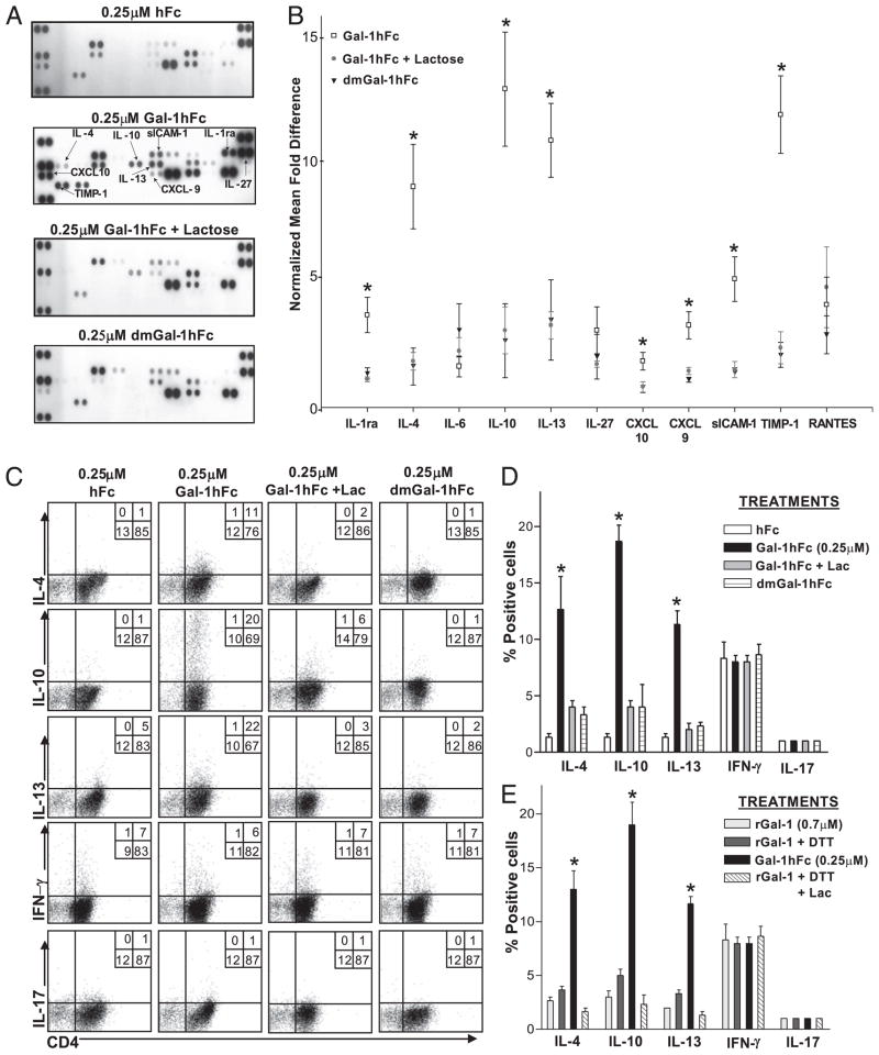
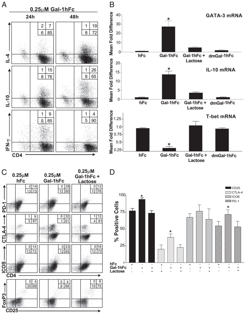
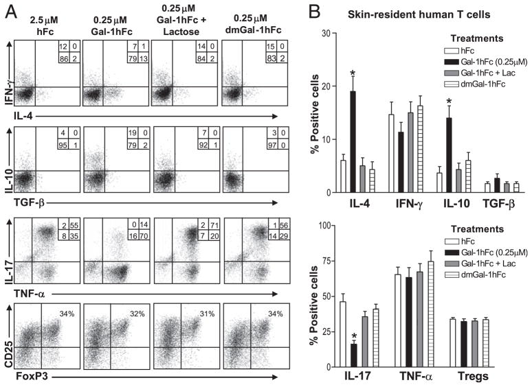
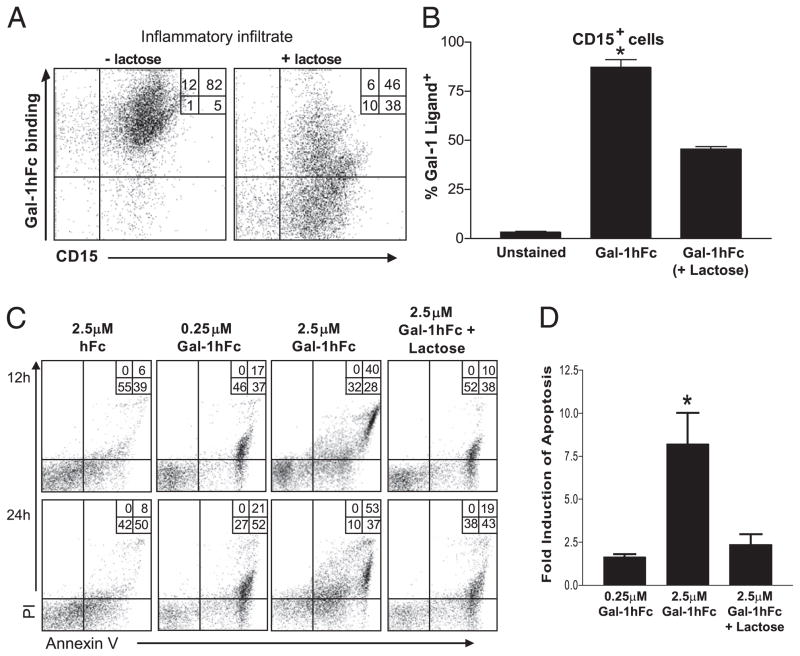
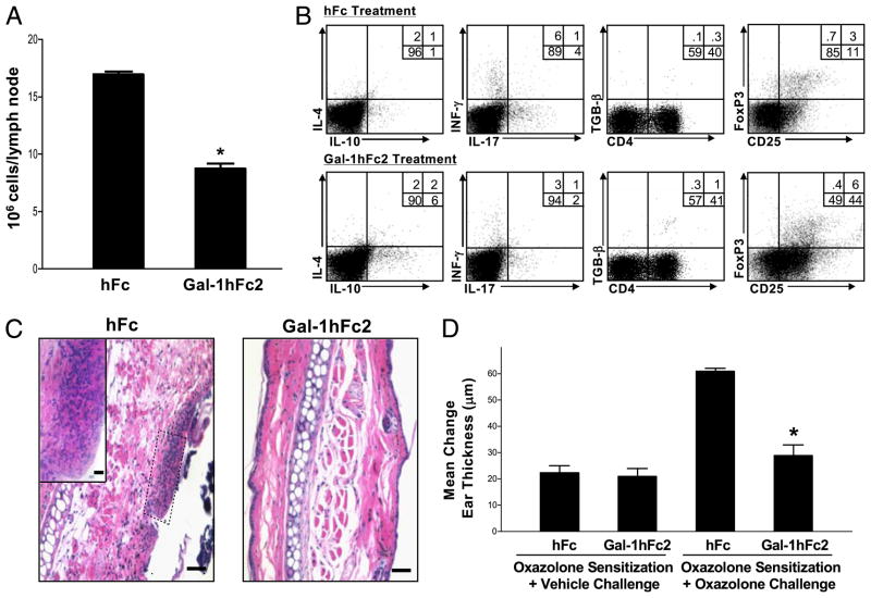
Galectin-1 (Gal-1), a β-galactoside-binding lectin, plays a profound role in modulating adaptive immune responses by altering the phenotype and fate of T cells. Experimental data showing recombinant Gal-1 (rGal-1) efficacy on T cell viability and cytokine production, nevertheless, is controversial due to the necessity of using stabilizing chemicals to help retain Gal-1 structure and function. To address this drawback, we developed a mouse Gal-1 human Ig chimera (Gal-1hFc) that did not need chemical stabilization for Gal-1 ligand recognition, apoptosis induction, and cytokine modulation in a variety of leukocyte models. At high concentrations, Gal-1hFc induced apoptosis in Gal-1 ligand(+) Th1 and Th17 cells, leukemic cells, and granulocytes from synovial fluids of patients with rheumatoid arthritis. Importantly, at low, more physiologic concentrations, Gal-1hFc retained its homodimeric form without losing functionality. Not only did Gal-1hFc-binding trigger IL-10 and Th2 cytokine expression in activated T cells, but members of the CD28 family and several other immunomodulatory molecules were upregulated. In a mouse model of contact hypersensitivity, we found that a non-Fc receptor-binding isoform of Gal-1hFc, Gal-1hFc2, alleviated T cell-dependent inflammation by increasing IL-4(+), IL-10(+), TGF-β(+), and CD25(high)/FoxP3(+) T cells, and by decreasing IFN-γ(+) and IL-17(+) T cells. Moreover, in human skin-resident T cell cultures, Gal-1hFc diminished IL-17(+) T cells and increased IL-4(+) and IL-10(+) T cells. Gal-1hFc will not only be a useful new tool for investigating the role of Gal-1 ligands in leukocyte death and cytokine stimulation, but for studying how Gal-1-Gal-1 ligand binding shapes the intensity of immune responses.


Figures

FIGURE 1
FIGURE 1
Construction and purification of Gal-1hFc and its mutants. A, Mouse Gal-1 cDNA containing native signal sequence (SS) or IL-2-SS was ligated in-frame into commercially available vector encoding the Fc region of IgG1 (pFUSE-Fc1) or the non-Fc receptor-binding mutant (pFUSE-Fc2), respectively. Purified plasmid DNA was transfected into J558L mouse plasmacytoma cells, drug selected and grown in serum-free medium. Gal-1hFc was purified by protein-G affinity chromatography. B, Schematic representation of Gal-1hFc in its reduced and nonreduced forms. C, Purified Gal-1hFc and its mutants were analyzed by SDS-PAGE and Western blotting with anti-human Fc or anti-mouse Gal-1 mAbs. D, Gal-1hFc or hFc was incubated on a covalent printed glycan array (version 4.0) developed by Core H investigators of the Consortium for Functional Glycomics. Mean fluorescence intensities of Gal-1hFc binding were normalized by dividing Gal-1hFc fluorescence intensities by control hFc-binding intensities and graphed as mean fold difference. The top 20 normalized glycans are listed.
FIGURE 2
FIGURE 2
Carbohydrate-binding activity of Gal-1hFc and its mutants to hematopoietic and nonhematopoietic cells by flow cytometry, Western blotting, and immunohistochemistry. A, Gal-1hFc binding to Wehi-3 cells or cells treated with Vibrio cholerae sialidase (0.2 U/ml) for 30 min at 37°C was assessed in the presence or absence of 50 mM lactose. B, Gal-1hFc binding was assayed on PC-3 cells and on PC-3 α1,3 fucosyltransferase 7 (FT7) transfectants (24). C and D, Gal-1hFc, mGal-1hFc, and dmGal-1hFc binding to HL-60 cells was assayed in the presence or absence of 50 mM lactose or sucrose. E, Lysates (30 μg/lane) from naive Th cells or polarized Th1 cells isolated from wt or CD43−/− mice were subjected to reducing 4–20% SDS-PAGE gels, blotted with Gal-1hFc, anti-CD43 mAb (1B11), or anti–β-actin mAb and then with respective AP-secondary Ab. F, KG1a cell lysate (30 μg/lane) and anti-CD43 and isotype control immunoprecipitates from KG1a cells were separated by 4–20% reducing SDS-PAGE gradient gels and then blotted with Gal-1hFc and AP–anti-hFc. G, Lymphocytes from LNs draining DNFB-sensitized or naive skin were analyzed by flow cytometry with Gal-1hFc, and anti-CD4 and -CD69 mAbs. Lactose (+ lac) was added to assay and washing buffers to control for carbohydrate-mediated binding. H, Sections of paraffin-embedded, formalin-fixed HL-60 or Wehi-3 cells were immunostained with 10 μg/ml Gal-1hFc or controls (hFc and dmGal-1hFc). Scale bars, 20 μm. Original magnification ×20. All data are representative of at least three experiments.
FIGURE 3
FIGURE 3
Comparative analysis of apoptosis induction in promyeloleukemic HL-60 cells using rGal-1 and Gal-1hFc. A, HL-60 cells were incubated with rGal-1 in the presence or absence of DTT and/or 50 mM lactose. Cell death analysis (Annexin V staining/PI uptake) was evaluated by flow cytometry at 4 and 24 h after incubation. B, Graphical representation of data from three different experiments depicting mean Annexin V+ staining/PI+ uptake values (±SD). *Statistically significant difference compared with no DTT control, p ≤ 0.01. C, HL-60 cells were incubated with Gal-1hFc or dmGal-1hFc in the presence or absence of 50 mM lactose. D, Graphical representation of data from three different experiments depicting mean Annexin V+ staining/PI+ uptake values (±SD) at 4 and 24 h after incubation. *Statistically significant difference compared with hFc control, p ≤ 0.01.
FIGURE 4
FIGURE 4
Galectin-1 Gal-1hFc induces apoptosis in Th1 and Th17 cell subsets. A, Gal-1hFc binding was assessed on mouse ex vivo polarized Th cell subsets, and Th cell phenotype was assayed by intracellular cytokine staining for IL-4, IFN-γ, and IL-17. dmGal-1hFc and Gal-1hFc binding was performed in the presence or absence of lactose. B, Graphical representations of data from three experiments showing apoptosis (Annexin V+ and PI+) in Th1, Th2, and Th17 cell subsets after a 24-h treatment with 2.5 μM Gal-1hFc in the presence or absence of 50 mM lactose or dmGal-1hFc. *Statistically significant difference compared with hFc control, p ≤ 0.01. C, Representative FACS plots of Gal-1hFc–mediated apoptosis (Annexin V+ and PI+) on Th cell subsets are shown.
FIGURE 5
FIGURE 5
Gal-1hFc stimulates the secretion of immunoregulatory molecules and alters Th cell differentiation. A, Naive Th cells were isolated by immunomagnetic beads from mouse spleens and activated for 48 h with anti-CD3/CD28, and further incubated for an additional 24 h with Gal-1hFc (±50 mM lactose), control hFc, or dmGal-1hFc. Supernatants were collected and analyzed for expression of 40 cytokines with a mouse cytokine panel array kit and quantified by OD, and mean densities were normalized to hFc-treated group. The complete list of cytokines and their spatial arrangement in the array are shown in Supplemental Fig. S2C. B, Graphic representation of data from three experiments is shown as normalized mean fold difference. *Statistically significant difference compared with lactose control, p ≤ 0.01. C, Activated Th0 cells incubated with Gal-1hFc (0.25 μM) or controls for 24 h were stimulated with PMA/ionomycin in the presence of brefeldin A for 6 h and then stained with anti–IL-4, –IL-10, –IL-13, –IFN-γ, and –IL-17 mAbs, and analyzed by flow cytometry. D, Graphic representations of data from three experiments are shown. *Statistically significant difference compared with hFc control, p ≤ 0.01. E, Activated Th0 cells were incubated with 0.7 μM rGal-1 with or without 80 μM DTT (± lactose [Lac]) for 24 h, stimulated with PMA/ionomycin in the presence of brefeldin A for 6 h, and then stained with anti–IL-4, –IL-10, –IL-13, –IFN-γ, and –IL-17 mAbs, and analyzed by flow cytometry. Data from three independent experiments are shown. *Statistically significant difference compared with rGal-1, p ≤ 0.01. Representative FACS plots are shown in Supplemental Fig. 2D.
FIGURE 6
FIGURE 6
Gal-1hFc alters Th cell differentiation, cytokine production, and expression of regulatory surface molecules. A, Naive mouse Th cells activated with anti-CD3/28 were treated with 0.25 μM Gal-1hFc; and IL-4, IL-10, and IFN-γ for 24 or 48 h were assessed by intracellular cytokine FACS staining. B, Transcriptional activity of GATA-3, IL-10, and T-bet mRNA was analyzed 8 h after incubation with Gal-1hFc by quantitative RT-PCR. Data are expressed as relative mRNA levels normalized to hFc treatment. *Statistically significant difference compared with hFc control, p ≤ 0.01. C, Naive mouse Th cells activated with anti-CD3/28 were incubated for 24 h with 0.25 μM hFc or Gal-1hFc (±50 mM lactose), or stained with anti–CTLA-4, –PD-1, -ICOS, and -CD25 mAbs, and analyzed by flow cytometry. D, Graphic representation of data from three independent experiments. *Statistically significant difference compared with hFc control, p ≤ 0.01.
FIGURE 7
FIGURE 7
Gal-1hFc modulates effector molecules in skin-resident human T cells. A, Human skin-resident effector memory T cells were incubated in the presence of Gal-1hFc or molecular controls for 24 h and stained with anti-CD25, -FoxP3, –IFN-γ, –IL-4, –IL-10, –TGF-β, –IL-17, and –TNF-α mAbs, and analyzed by flow cytometry. T cells were first gated on CD3+ and CD8 cells. B, Graphic representations of data taken from six independent donors. *Statistically significant difference compared with hFc control, p ≤ 0.01.
FIGURE 8
FIGURE 8
Gal-1hFc induces apoptosis of granulocytic infiltrates from synovial fluid of patients with RA. A, Inflammatory cells from synovial fluid of patients with RA were stained with Gal-1hFc and anti-CD15 mAb (±50 mM lactose), and analyzed by flow cytometry. B, Data from four independent donors are shown. C, CD15+ cells from synovial fluid samples were incubated with 0.25 or 2.5 μM Gal-1hFc (±lactose). Annexin V+ and PI+ cells were evaluated after 12 and 24 h after incubation. D, Graphed data are from a set of four independent donors. *Statistically significant difference compared with lactose control, p ≤ 0.01.
FIGURE 9
FIGURE 9
Galectin-1 human Ig chimera 2 (Gal-1hFc2) modulates cytokine production in T cells draining Ag-sensitized skin and alleviates Ag-dependent inflammation. A, Inguinal LNs draining oxazolone-sensitized skin from mice treated with hFc or Gal-1hFc2 (2.3 mg/kg) were harvested on day 6 after sensitization, minced, and analyzed for cellularity by trypan blue exclusion. Data are expressed as mean 106 cells/LN (±SD). *Statistically significant difference compared with hFc control, p ≤ 0.01. B, Lymphocytes from inguinal LNs were then restimulated for 6 h with PMA/ionomycin/brefeldin A and analyzed for IL-4, IL-10, IL-17, IFN-γ, TGF-β, FoxP3, and CD25 expression by flow cytometry. C, Ears from mice treated with hFc or Gal-1hFc2 were fixed in 10% formalin and stained with H&E. Scale bars, 20 μM. Original magnification ×10; inset, original magnification ×40. D, Change in ear thickness was ascertained 24 h after vehicle alone or oxazolone challenge. All experiments were repeated three times and consisted of three mice per group. *Statistically significant difference compared with hFc-treated mice receiving oxazolone sensitization and challenge, p ≤ 0.01.

Similar articles

Cited by

References

    1. Ilarregui JM, Croci DO, Bianco GA, Toscano MA, Salatino M, Vermeulen ME, Geffner JR, Rabinovich GA. Tolerogenic signals delivered by dendritic cells to T cells through a galectin-1-driven immunoregulatory circuit involving interleukin 27 and interleukin 10. Nat Immunol. 2009;10:981–991. - PubMed
    1. Garín MI, Chu CC, Golshayan D, Cernuda-Morollón E, Wait R, Lechler RI. Galectin-1: a key effector of regulation mediated by CD4+ CD25+ T cells. Blood. 2007;109:2058–2065. - PubMed
    1. Motran CC, Molinder KM, Liu SD, Poirier F, Miceli MC. Galectin-1 functions as a Th2 cytokine that selectively induces Th1 apoptosis and promotes Th2 function. Eur J Immunol. 2008;38:3015–3027. - PMC - PubMed
    1. Toscano MA, Bianco GA, Ilarregui JM, Croci DO, Correale J, Hernandez JD, Zwirner NW, Poirier F, Riley EM, Baum LG, Rabinovich GA. Differential glycosylation of TH1, TH2 and TH-17 effector cells selectively regulates susceptibility to cell death. Nat Immunol. 2007;8:825–834. - PubMed
    1. Karmakar S, Stowell SR, Cummings RD, McEver RP. Galectin-1 signaling in leukocytes requires expression of complex-type N-glycans. Glycobiology. 2008;18:770–778. - PMC - PubMed

Publication types

MeSH terms