. 2010 Dec 16;116(25):5622-30.
doi: 10.1182/blood-2010-05-285866. Epub 2010 Sep 15.

8-Aminoadenosine inhibits Akt/mTOR and Erk signaling in mantle cell lymphoma

Affiliations

8-Aminoadenosine inhibits Akt/mTOR and Erk signaling in mantle cell lymphoma

Jennifer B Dennison et al. Blood. .

Abstract

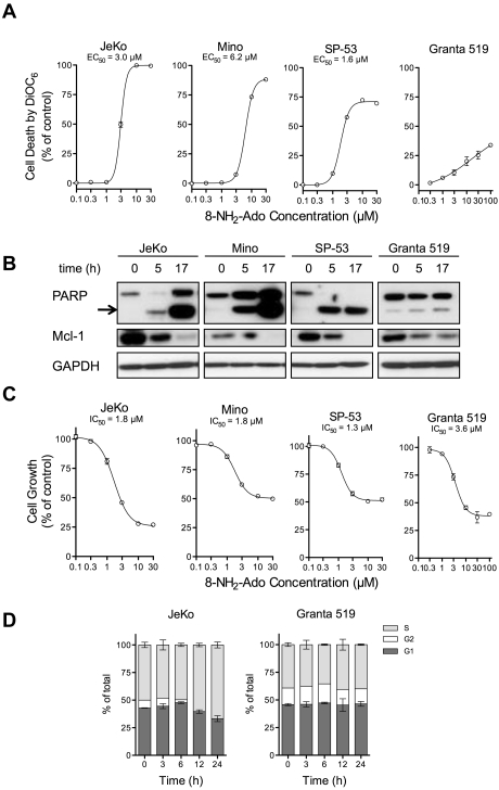
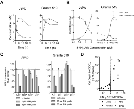
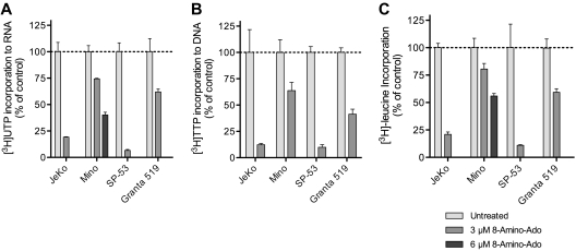
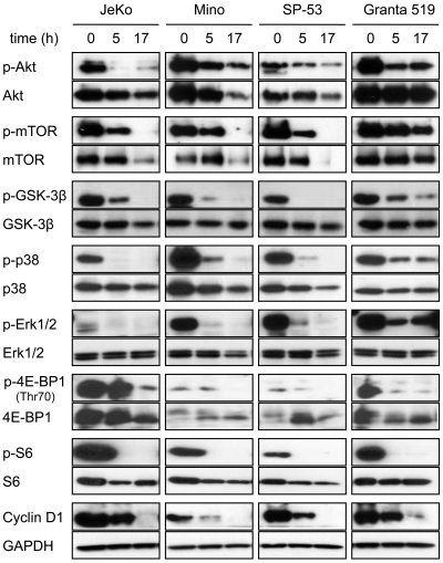
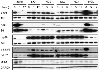
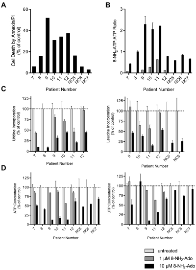
8-Aminoadenosine (8-NH(2)-Ado), a ribosyl nucleoside analog, in preclinical models of multiple myeloma inhibits phosphorylation of proteins in multiple growth and survival pathways, including Akt. Given that Akt controls the activity of mammalian target of rapamycin (mTOR), we hypothesized that 8-NH(2)-Ado would be active in mantle cell lymphoma (MCL), a hematological malignancy clinically responsive to mTOR inhibitors. In the current study, the preclinical efficacy of 8-NH(2)-Ado and its resulting effects on Akt/mTOR and extracellular-signal-regulated kinase signaling were evaluated using 4 MCL cell lines, primary MCL cells, and normal lymphocytes from healthy donors. For all MCL cell lines, 8-NH(2)-Ado inhibited growth and promoted cell death as shown by reduction of thymidine incorporation, loss of mitochondrial membrane potential, and poly (adenosine diphosphate-ribose) polymerase cleavage. The efficacy of 8-NH(2)-Ado was highly associated with intracellular accumulation of 8-NH(2)-adenosine triphosphate (ATP) and loss of endogenous ATP. Formation of 8-NH(2)-ATP was also associated with inhibition of transcription and translation accompanied by loss of phosphorylated (p-)Akt, p-mTOR, p-Erk1/2, p-phosphoprotein (p)38, p-S6, and p-4E-binding protein 1. While normal lymphocytes accumulated 8-NH(2)-ATP but maintained their viability with 8-NH(2)-Ado treatment, primary lymphoma cells accumulated higher concentrations of 8-NH(2)-ATP, had increased loss of ATP, and underwent apoptosis. We conclude that 8-NH(2)-Ado is efficacious in preclinical models of MCL and inhibits signaling of Akt/mTOR and Erk pathways.


Figures

Figure 1
Figure 1
8-NH2-Ado promotes cell death and inhibits cell growth but with only minor cell-cycle effects in MCL cell lines. (A) Cell death after 24 hours of continuous incubation was quantified by loss of mitochondrial membrane potential as measured by DiOC6 staining, at various concentrations of 8-NH2-Ado. For each cell line, the dose-response curve was estimated by nonlinear regression using a variable Hill Slope model. The EC50 was defined as the drug concentration required to achieve 50% of the maximum response. The standard errors of the estimated EC50 values for JeKo, Mino, and SP-53 were less than 3%; the EC50 value could not be accurately estimated for Granta 519. (B) Cell death was also evaluated by PARP cleavage. The arrow denotes cleaved PARP. The cells were continuously incubated with 3μM 8-NH2-Ado for 24 hours. (C) The growth inhibition of MCL cells was determined after 24 hours of continuous exposure to multiple concentrations of 8-NH2-Ado. The cell concentrations were quantified using a particle count and size analyzer (Beckman Coulter). The IC50 values were estimated by nonlinear regression using a variable Hill Slope model. The IC50 value was defined as the drug concentration required to achieve 50% of the maximum growth inhibition. The standard errors for the IC50 value estimates were less than 8% for all cell lines. (D) The cell-cycle effects were determined by flow cytometry for JeKo and Granta 519 after continuous treatment with 3μM 8-NH2-Ado at various time points up to 24 hours. Independent experiments were performed in triplicate shown with SD error bars.
Figure 2
Figure 2
Cell death and growth inhibition with 8-NH2-Ado are associated with accumulation of intracellular 8-NH2-ATP and reduction of ATP and UTP concentrations. (A) Three micromolar 8-NH2-Ado accumulated as 8-NH2-ATP and depleted ATP after continuous treatment for up to 24 hours. (B) 8-NH2-ATP accumulation and depletion of ATP was dose-dependent after 24 hours for MCL cell lines JeKo and Granta 519. (C) ATP and UTP pool concentrations were substantially depleted after 24 hours of continuous 3μM 8-NH2-Ado treatment. For panels A through C, experiments were performed with JeKo and Granta 519 cells. (D) Cell death after 24 hours of 8-NH2-Ado treatment at multiple concentrations was positively associated with 8-NH2-ATP:ATP ratios for the 4 MCL cell lines. Independent experiments were performed in triplicate shown with SD error bars.
Figure 3
Figure 3
8-NH2-Ado treatment of MCL cell lines reduces the rates of macromolecule synthesis. 8-NH2-Ado inhibited the rates of (A) transcription as quantified by uridine incorporation, (B) DNA synthesis as quantified by thymidine incorporation, and (C) translation as quantified by leucine incorporation. MCL cell lines were treated continuously with 3μM 8-NH2-Ado for 24 hours. Because 3μM 8-NH2-Ado was below its EC50 (Figure 1), Mino cells were also treated with 6μM 8-NH2-Ado for uridine and leucine incorporation experiments. All sample values were normalized to cell count. For [3H]uridine and [3H]thymidine incorporation values, the total counts of the acid-insoluble pellet were also normalized to the specific activity of UTP and TTP in the acid-soluble fraction at the end of the 45-minute chase. Independent experiments were performed in triplicate shown with SD error bars.
Figure 4
Figure 4
8-NH2-Ado inhibits Akt and Erk signaling pathways of MCL cell lines. JeKo, Mino, SP-53, and Granta 519 cells were incubated continuously with 3μM 8-NH2-Ado for 0, 5, and 17 hours. Cell lysates (30 μg) were immunoblotted for phospho- and total protein levels as shown. GAPDH was used as a loading control.
Figure 5
Figure 5
Actions of 8-NH2-Ado on Akt and Erk signaling pathways of peripheral blood monocytes. Freshly isolated PBMCs in 10% fetal bovine serum (FBS), RPMI were incubated continuously with 3μM 8-NH2-Ado for 0, 5, and 17 hours. Lymphoma cells from a lymphoma patient (MCL) were collected from the peripheral blood and allowed to proliferate in culture (20% FBS in Dulbecco modified Eagle medium). Once sufficient cell counts were obtained, the lymphoma cells were incubated continuously with 3μM 8-NH2-Ado for 0, 5, and 17 hours. JeKo cell lysates (Figure 4) were used for comparison. Cell lysates (30 μg) were immunoblotted for phospho- and total protein levels as shown. GAPDH was used as a loading control.
Figure 6
Figure 6
Differential effects of 8-NH2-Ado on primary lymphoma cells and normal PBMCs. Freshly isolated cells were exposed to 1μM (gray bars) or 10μM (black bars) 8-NH2-Ado continuously for up to 48 hours. PBMC data from healthy donors are presented as controls. (A) Cell death by Annexin-V staining was normalized to untreated control cells. (B) The combined relative 8-NH2-Ado accumulation and ATP loss were represented by 8-NH2-ATP:ATP ratios. (C) Inhibition of transcription (left) and translation (right) was quantified by [3H]uridine and [3H]leucine incorporation. (D) The reduction in ATP (left) and UTP (right) levels were determined by HPLC analyses. When cell numbers were sufficient, independent experiments were performed in triplicate shown with SD error bars. All cells were incubated with 1 or 10μM 8-NH2-Ado for 48 hours in 10% human serum/RPMI with the following exceptions: patient 9 cells were incubated for 24 hours in 10% FBS/RPMI, and patient 12 cells were incubated for 24 hours. Patient numbers 7, 8, 9, and 12 were diagnosed with MCL. Patient 10 was diagnosed with marginal zone B-cell lymphoma, and patient 11 was diagnosed with splenic marginal zone B-cell lymphoma in transformation.

References

    1. Medeiros LJ, Van Krieken JH, Jaffe ES, Raffeld M. Association of Bcl-1 rearrangements with lymphocytic lymphoma of intermediate differentiation. Blood. 1990;76(10):2086–2090. - PubMed
    1. Motokura T, Bloom T, Kim HG, et al. A novel cyclin encoded by a Bcl1-linked candidate oncogene. Nature. 1991;350(6318):512–515. - PubMed
    1. Stilgenbauer S, Winkler D, Ott G, et al. Molecular characterization of 11q deletions points to a pathogenic role of the ATM gene in mantle cell lymphoma. Blood. 1999;94(9):3262–3264. - PubMed
    1. Rizzatti EG, Falcao RP, Panepucci RA, et al. Gene expression profiling of mantle cell lymphoma cells reveals aberrant expression of genes from the PI3K-AKT, WNT and TGFbeta; signalling pathways. Br J Haematol. 2005;130(4):516–526. - PubMed
    1. Rudelius M, Pittaluga S, Nishizuka S, et al. Constitutive activation of Akt contributes to the pathogenesis and survival of mantle cell lymphoma. Blood. 2006;108(5):1668–1676. - PMC - PubMed

Publication types

MeSH terms