. 2010 Dec;299(6):C1450-60.
doi: 10.1152/ajpcell.00029.2010. Epub 2010 Sep 15.

Syntaxin 3 is necessary for cAMP- and cGMP-regulated exocytosis of CFTR: implications for enterotoxigenic diarrhea

Affiliations

Syntaxin 3 is necessary for cAMP- and cGMP-regulated exocytosis of CFTR: implications for enterotoxigenic diarrhea

Anne Collaco et al. Am J Physiol Cell Physiol. 2010 Dec.

Abstract

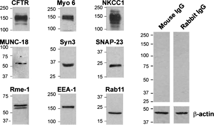
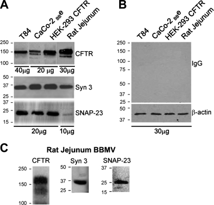
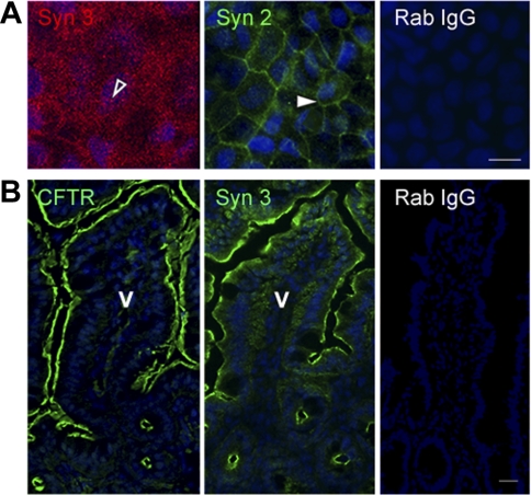
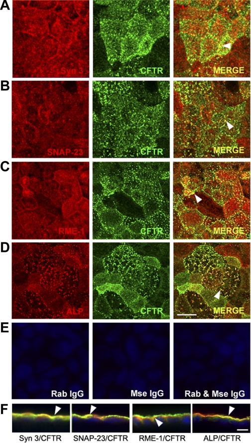
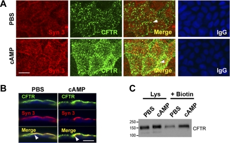
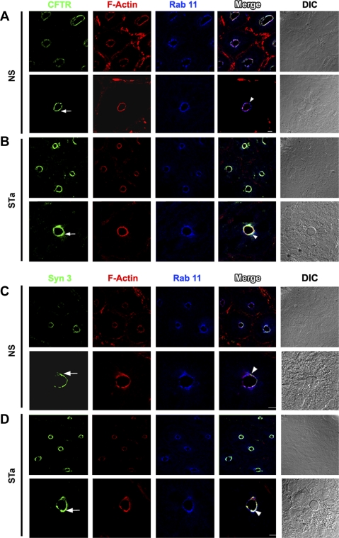
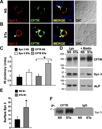
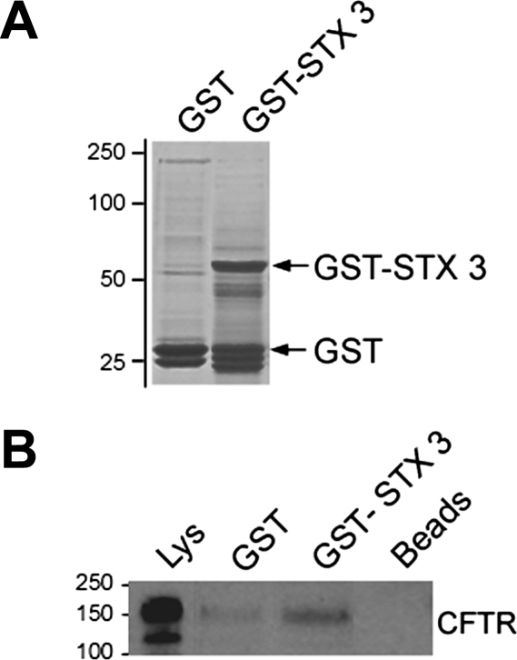
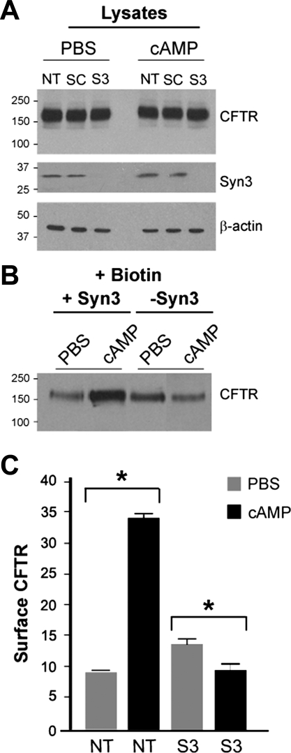
Enterotoxins elaborated by Vibrio cholerae and Escherichia coli cannot elicit fluid secretion in the absence of functional cystic fibrosis transmembrane conductance regulator (CFTR) chloride channels. After enterotoxin exposure, CFTR channels are rapidly recruited from endosomes and undergo exocytic insertion into the apical plasma membrane of enterocytes to increase the number of channels on the cell surface by at least fourfold. However, the molecular machinery that orchestrates exocytic insertion of CFTR into the plasma membrane is largely unknown. The present study used immunofluorescence, immunoblotting, surface biotinylation, glutathione S-transferase (GST) pulldown assays, and immunoprecipitation to identify components of the exocytic soluble N-ethylmaleimide (NEM)-sensitive factor attachment receptor (SNARE) vesicle fusion machinery in cyclic nucleotide-activated exocytosis of CFTR in rat jejunum and polarized intestinal Caco-2(BB)e cells. Syntaxin 3, an intestine-specific SNARE, colocalized with CFTR on the apical domain of enterocytes in rat jejunum and polarized Caco-2(BB)e cells. Coimmunoprecipitation and GST binding studies confirmed that syntaxin 3 interacts with CFTR in vivo. Moreover, heat-stable enterotoxin (STa) activated exocytosis of both CFTR and syntaxin 3 to the surface of rat jejunum. Silencing of syntaxin 3 by short hairpin RNA (shRNA) interference abrogated cyclic nucleotide-stimulated exocytosis of CFTR in cells. These observations reveal a new and important role for syntaxin 3 in the pathophysiology of enterotoxin-elicited diarrhea.


Figures

Fig. 1.
Fig. 1.
Western blot analysis of endogenous cystic fibrosis transmembrane conductance regulator (CFTR) and apical recycling, exocytic, and ion transport proteins in polarized Caco-2BBe cells. Caco-2BBe cell lysates (20 μg) were resolved by SDS-PAGE, and proteins were detected by Western blot. Bands show endogenous expression of endocytic (EEA-1, myosin VI) and apical recycling (rme-1, rab11) proteins and exocytic proteins involved in vesicle fusion (SNAP 23, munc-18, syntaxin 3), basolateral chloride entry transport (NKCC1), and apical exit (CFTR). Control immunoblots probed with relevant IgG and β-actin loading are shown. Molecular mass standards (kDa) are indicated.
Fig. 2.
Fig. 2.
Western blot analysis of soluble N-ethylmaleimide (NEM)-sensitive factor attachment receptor (SNARE) proteins and CFTR in cultured cells and rat jejunum. Brush border membrane vesicle (BBMV) preparations and cell lysates (10–40 μg protein) were resolved by SDS-PAGE, and proteins were detected by immunoblotting. A: bands show endogenous expression of CFTR, syntaxin 3 (Syn 3), and SNAP-23 in T84, Caco-2BBe, and HEK-293 cells and rat jejunum. B: control immunoblot probed with relevant IgG antibodies and β-actin loading controls are shown. C: bands show CFTR, syntaxin 3, and SNAP-23 detected in BBMV preparations from rat jejunum. Molecular mass standards (kDa) are indicated.
Fig. 3.
Fig. 3.
Polarized distribution of syntaxin 2 and 3 in the intestine. Confluent polarized Caco-2BBe cells (A) and cryostat sections of mouse jejunum (B) were fixed and immunofluorescence performed as described in materials and methods. A: en face image taken at the level of the brush border shows the apical distribution of syntaxin 3 (open arrowhead). Image taken at basal level shows distribution of syntaxin 2 (Syn 2, solid arrowhead). B: staining for CFTR (green) and syntaxin 3 (green) in the apical domain of crypt and villus sections of mouse jejunum. Control labeling of cells (A) and tissue sections (B) with rabbit IgG (Rab IgG) antibody is shown. Hoechst nuclear stain labels the nuclei (blue) V, villus. Scale bar, 10 μm.
Fig. 4.
Fig. 4.
Subcellular distribution of endogenous CFTR and apical recycling and exocytic proteins in polarized Caco-2BBe cells. Confluent polarized Caco-2BBe cells were fixed, double-label immunofluorescence was performed, and cells were examined by confocal microscopy. A–D: en face images taken just above the level of the brush border show the distribution of syntaxin 3 (red, A), SNAP-23 (red, B), rme-1 (red, C), alkaline phosphatase (ALP, red, D), and CFTR (green, A–D). Merged images show colocalization (merge, yellow, arrowhead). E: control labeling with rabbit (Rab) and/or mouse (Mse) IgG antibodies. F: merged images of xz vertical sections of immunolabeled cells. Arrowheads indicate colocalization. Hoechst nuclear stain labels nuclei blue. Scale bar, 10 μm.
Fig. 5.
Fig. 5.
cAMP stimulates apical recruitment of syntaxin 3 and CFTR in Caco-2BBe cells. Confluent monolayers of Caco-2BBe cells were treated with PBS or 1 mM cAMP. Cells were fixed, immunolabeled, and examined by confocal microscopy. A: en face views show the distribution of syntaxin 3 (red) and CFTR (green), and merged images show areas of colocalization (yellow) in PBS (top)- or cAMP (bottom)-treated cells. Control staining with relevant IgG antibodies is shown. B: images of vertical xz sections of immunolabeled cells show CFTR (green) and syntaxin 3 (red), and merged images (yellow) show colocalization (arrowheads). Scale bar, 10 μm. C: after PBS or cAMP treatment surface proteins were detected by sulfo-NHS-SS-biotin labeling and analyzed by Western blot as described in materials and methods. Cell lysates (Lys, 20 μg protein) and equivalent loads of surface biotinylated (+Biotin) proteins were resolved by SDS-PAGE and immunoblotted to detect CFTR. Molecular mass standards (kDa) are indicated.
Fig. 6.
Fig. 6.
Apical distribution of CFTR, syntaxin 3, and rab11 in rat jejunum after heat-stable enterotoxin (STa). Cryostat sections of rat jejunum treated with normal saline (NS) or STa were immunolabeled and examined by confocal microscopy as described in materials and methods. Low- and high-magnification images of crypt sections are shown. Images of immunolabeled sections from rat jejunum after NS (A and C) or STa (B and D) show the distribution of CFTR (green, arrow, A and B), F-actin (red, A–D), rab11 (blue, A–D), and syntaxin 3 (green, arrow, C and D). Merged images show areas of colocalization (white; arrowhead). DIC, differential interference contrast. Scale bars, 10 μm and 100 μm.
Fig. 7.
Fig. 7.
STa stimulates recruitment of CFTR and syntaxin 3 to the surface of rat jejunum. Cryostat sections of NS- or STa-treated rat jejunum were immunolabeled and examined by confocal microscopy. A and B: high-magnification images of crypt from NS (A)- or STa (B)-treated tissues show the apical distribution of syntaxin 3 (red) and CFTR (green) and merge (yellow). Increased apical staining for syntaxin 3 (B, arrowhead) and CFTR (green, arrowhead) after STa is shown. Merged images show areas of colocalization of CFTR and syntaxin 3 in NS (A, arrow)- and STa (B, arrowhead)-treated jejunum. Nuclei are stained with Hoechst stain (blue), Scale bar, 10 μm. C: quantification of fluorescence intensity (FI) of syntaxin 3 and CFTR label in the apical domain of NS- and STa-treated rat jejunum. Data represent means + SE (n > 10). *P < 0.01. D: after NS or STa treatment, surface proteins were labeled with sulfo-NHS-SS biotin in vivo, cells were lysed, and proteins were bound to streptavidin-agarose. Equivalent (20 μg) loads of protein from cell lysates and surface biotinylated samples were resolved by SDS-PAGE and immunoblotted to detect CFTR, syntaxin 3, and alkaline phosphatase. E: quantification (mean pixel values) of surface labeled (B+) syntaxin 3 (Syn 3) in NS- and STa-treated enterocytes. Data represent means + SE (n > 4). *P = 0.05. F: Western blot of CFTR coimmunoprecipitates from NS- and STa-treated jejunum detects increased syntaxin 3 in STa-treated jejunum. Equivalent starting proteins from tissue lysates were immunoprecipitated with either anti-CFTR or control rabbit IgG antibodies and bound to protein A beads, and samples were separated by SDS-PAGE and immunoblotted with anti-syntaxin 3 antibodies. Molecular mass standards (kDa) are indicated.
Fig. 8.
Fig. 8.
Glutathione S-transferase (GST) pulldown assay detects CFTR interaction with syntaxin 3 in rat jejunum. GST or GST + syntaxin 3 (GST-STX3) (200 μg) of were bound to glutathione Sepharose beads and incubated with rat jejunum lysates. Lysate and GST samples were resolved on SDS-PAGE gels and stained with Coomassie blue (A) or transferred to polyvinylidene difluoride nitrocellulose (B) and immunoblotted with anti-CFTR antibody. Molecular mass standards (kDa) are indicated.
Fig. 9.
Fig. 9.
Short hairpin RNA (shRNA) silencing of syntaxin 3 reduces surface CFTR in cAMP-stimulated Caco-2BBe cells. A: immunoblot analysis of Caco-2BBe cell lysates from control [nontransduced (NT) or scrambled shRNA (SC)] or after transduction with lentiviral particles containing syntaxin 3 (S3). Cells were treated with PBS or 1 mM DbcAMP (cAMP) for 30 min. Lysates (20 μg) were separated on SDS-PAGE gels and analyzed by Western blot using CFTR, syntaxin 3, or β-actin antibodies. Syntaxin 3 shRNA reduced syntaxin 3 expression by ∼100%. β-Actin loading control bands are shown. B: surface-biotinylated CFTR in PBS- or cAMP-stimulated cells transduced with syntaxin 3 shRNA. After PBS or 1 mM DbcAMP treatment, surface proteins were labeled with sulfo-NHS-SS-biotin. Cells were lysed, and proteins were bound to streptavidin-agarose. Equivalent biotinylated protein samples were resolved by SDS-PAGE and immunoblotted to detect CFTR. C: quantification (mean pixel values) of surface CFTR in control and syntaxin 3 shRNA (S3)-transduced Caco-2BBe cells. Data represent means + SE (n > 3).*P = 0.05. Molecular mass standards (kDa) are indicated.

Similar articles

Cited by

References

    1. Ameen N, Alexis J, Salas P. Cellular localization of the cystic fibrosis transmembrane conductance regulator in mouse intestinal tract. Histochem Cell Biol 114: 69– 75, 2000 - PubMed
    1. Ameen N, Apodaca G. Defective CFTR apical endocytosis and enterocyte brush border in myosin VI-deficient mice. Traffic 8: 998– 1006, 2007 - PubMed
    1. Ameen NA, Marino C, Salas PJ. cAMP-dependent exocytosis and vesicle traffic regulate CFTR and fluid transport in rat jejunum in vivo. Am J Physiol Cell Physiol 284: C429– C438, 2003 - PubMed
    1. Ameen NA, Martensson B, Bourguinon L, Marino C, Isenberg J. CFTR channel insertion to the apical surface in rat duodenal villus epithelial cells is upregulated by VIP in vivo. J Cell Sci 112: 887– 894, 1999 - PubMed
    1. Ameen NA, van Donselaar E, Posthuma G, de Jonge H, McLaughlin G, Geuze HJ, Marino C, Peters PJ. Subcellular distribution of CFTR in rat intestine supports a physiologic role for CFTR regulation by vesicle traffic. Histochem Cell Biol 114: 219– 228, 2000 - PubMed

Publication types