Case Reports
. 2010 Sep 16;467(7313):318-22.
doi: 10.1038/nature09328.

Transfusion independence and HMGA2 activation after gene therapy of human β-thalassaemia

Affiliations
Case Reports

Transfusion independence and HMGA2 activation after gene therapy of human β-thalassaemia

Marina Cavazzana-Calvo et al. Nature. .

Abstract

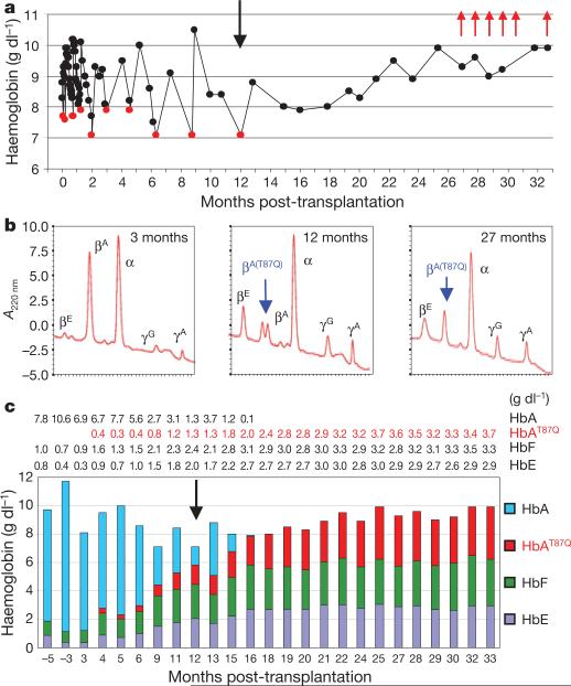
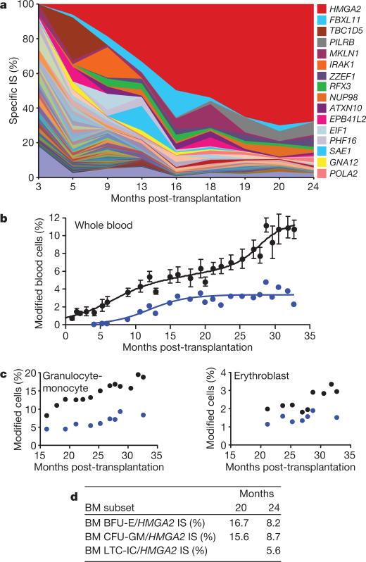
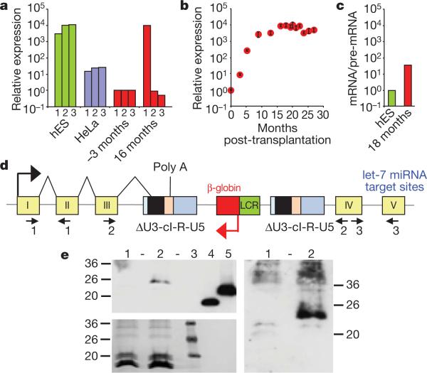
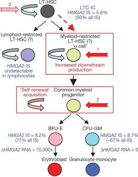
The β-haemoglobinopathies are the most prevalent inherited disorders worldwide. Gene therapy of β-thalassaemia is particularly challenging given the requirement for massive haemoglobin production in a lineage-specific manner and the lack of selective advantage for corrected haematopoietic stem cells. Compound β(E)/β(0)-thalassaemia is the most common form of severe thalassaemia in southeast Asian countries and their diasporas. The β(E)-globin allele bears a point mutation that causes alternative splicing. The abnormally spliced form is non-coding, whereas the correctly spliced messenger RNA expresses a mutated β(E)-globin with partial instability. When this is compounded with a non-functional β(0) allele, a profound decrease in β-globin synthesis results, and approximately half of β(E)/β(0)-thalassaemia patients are transfusion-dependent. The only available curative therapy is allogeneic haematopoietic stem cell transplantation, although most patients do not have a human-leukocyte-antigen-matched, geno-identical donor, and those who do still risk rejection or graft-versus-host disease. Here we show that, 33 months after lentiviral β-globin gene transfer, an adult patient with severe β(E)/β(0)-thalassaemia dependent on monthly transfusions since early childhood has become transfusion independent for the past 21 months. Blood haemoglobin is maintained between 9 and 10 g dl(-1), of which one-third contains vector-encoded β-globin. Most of the therapeutic benefit results from a dominant, myeloid-biased cell clone, in which the integrated vector causes transcriptional activation of HMGA2 in erythroid cells with further increased expression of a truncated HMGA2 mRNA insensitive to degradation by let-7 microRNAs. The clonal dominance that accompanies therapeutic efficacy may be coincidental and stochastic or result from a hitherto benign cell expansion caused by dysregulation of the HMGA2 gene in stem/progenitor cells.


Figures

Figure 1
Figure 1. Conversion to transfusion independence
a, Total Hb concentrations in whole blood. Red dots, transfusion time points; black vertical arrow, the last time the patient was transfused; red arrows, phlebotomies (200 ml each) to remove excess iron. b, HPLC blood globin chain profiles. Note that βA only derives from blood transfusions. c, Contribution of each Hb species, quantified by HPLC, to total blood Hb concentrations (in g dl–1). Actual numbers for each Hb species are indicated above the chart.
Figure 2
Figure 2. Genome-wide integration site (IS) distribution and HMGA2 IS clonal dominance
a, Relative abundance of vector-bearing cell clones in NBCs, as analysed by DNA pyrosequencing of LM-PCR products (one colour per IS, with the predominant HMGA2 IS in red). b, c, Mean percentages of vector-bearing NBCs at any IS (black dots, ±s.d.) versus the specific HMGA2 IS (blue dots), as quantified by qPCR in NBCs (b) and in specific cell fractions (c). d, Percentages of vector-bearing BFU-Es, CFU-GMs and LTC-ICs at the HMGA2 IS in bone marrow (BM) harvests.
Figure 3
Figure 3. Elevated, erythroid-specific expression of truncated HMGA2 transcripts
a, Staggered RT-qPCR of HMGA2 transcripts to detect the junctions between exons I–II (1), III–IV (2) and IV–V (3) before (–3 months) and after (16 months) transplantation. HeLa and human embryonic stem (hES) cells express full-length HMGA2 mRNA. b, Kinetics of HMGA2 transcripts (mean ± s.d.). c, Ratios of spliced versus unspliced HMGA2 RNA by RT-qPCR specific for either exon I–intron I or exon I–exon II junctions. d, Diagram of the vector at the HMGA2 IS, showing abnormal splicing and polyadenylation/cleavage of the truncated transcript within the vector provirus. ΔU3, self-inactivating U3; cI, cHS4. e, Western blot analysis with a polyclonal antibody against human HMGA2 and with the enhanced chemiluminescence (ECL, top left) or ECL advance (right) western blot detection systems. Numbers along the left and right edges are the weights (in kilodaltons) of the molecular markers. Bottom left shows Ponceau staining. Numbers along the top indicate blood progenitors from a β-thalassaemic control individual (1), patient P2's blood progenitors 27 months post-transplantation showing a band consistent with truncated fusion protein of 135 amino acids (2), recombinant truncated (73 amino acids) (4) and full-length (109 amino acids) (5) human HMGA2. Molecular mass markers are shown in (3). HMGA2 protein was not detected in CD15+ cells.
Figure 4
Figure 4. Homeostatic myeloid-biased cell expansion
Assuming that HMGA2 dysregulation was causative in the onset of clonal dominance, cell expansion is likely to occur by transient HMGA2 expression upstream of the erythroid/granulocyte-monocyte bifurcation (by lineage priming and/or loss of let-7 microRNA control). This is because the HMGA2 IS is represented in erythroblasts, granulocyte-monocyte and LTC-IC cells in similar proportions, whereas HMGA2 expression is only detected in erythroid cells. Because the HMGA2 IS is undetectable in lymphocytes, long-term (LT) homeostatic cell expansion is myeloid-biased. The HMGA2 IS initiating cell is likely to be a myeloid-biased LT-HSC/α cell with increased downstream cell production (1, in red) or a common myeloid progenitor with acquired self-renewal capability (2, in red).

Comment in

References

    1. Fucharoen S, Winichagoon P. Clinical and hematologic aspects of hemoglobin E β-thalassemia. Curr. Opin. Hematol. 2000;7:106–112. - PubMed
    1. Olivieri NF, et al. Studies in haemoglobin E β-thalassaemia. Br. J. Haematol. 2008;141:388–397. - PubMed
    1. Grosveld F, van Assendelft GB, Greaves DR, Kollias G. Position-independent, high-level expression of the human β-globin gene in transgenic mice. Cell. 1987;51:975–985. - PubMed
    1. Tuan D, London IM. Mapping of DNase I-hypersensitive sites in the upstream DNA of human embryonic ε-globin gene in K562 leukemia cells. Proc. Natl Acad. Sci. USA. 1984;81:2718–2722. - PMC - PubMed
    1. May C, et al. Therapeutic haemoglobin synthesis in β-thalassaemic mice expressing lentivirus-encoded human β-globin. Nature. 2000;406:82–86. - PubMed

Publication types

MeSH terms