. 2010 Sep 16;467(7313):333-7.
doi: 10.1038/nature09356.

Unexpected requirement for ELMO1 in clearance of apoptotic germ cells in vivo

Affiliations

Unexpected requirement for ELMO1 in clearance of apoptotic germ cells in vivo

Michael R Elliott et al. Nature. .

Abstract

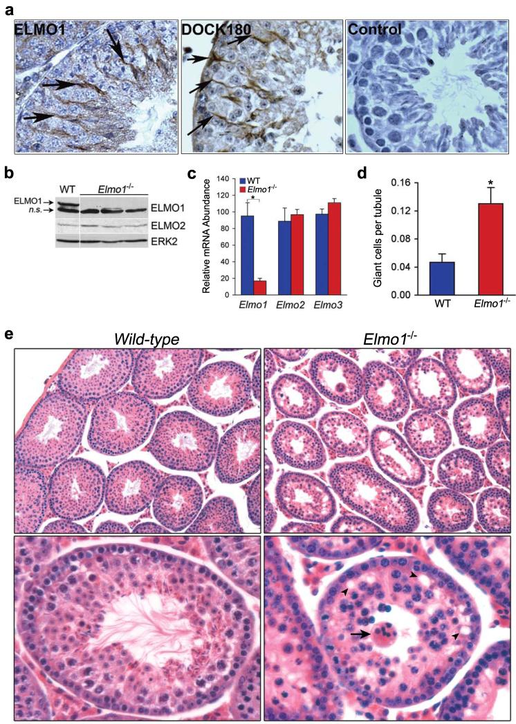
Apoptosis and the subsequent clearance of dying cells occurs throughout development and adult life in many tissues. Failure to promptly clear apoptotic cells has been linked to many diseases. ELMO1 is an evolutionarily conserved cytoplasmic engulfment protein that functions downstream of the phosphatidylserine receptor BAI1, and, along with DOCK1 and the GTPase RAC1, promotes internalization of the dying cells. Here we report the generation of ELMO1-deficient mice, which we found to be unexpectedly viable and grossly normal. However, they had a striking testicular pathology, with disrupted seminiferous epithelium, multinucleated giant cells, uncleared apoptotic germ cells and decreased sperm output. Subsequent in vitro and in vivo analyses revealed a crucial role for ELMO1 in the phagocytic clearance of apoptotic germ cells by Sertoli cells lining the seminiferous epithelium. The engulfment receptor BAI1 and RAC1 (upstream and downstream of ELMO1, respectively) were also important for Sertoli-cell-mediated engulfment. Collectively, these findings uncover a selective requirement for ELMO1 in Sertoli-cell-mediated removal of apoptotic germ cells and make a compelling case for a relationship between engulfment and tissue homeostasis in vivo.


Figures

Figure 1
Figure 1. Disrupted seminiferous tubule architecture in ELMO1-deficient mice
a, Immunolocalization of ELMO1 and DOCK180 in Sertoli cells (arrows) of adult C57BL/6 mice testes. Control, secondary antibody only. b, Immunoblotting of lysates from testes of adult mice with the indicated Elmo1 genotypes. A non-specific (n.s) band served as an internal loading control. c, mRNA levels of Elmo1, 2 and 3 in Sertoli cells relative to Gapdh and normalized to WT for each gene. *p=0.01, n=3 mice per group. d, Average number of giant cells per tubule in testes of 6-week old mice, *p=0.03, n=3 or 4 per group. e, H & E sections of testes of mice with the indicated Elmo1 genotypes. Magnification, 10× (upper panels); 25× (lower panels). Arrow, a giant cell; arrowhead, vacuolar spaces in the epithelium. Error bars indicate s.e.m.
Figure 2
Figure 2. Requirement for ELMO1 in apoptotic cell clearance in vivo and in vitro
a, Detection of apoptotic cells in the testes of wild-type and Elmo1−/− mice by IHC (apostain, brown) viewed under 10× (left) and 25× (right) magnification. Arrow, apoptotic cells. b, Average number of apoptotic nuclei per tubule. *p=0.04, n=3 or 5 mice per group. c, Total number of Sertoli cells isolated from testes of individual mice, n=4. d, e, Fluorescence microscopy of WT and Elmo1−/− Sertoli cells. d, Staining with antibodies against the Sertoli cell marker anti-Müllerian hormone (anti-Amh, green) or control IgG (inset). e, Sertoli cells were fed 2.1μm carboxylate beads (red) or apoptotic mouse spermatic germ cells labeled with CypHer 5 (red). Green, phalloidin staining. Arrows, cells with engulfed targets; arrowheads, individual engulfed targets. Blue, DAPI-stained nuclei. Scale bar, 50μm. f-h, In vitro phagocytosis by WT and Elmo1−/− Sertoli cells. Uptake of fluorescent 2.1μm carboxylate bead uptake (f) and CypHer 5-labeled apoptotic germ cells (h) was quantified by flow cytometry, n=4 mice per group, *p<0.005. g, Representative flow cytometry histograms of data in h.
Figure 3
Figure 3. ELMO1 functions in Sertoli cell-mediated apoptotic germ cell clearance
a, Detection of apoptotic cells (apostain, brown) in testes of 6-8 week Amh-Cre/Elmo1F/W and Amh-Cre/Elmo1F/F mice (arrow, apoptotic cell). b, qPCR of Elmo1 and Elmo2 mRNA levels in Sertoli cells from Amh-Cre+ mice with the indicated genotypes. c, Average number of apoptotic cells per tubule. *p=0.04, 4-6 mice per group. d, Total sperm output in 8 week Amh-Cre+ Elmo1F/F mice with littermate controls. *p=0.007, n=6-8 testis per group. e, Phagocytosis by TM4 Sertoli cells expressing GFP, GFP-tagged wild-type or dominant-negative ELMO1 (T625) expression constructs. f, RAC1-GTP levels in TM4 Sertoli cells transfected with the indicated constructs. RAC1-GTP relative to actin was quantified by densitometry. g, Uptake of 2.1μm carboxylated targets by TM4 Sertoli cells following siRNA knockdown of RAC1. Inset, immunoblot of lysates after RAC1 knockdown. DOCK180, loading control.
Figure 4
Figure 4. BAI1 participates as an engulfment receptor in Sertoli cell-mediated engulfment
a, Phosphatidylcholine (PC, blue) or phosphatidylserine (PtdSer, red) microbubble uptake by TM4 Sertoli cells. *p < 0.05, n=4. b, Levels of PtdSer recognition receptors by qPCR from the indicated murine tissues. # indicates mRNA below detectable level. c, Phagocytosis by TM4 Sertoli cells transfected with control or BAI1 siRNA. *p<0.05. d, Left, schematic of microinjection into rete testis. Rete testis and efferent ducts prior to (top right) and 24 h after injection of fluorescent microspheres under light (middle) and fluorescence (lower) microscopy of whole mounted seminiferous. Dashed lines, tubule wall borders. Arrow, cluster of microspheres internalized by Sertoli cells. e, 15P-1 Sertoli cells on Matrigel incubated with fluorescent targets along with GST or BAI1-TSR protein. f-g, Intra-testicular injection of GST or BAI1-TSR into 6-8 week old C57BL/6 mice. f, Apoptotic cells detected by IHC (apostain, brown) after 24 hr. Magnification, 12.5× (upper panels) and 25× (lower panels). Arrow, apoptotic cells. g, Sperm counts normalized to testis weight. n=3, *p<0.05.

Similar articles

Cited by

References

    1. Henson PM. Dampening inflammation. Nat Immunol. 2005;6:1179–1181. - PubMed
    1. Nagata S, Hanayama R, Kawane K. Autoimmunity and the clearance of dead cells. Cell. 140:619–630. - PubMed
    1. Gregory CD, Pound JD. Results of Defective Clearance of Apoptotic Cells: Lessons from Knock-out Mouse Models. In: Krysko DV, Vandenabeele P, editors. Phagocytosis of Dying Cells. Vol. 1. Springer Science; 2009. pp. 271–298.
    1. Zhou Z, Caron E, Hartwieg E, Hall A, Horvitz HR. The C. elegans PH domain protein CED-12 regulates cytoskeletal reorganization via a Rho/Rac GTPase signaling pathway. Dev Cell. 2001;1:477–489. - PubMed
    1. Wu YC, Tsai MC, Cheng LC, Chou CJ, Weng NY. C. elegans CED-12 acts in the conserved crkII/DOCK180/Rac pathway to control cell migration and cell corpse engulfment. Dev Cell. 2001;1:491–502. - PubMed

Publication types

MeSH terms