

Automated pattern-guided principal component analysis vs expert-based immunophenotypic classification of B-cell chronic lymphoproliferative disorders: a step forward in the standardization of clinical immunophenotyping
- PMID: 20844562
- PMCID: PMC3035971
- DOI: 10.1038/leu.2010.160
Automated pattern-guided principal component analysis vs expert-based immunophenotypic classification of B-cell chronic lymphoproliferative disorders: a step forward in the standardization of clinical immunophenotyping
Erratum in
- Leukemia. 2011 Feb;25(2):385
Abstract
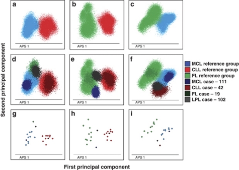
Immunophenotypic characterization of B-cell chronic lymphoproliferative disorders (B-CLPD) is becoming increasingly complex due to usage of progressively larger panels of reagents and a high number of World Health Organization (WHO) entities. Typically, data analysis is performed separately for each stained aliquot of a sample; subsequently, an expert interprets the overall immunophenotypic profile (IP) of neoplastic B-cells and assigns it to specific diagnostic categories. We constructed a principal component analysis (PCA)-based tool to guide immunophenotypic classification of B-CLPD. Three reference groups of immunophenotypic data files-B-cell chronic lymphocytic leukemias (B-CLL; n = 10), mantle cell (MCL; n = 10) and follicular lymphomas (FL; n = 10)--were built. Subsequently, each of the 175 cases studied was evaluated and assigned to either one of the three reference groups or to none of them (other B-CLPD). Most cases (89%) were correctly assigned to their corresponding WHO diagnostic group with overall positive and negative predictive values of 89 and 96%, respectively. The efficiency of the PCA-based approach was particularly high among typical B-CLL, MCL and FL vs other B-CLPD cases. In summary, PCA-guided immunophenotypic classification of B-CLPD is a promising tool for standardized interpretation of tumor IP, their classification into well-defined entities and comprehensive evaluation of antibody panels.
Figures
References
-
- Bottcher S, Ritgen M, Buske S, Gesk S, Klapper W, Hoster E, et al. Minimal residual disease detection in mantle cell lymphoma: methods and significance of four-color flow cytometry compared to consensus IGH-polymerase chain reaction at initial staging and for follow-up examinations. Haematologica. 2008;93:551–559. - PubMed
-
- Rawstron AC, Villamor N, Ritgen M, Bottcher S, Ghia P, Zehnder JL, et al. International standardized approach for flow cytometric residual disease monitoring in chronic lymphocytic leukaemia. Leukemia. 2007;21:956–964. - PubMed
-
- Ashman M, Sachdeva N, Davila L, Scott G, Mitchell C, Cintron L, et al. Influence of 4- and 6-color flow cytometers and acquisition/analysis softwares on the determination of lymphocyte subsets in HIV infection. Cytometry. 2007;72:380–386. - PubMed
-
- Braylan RC, Orfao A, Borowitz MJ, Davis BH. Optimal number of reagents required to evaluate hematolymphoid neoplasias: results of an international consensus meeting. Cytometry. 2001;46:23–27. - PubMed
