. 2010 Sep 9;6(9):e1001096.
doi: 10.1371/journal.ppat.1001096.

Cyclin-dependent kinase activity controls the onset of the HCMV lytic cycle

Affiliations

Cyclin-dependent kinase activity controls the onset of the HCMV lytic cycle

Martin Zydek et al. PLoS Pathog. .

Abstract

The onset of human cytomegalovirus (HCMV) lytic infection is strictly synchronized with the host cell cycle. Infected G0/G1 cells support viral immediate early (IE) gene expression and proceed to the G1/S boundary where they finally arrest. In contrast, S/G2 cells can be infected but effectively block IE gene expression and this inhibition is not relieved until host cells have divided and reentered G1. During latent infection IE gene expression is also inhibited, and for reactivation to occur this block to IE gene expression must be overcome. It is only poorly understood which viral and/or cellular activities maintain the block to cell cycle or latency-associated viral IE gene repression and whether the two mechanisms may be linked. Here, we show that the block to IE gene expression during S and G2 phase can be overcome by both genotoxic stress and chemical inhibitors of cellular DNA replication, pointing to the involvement of checkpoint-dependent signaling pathways in controlling IE gene repression. Checkpoint-dependent rescue of IE expression strictly requires p53 and in the absence of checkpoint activation is mimicked by proteasomal inhibition in a p53 dependent manner. Requirement for the cyclin dependent kinase (CDK) inhibitor p21 downstream of p53 suggests a pivotal role for CDKs in controlling IE gene repression in S/G2 and treatment of S/G2 cells with the CDK inhibitor roscovitine alleviates IE repression independently of p53. Importantly, CDK inhibiton also overcomes the block to IE expression during quiescent infection of NTera2 (NT2) cells. Thus, a timely block to CDK activity not only secures phase specificity of the cell cycle dependent HCMV IE gene expression program, but in addition plays a hitherto unrecognized role in preventing the establishment of a latent-like state.


Conflict of interest statement

The authors have declared that no competing interests exist.

Figures

Figure 1
Figure 1. Block of HCMV major immediate early (MIE) gene expression in S/G2 is neither virus strain nor cell type-dependent.
(A) Asynchronously proliferating HEL fibroblasts were infected with the indicated HCMV strains at high multiplicity of infection (MOI = 5). Three hours post infection (hpi) cells were harvested and analyzed for DNA content and MIE gene expression by flow cytometry. Shown are dot plots where cells were divided into four subpopulations: upper left quadrant - MIE-positive G1 cells (DNA content  = 2n), lower left quadrant - MIE-negative G1 cells, upper right quadrant – MIE-positive S/G2 cells (DNA content >2n), lower right quadrant – MIE-negative S/G2 cells. The relative proportion of each subpopulation is given in percent of total cells. In addition, the percentage of MIE-positive cells in the G1 compartment (both left quadrants) and the S/G2 compartment (right quadrants) is given as a separate information. These latter values are framed in the small boxes at the top corners of each diagram. (B, C) The indicated cell lines were infected with HCMV-AD169 (U373 cells, MOI = 5) or HCMV-TB40/e (HEK293, HeLa, HUVEC, MOI = 1) and harvested at 5 hpi. Cell cycle position was determined by propidium iodide staining (B) or EdU labelling (C), IE expression by IE1/IE2-specific antibody. (B) Flow cytometry data were analyzed as described above. (C) Cells were divided into MIE+EdU (upper left), MIEEdU (lower left), MIE+EdU+ (upper right) and MIEEdU+ (lower right) fractions. The proportion of each fraction was indicated in percent of total cell number. The small boxes in the top corners of the diagrams display the percentage of MIE-positive cells in the EdU+ fractions (left corner) and the EdU fractions (right corner) respectively.
Figure 2
Figure 2. Inhibition of cellular DNA synthesis has no immediate but a delayed effect on the start of MIE gene expression in S/G2.
(A) Experimental setup. Proliferating HEL fibroblasts were treated with aphidicolin (Aph) or hydroxyurea (HU) and infected with HCMV-AD169 at the indicated times after drug addition (0 h). At 5 hpi cells were harvested and subjected to propidium iodide and IE1/IE2-specific antibody staining, followed by flow cytometry. (B) Shown are dot plots where the numbers indicate the relative proportion of each quadrant in percent of total cells and (in the small boxes) the percentage of MIE-positive cells within the G1 and S/G2 fractions only, as described in the legend to figure 1.
Figure 3
Figure 3. The long-term cellular response to genotoxic stress makes S/G2 cells permissive for MIE gene expression.
(A) Experimental setup. Proliferating HEL fibroblasts were treated with sublethal doses of doxorubicin or UV-C radiation. At the indicated times after treatment, cells were infected with HCMV-AD169 and harvested 4 h later. (B) Flow cytometry analysis of DNA content and MIE gene expression, as described in the legend to figure 1.
Figure 4
Figure 4. Infection at late times after genotoxic stress allows the HCMV replicative cycle to proceed in S/G2 cells with similar kinetics as in normal G1 cells.
Proliferating HEL fibroblasts were incubated for 60 min with EdU to label S phase cells. After removal of EdU, cells were treated with doxorubicin (+doxo) or left untreated. 24 h post doxorubicin treatment, or – in the case of untreated cells – immediately after EdU labelling, cells were infected with HCMV-AD169. Cells were harvested at the indicated times post infection and analyzed for EdU incorporation and HCMV MIE (IE1/2), early (gB) and late (pp28) gene expression by flow cytometry. The percentage of cells staining positive for the different viral gene products was displayed as described in the legend to figure 1C.
Figure 5
Figure 5. P53 is required for the checkpoint-dependent rescue of MIE gene expression in S/G2.
HEL fibroblasts were stably transduced with lentiviruses expressing p53 or GFP-specific shRNAs. The resulting knock-down (KD) cells and a mock-infected control were treated with aphidicolin or doxorubicin as indicated. Immediately after treatment (0 h) or 24 h later cells were infected with HCMV-AD169 (A). Cells were harvested 4 h post infection and analyzed for expression of p53, p21 and GAPDH by immunoblotting (B) and for MIE expression versus DNA content by flow cytometry (C).
Figure 6
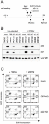
Figure 6. The positive effect of proteasome inhibition on MIE gene expression in S/G2 is mediated by p53 stabilization.
(A) Experimental setup. Proliferating HEL fibroblasts and the indicated knock-down derivatives were first incubated with EdU to label S-phase cells. After removal of EdU, cells were infected with HCMV-AD169 or left uninfected. Together with the virus, the proteasome inhibitor MG132 was added to the cells and was not removed before harvest at 8 hpi. A solvent control (DMSO) was included. (B) Cells were examined for expression of the indicated proteins by immunoblot analysis. (C) Flow cytometry analysis of MIE gene expression and EdU incorporation. The percentage of MIE-positive cells in the EdU-positive and EdU-negative fractions respectively were displayed as described in the legend to figure 1C.
Figure 7
Figure 7. DNA damage-dependent rescue of MIE gene expression in S/G2 depends on the CDK inhibitor p21.
Proliferating HCT116 wild type (wt), p53−/− and p21−/− cells were treated with doxorubicin and infected thereafter with HCMV-TB40/e at an MOI of 1. Infection was carried at 0 or 24 h post doxorubicin treatment (A). Cells were harvested at 7 hpi and analyzed for DNA content and MIE gene expression using flow cytometry (B), as described in the legend to figure 1.
Figure 8
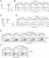
Figure 8. Roscovitine treatment efficiently abrogates the block of MIE gene expression in S/G2.
(A) Experimental setup of B. Proliferating HEL fibroblasts were infected with HCMV-AD169. Roscovitine, at the indicated concentrations, was added to the cells at the time of infection. It was left on the cells until harvest at 4 hpi. (C) Experimental setup of D. This time, roscovitine treatment was started 30 min before the fibroblasts were infected with HCMV. Treatment was stopped at 2 hpi and infected cells were maintained for further 3 h in regular growth medium. (B, D) MIE gene expression and DNA content of roscovitine treated cells were analyzed by flow cytometry. DMSO treated cells were also analyzed to control for solvent effects. (E, F) Proliferating U373 and HUVEC cells were incubated with EdU for 60 min to label cells undergoing DNA synthesis. After removal of EdU, cells were infected with HCMV and harvested 5 h later. Where indicated, roscovitine was transiently added to the cells according to the schedule described in C. After harvest, cells were analyzed for EdU incorporation and IE1/2-expression by flow cytometry, as detailed in the legend to figure 1.
Figure 9
Figure 9. The roscovitine-mediated rescue of MIE gene expression in S/G2 cells is p53 independent.
The indicated knock-down fibroblasts were treated with doxorubicin (+ Doxo) and infected by HCMV 24 later. Or cells were treated with 25 µM roscovitine (+ Rosco) and infected with HCMV shortly after the start of treatment, as detailed in figure 8C. Cells were harvested at 5 hpi and analyzed for MIE expression and DNA content, as described in the legend to figure 1.
Figure 10
Figure 10. CDK inhibition relieves the block of MIE gene expression in undifferentiated NT2 cells.
(A) Experimental setup. NT2 cells were infected with HCMV-TB40/e (MOI = 5) in the presence of the cyclin-dependent kinase (CDK) inhibitors roscovitine (Rosco), CVT313, SU9515, the histone deacetylase inhibitor trichostatine A (TSA) or solvent (DMSO). CDK inhibitors were used at concentrations from 10 to 50 µM (Rosco, CVT313) and from 2 to 10 µM (SU9516) as indicated. The different compounds were left on the cells for the indicated lengths of time. In addition, NT2 cells were infected after a 7-day exposure to retinoic acid (RA). All cells were harvested at 24 hpi. (B) Cells were stained for IE1/2 and Oct3/4 expression and analyzed by flow cytometry. Shown are dot plots where cells were divided into four subpopulations: MIEOct (lower left quadrant), MIE+Oct (upper left quadrant), MIE+Oct+ (upper right quadrant), MIEOct+ (lower right quadrant). The proportion of each subpopulation is given as percent of total cells. (C) Cells were stained for IE1/2 expression and DNA content and analyzed by flow cytometry. The depicted cells were divided into the following subpopulations: MIEG1 (lower left quadrant), MIE+G1 (upper left quadrant), MIE+S/G2/M (upper right quadrant) and MIE-S/G2/M (lower right quadrant) cells. The proportion of each subpopulation is given in percent of total cells.
Figure 11
Figure 11. Induction of MIE gene expression by CDK inhibition does not require nuclear localization of pp71.
(A) Undifferentiated NT2 cells, differentiated NT2 cells (treated with RA for 7 days) and mouse bone marrow (BM) cells were analyzed for Cyclin A2 expression by immunoblot analysis. GAPDH expression was analyzed to control for equal loading. (B) Undifferentiated and differentiated NT2 cells were infected with HCMV-TB40/e (MOI = 5). Where indicated, SU9516 at 10 µM final concentration was added together with the virus. At 6 hpi cells were analyzed for subcellular localization of pp71 (green) by immunofluorescence microscopy. Nuclei were stained with DAPI fluorochrome (blue). Non-infected cells (right panel) were analyzed to control for background staining. Scale bars (red)  = 10 µm.
Figure 12
Figure 12. Sequential regulation of the HCMV replication cycle by different CDKs.
After virus entry a cell cycle-regulated CDK is able to prevent the onset of IE gene expression. Once IE expression has started, CDK7 and 9 become an essential part of the viral transcription machinery. At early and late times of infection CDKs are needed for proper function and localization of viral substrates.

Similar articles

Cited by

References

    1. Mocarski ES, Shenk T, Pass RF. Cytomegaloviruses. In: Knipe DM, Howley PM, Griffin DE, Lamb RA, Martin MA, Roizman B, Straus SE, editors. Fields virology, 5th ed. Lippincott Williams & Wilkins, Philadelphia, PA; 2007. pp. 2701–2772.
    1. Meier JL, Stinski MF. Major immediate-early enhancer and its gene products. In Cytomegaloviruses: Molecular Biology and Immunology ed. In: Reddehase M, editor. Norfolk: Caister Academic Press; 2006. pp. 151–166.
    1. Bego M, Maciejewski J, Khaiboullina S, Pari G, St Jeor S. Characterization of an antisense transcript spanning the UL81-82 locus of human cytomegalovirus. J Virol. 2005;79:11022–11034. - PMC - PubMed
    1. Kondo K, Xu J, Mocarski ES. Human cytomegalovirus latent gene expression in granulocyte-macrophage progenitors in culture and in seropositive individuals. Proc Natl Acad Sci U S A. 1996;93:11137–11142. - PMC - PubMed
    1. Petrucelli A, Rak M, Grainger L, Goodrum F. Characterization of a novel Golgi apparatus-localized latency determinant encoded by human cytomegalovirus. J Virol. 2009;83:5615–5629. - PMC - PubMed

Publication types

MeSH terms

Substances