. 2010 Sep 9;6(9):e1001094.
doi: 10.1371/journal.ppat.1001094.

A novel family of Toxoplasma IMC proteins displays a hierarchical organization and functions in coordinating parasite division

Affiliations

A novel family of Toxoplasma IMC proteins displays a hierarchical organization and functions in coordinating parasite division

Josh R Beck et al. PLoS Pathog. .

Abstract

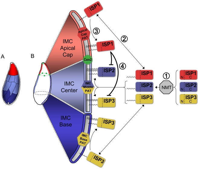
Apicomplexans employ a peripheral membrane system called the inner membrane complex (IMC) for critical processes such as host cell invasion and daughter cell formation. We have identified a family of proteins that define novel sub-compartments of the Toxoplasma gondii IMC. These IMC Sub-compartment Proteins, ISP1, 2 and 3, are conserved throughout the Apicomplexa, but do not appear to be present outside the phylum. ISP1 localizes to the apical cap portion of the IMC, while ISP2 localizes to a central IMC region and ISP3 localizes to a central plus basal region of the complex. Targeting of all three ISPs is dependent upon N-terminal residues predicted for coordinated myristoylation and palmitoylation. Surprisingly, we show that disruption of ISP1 results in a dramatic relocalization of ISP2 and ISP3 to the apical cap. Although the N-terminal region of ISP1 is necessary and sufficient for apical cap targeting, exclusion of other family members requires the remaining C-terminal region of the protein. This gate-keeping function of ISP1 reveals an unprecedented mechanism of interactive and hierarchical targeting of proteins to establish these unique sub-compartments in the Toxoplasma IMC. Finally, we show that loss of ISP2 results in severe defects in daughter cell formation during endodyogeny, indicating a role for the ISP proteins in coordinating this unique process of Toxoplasma replication.


Conflict of interest statement

The authors have declared that no competing interests exist.

Figures

Figure 1
Figure 1. mAb 7E8 stains an apical cap structure in mature Toxoplasma tachyzoites and forming daughter parasites.
AC. IFA labeling with 7E8 and anti-tubulin displaying parasites before the onset of endodyogeny (A), early in endodyogeny (B), and late in endodyogeny (C). (A) 7E8 labels a peripheral cone-shaped structure at the parasite apex ∼1.5 µm in length. A gap in staining exists at the apex of the cone (arrow). (B) During early endodyogeny, 7E8 labels small rings with a central hole at the apex of forming daughter parasites (arrows). (C) As endodyogeny proceeds, the 7E8 rings enlarge and elongate into the apical cap structures seen in mature tachyzoites. 7E8 also labels a single spot in many parasites, which resides near the base of the apical cap but is clearly distinct (arrow). This spot is present through the cell cycle as seen in (A) and (B). We denote it here in late endodyogeny to demonstrate that it is a distinct structure and not merely an early daughter bud. Red: mAb 7E8 detected by Alexa594-anti-mouse IgG. Green: anti-tubulin antibody detected by Alexa488-anti-rabbit IgG. Scale bar  = 5 µm. D. 7E8 staining is delimited at both its apex and base by TgCentrin2, which labels the preconoidal rings as well as a series of annuli further down the cell periphery in the apical end of the parasite. The 7E8 apical spot does not colocalize with TgCentrin2 annuli (inset arrows). TgCentrin2 also localizes to the centriole and the basal complex. Red: 7E8 antibody detected by Alexa488-anti-mouse IgG (pseudo-colored red for consistency in the color scheme). Green: mRFP-TgCentrin2 (pseudo-colored green). E. Western blot analysis of Toxoplasma lysates by 7E8 detects a single band at ∼18 kD. F. The 7E8 immunoaffinity purified 18 kD protein visualized by Coomassie-staining in an SDS-PAGE gel. The band was excised from the gel, digested by trypsin and the resulting peptide fragments identified by mass spectrometry. G. The 176 amino acid sequence of ISP1, the protein recognized by 7E8. Boxed regions indicated 7 tryptic peptides identified by MS/MS. Arrowheads denote exon boundaries.
Figure 2
Figure 2. ISP2 and ISP3 define two additional novel sub-compartments of the IMC.
A. BLAST analysis within the T. gondii genome identified two paralogs of ISP1 we denoted ISP2 and ISP3. The greatest sequence homology is present within the C-terminal portion of this family of proteins beginning at residue 71 of ISP1 while the N-terminal portion of each protein is more divergent. ISP1 and ISP2 show a higher sequence similarity with each other compared to ISP3. All family members have a conserved glycine at position two predicted to be myristoylated and a pair of conserved cysteines predicted to be palmitoylated within the first 10 residues (boxed). ISP2 contains an additional cysteine at position 5 that is predicted to be palmitoylated (boxed). The gene models for ISP1-3 were confirmed by cDNA sequencing. Nucleotide sequences are available in GenBank under the accession numbers HQ012577-HQ012579. BC. ISP2 and ISP3 were expressed with a C-terminal HA epitope-tag under the control of their endogenous promoter in parasites expressing mRFP-TgCentrin2. (B) ISP2 localizes to a novel, peripheral sub-compartment beginning at the basal border of the ISP1 apical cap and extending approximately two-thirds the length of the parasite. ISP2 is not found in the basal third of the parasite periphery. (C) ISP3 staining overlaps with ISP2 but extends further to the base of the parasite where there is a small gap in staining, indicating an association with the IMC. The boundary between the ISP1 apical cap and the sub-compartments labeled by ISP2 and ISP3 is occupied by a ring of TgCentrin2 annuli. Blue: anti-HA antibody detected by Alexa350-anti-rabbit IgG. Red: 7E8 antibody detected by Alexa488-anti-mouse IgG (pseudo-colored red). Green: mRFP-TgCentrin2 (pseudo-colored green). D. The base of the ISP2 compartment terminates in a jagged edge (arrows). A trace of the ISP2 compartment boundary (solid line) was performed to illustrate this feature within the whole parasite (dashed line). Red: anti-HA antibody detected by Alexa594-anti-mouse IgG. E. Different stages of daughter budding were observed in parasites stably expressing ISP3-HA. With the beginning of endodyogeny, the maternal ISP3 signal decreases as the signal increases in daughter parasites. By mid-endodyogeny, ISP3 has disappeared completely from the maternal cell periphery. The parasites used in these images are Δisp1, thus ISP3 targets throughout the IMC, including the apical cap (see Figure 7). Red: anti-HA antibody detected by Alexa594-anti-mouse IgG. Green: anti-tubulin antibody detected by Alexa488-anti-rabbit IgG. All scale bars  = 5 µm.
Figure 3
Figure 3. ISPs associate with the IMC but are not imbedded in the underlying protein meshwork.
AB. Extracellular parasites expressing ISP2-HA (A) or ISP3-HA (B) were incubated 4 hrs with or without 20 nM Clostridum septicum alpha-toxin. In untreated cells, the plasma membrane marker SAG1 cannot be resolved from the IMC membranes. Following alpha-toxin treatment, a dramatic swelling of the plasma membrane occurs, separating it from the underlying IMC and enabling resolution of these two membrane systems. Each ISP family member clearly segregates with the cell body and not the distended plasma membrane, indicating an association with the IMC. Red: 7E8 antibody detected by Alexa594-anti-mouse IgG. Green: anti-HA antibody detected by Alexa488-anti-rat IgG. Blue: anti-SAG1 antibody detected by Alexa350-anti-rabbit IgG. C. Parasites were extracted with 0.5% NP-40 and separated into total (T), soluble (S) and pellet (P) fractions. Extracts were subjected to SDS-PAGE, blotted and probed with antibodies as indicated. As expected, the detergent resistant IMC protein meshwork containing IMC1 remains in the pellet under these conditions. In contrast, ISP1-3 are resolved into the soluble fraction, similar to the soluble control protein ROP1, demonstrating that these proteins are not embedded in the protein meshwork of the IMC.
Figure 4
Figure 4. ISP1 and ISP3 are targeted to early daughter buds in the absence of parasite microtubules.
A. Parasites were treated with 2.5 µM oryzalin for 40 hrs. In the absence of microtubules, ISP1 labels numerous ring structures (inset) within the center of the cell reminiscent of early daughter buds. Unpolymerized parasite tubulin is dispersed throughout the cytoplasm. Red: mAb 7E8 detected by Alexa594-anti-mouse IgG. Green: YFP-αTubulin. B. Parasites expressing ISP2-HA were treated with 2.5 µM oryzalin for 40 hrs. ISP2 is localized in patches at the cell periphery and does not appear to associate with the ISP1 labeled rings (inset). Red: mAb 7E8 detected by Alexa594-anti-mouse IgG. Green: anti-HA antibody detected by Alexa488-anti-rabbit IgG. C. Parasites expressing ISP3-HA were treated with 2.5 µM oryzalin for 40 hrs. ISP3 is also localized to the ring structures labeled by ISP1 (inset arrows), although ISP1 and 3 rings do not always perfectly colocalize. Red: mAb 7E8 detected by Alexa594-anti-mouse IgG. Green: anti-HA antibody detected by Alexa488-anti-rabbit IgG. D. Parasites expressing ISP1-HA were treated with 0.5 µM oryzalin for 30 hrs. IMC1 labels partially formed parasites and sheets of IMC1 at the cell periphery and ISP1 apical cap staining can be observed at the apex of some of these structures. However, the majority of ISP1 signal is still localized to rings within the center of the cell under these less stringent conditions. These rings do not associate with IMC1 stained structures (insets). A 3D projection of this image is presented in Video S2. Red: anti-HA antibody detected by Alexa594-anti-rabbit IgG. Green: anti-IMC1 antibody detected by Alexa488-anti-mouse IgG. Scale bars  = 5 µm. Inset scale bars  = 1 µm.
Figure 5
Figure 5. An N-terminal domain is sufficient for IMC sub-compartment targeting of ISP1 and 3 but not ISP2.
A. The C-terminal portion of ISP1 (residues 64–176), which bears the greatest sequence homology with other ISP family members, was expressed with a C-terminal YFP tag. The ISP164–176-YFP protein is dispersed throughout the cytosol and nucleus, demonstrating that the first 63 residues of ISP1 are necessary for IMC apical cap targeting. Green: ISP164–176-YFP. Red: anti-IMC1 antibody detected by Alexa594-anti-mouse IgG. BC. Residues 1–65 (containing the putative acylation sequence and divergent N-terminal region) or 1–29 (containing the putative acylation sequence) of ISP1 were expressed with a C-terminal YFP fusion. Both proteins target in an identical manner to endogenous ISP1, demonstrating that the first 29 residues are sufficient for IMC apical cap targeting (cap shown in inset). Green: ISP11–65-YFP or ISP11–29-YFP. D. Residues 1–36 of ISP3 were expressed with a C-terminal YFP fusion. The ISP31–36-YFP protein targets in an identical manner to endogenous ISP3, including exclusion from the apical cap demonstrated by non-overlapping signal with ISP1 (inset), showing that these residues are sufficient for proper ISP3 sub-compartment targeting within the IMC. Green: ISP31–36-YFP. Red: mAb 7E8 detected by Alexa594-anti-mouse IgG. E. Targeting of full length ISP2-HA is restricted to the central IMC sub-compartment identical to endogenous ISP2 as shown by non-overlapping signal with ISP1 in the apical cap (inset) and lack of signal in the basal IMC sub-compartment (bracket). F. Residues 1–41 of ISP2 were expressed with a C-terminal HA tag. The ISP21–41-HA protein targets to all three sub-compartments of the IMC, as shown by overlap with endogenous ISP1 in the apical cap (inset) and signal within the basal IMC sub-compartment (bracket). A small gap is visible at the extreme apex and base of the ISP21–41-HA staining, indicating this protein is still targeting to the IMC. Identical results were seen using YFP in place of HA (data not shown). Green: anti-HA antibody detected by Alexa488-anti-rabbit IgG. Red: mAb 7E8 detected by Alexa594-anti-mouse IgG.
Figure 6
Figure 6. Mutation of ISP1 residues predicted for acylation results in ISP1 mistargeting.
Mutations of residues predicted for myristoylation or palmitoylation were generated in an HA epitope-tagged copy of ISP1 and expressed in parasites under the endogenous promoter. Wild-type (WT) ISP1-HA targets in an identical fashion to endogenous ISP1. A severe targeting defect occurs in ISP1(G2A) with the mutant protein dispersed throughout the cell in a punctate fashion. Mutation of individual cysteines predicted for palmitoylation (C7S and C8S) produces no significant defect in targeting, but coordinated mutation of these cysteines results in gross mistargeting of ISP1(C7,8S) throughout the cell in a punctate fashion with a signal accumulation just apical of the nucleus (arrows). Red: anti-HA antibody detected by Alexa594-anti-mouse IgG. Green: anti-tubulin antibody detected by Alexa488-anti-rabbit IgG. Blue: Hoechst stain.
Figure 7
Figure 7. ISP2 and ISP3 are relocalized to the apical cap in the absence of ISP1.
A. Schematic showing the ISP1 knockout strategy. Double homologous recombination results in the replacement of ISP1 with the selectable marker HPT and the loss of the downstream marker GFP. An additional round of homologous recombination removes HPT to exclude any polar or selectable marker effects. B. Loss of ISP1 is demonstrated by the absence of 7E8 staining by IFA in Δisp1 parasites. Red: mAb 7E8 detected by Alexa594-anti-mouse IgG. Green: anti-tubulin antibody detected by Alexa488-anti-rabbit IgG. C. Western blot analysis detects ISP1 in parental strain but not in Δisp1 parasites. ROP1 serves as a loading control. D. ISP2 localization in wild-type parasites is non-overlapping with ISP1 and ends sharply at the basal boundary of the apical cap normally occupied by ISP1. A ring of TgCentrin2 annuli resides at this boundary (arrowheads). In Δisp1 parasites, ISP2 relocalizes above the TgCentrin2 boundary, filling the apical cap. E. ISP3 is also relocalized to the apical cap in Δisp1 parasites as assessed by the co-marker TgCentrin2. Red: mRFP-TgCentrin2. Green: anti-HA antibody detected by Alexa488-anti-rabbit IgG.
Figure 8
Figure 8. Exclusion of ISP2 and ISP3 is mediated by the C-terminal domain of ISP1.
A. Full length ISP1 with a C-terminal YFP fusion was expressed in Δisp1 parasites together with ISP2-HA. ISP1-YFP targets correctly to the apical cap and reestablishes normal localization of ISP2, excluding it from this region of the IMC (insets). Green: ISP1-YFP. Red: anti-HA antibody detected by Alexa594-anti-mouse IgG. B. ISP11–65-YFP was expressed in Δisp1 parasites together with ISP2-HA. While ISP11–65-YFP targets correctly, ISP2 continues to relocalize to the apical cap in the presence of this truncated ISP1 protein (insets), demonstrating that the ISP1 C-terminal region (residues 66–176) is necessary for exclusion of ISP2 from the apical cap. Green: ISP11–65-YFP fusion. Red: anti-HA antibody detected by Alexa594-anti-mouse IgG. C. A chimeric protein consisting of the ISP1 N-terminus (residues 1–65) and the ISP2 C-terminus (residues 43–160) fused to YFP was expressed in Δisp1 parasites together with ISP2-HA. This chimeric protein targets to the apical cap but does not prevent relocalization of ISP2-HA into the cap, as seen by the overlap of the two signals (insets). Green: ISP1N/2C-YFP. Red: anti-HA antibody detected by Alexa594-anti-mouse IgG. D. A chimeric protein consisting of the ISP2 N-terminus and the ISP1 C-terminus fused to YFP (ISP2N/1C-YFP) was expressed in wild-type parasites. This chimeric protein targets to the apical cap and central IMC compartments. Green: ISP2N/1C-YFP. Red: anti-tubulin antibody detected by Alexa594-anti-rabbit IgG. EF. The exclusion activity of the ISP1 C-terminal domain against ISP2/3 can function in other IMC sub-compartments. (E) ISP2N/1C-YFP was transiently expressed in Δisp1 parasites stably expressing ISP2-HA. ISP2 is relocalized to the base sub-compartment of the IMC in parasites expressing this chimera. ISP2 is not present in the base IMC sub-compartment in parasites that are not expressing the chimeric protein (brackets). (F) ISP2N/1C-YFP was transiently expressed in Δisp1 parasites stably expressing ISP3-HA. In the presence of the chimeric protein, ISP3 is concentrated in the base sub-compartment of the IMC (brackets). ISP3 is evenly distributed throughout the IMC of parasites that are not expressing the chimeric protein. Green: ISP2N/1C-YFP. Red: anti-HA antibody detected by Alexa594-anti-mouse IgG. All scale bars  = 5 µm.
Figure 9
Figure 9. Disruption of ISP2 results in a severe loss of parasite fitness and defects in daughter cell formation.
A. Western blot analysis using polyclonal anti-ISP2 confirms the loss of ISP2 in Δisp2 parasites. ROP1 serves as a loading control. B. A competition growth assay reveals a severe fitness loss in Δisp2 parasites. Parent and Δisp2 parasites were mixed in culture and passaged. At each passage, the composition of the mixed culture was evaluated by IFA. Although Δisp2 parasites initially comprised >80% of the culture, they were rapidly out competed by the parental strain and essentially lost from the culture within four passages. Values represent means ± 3 standard deviations. C. Parasites lacking ISP2 assemble >2 daughters per round of endodyogeny. ISP1 was used as a marker for daughter buds. The top left parasite in this vacuole is assembling four daughters while the other three parasites are assembling five daughters each. While the top left parasite has divided its nucleus and is now budding two daughters around each of two separate nuclei, the other three parasites appear to each be budding five daughters around a single polyploid nucleus. Red: mAb 7E8 detected by Alexa594-anti-mouse IgG. Green: anti-tubulin antibody detected by Alexa488-anti-rabbit IgG. Blue: Hoechst stain. D. Quantification of the >2 daughters phenotype in Δisp2 parasites. Parasites undergoing endodyogeny were counted and scored for the percentage of vacuoles in which parasites were assembling >2 daughters. Most vacuoles contain one or more parasites assembling >2 daughters in the Δisp2 strain. Values represent means ± SD, n = 3, from a representative experiment. *P<0.001. EF. Parasites lacking ISP2 can perform karyokinesis before budding. (E) ISP1 is an early marker for bud formation visible before nuclear segregation during endodyogeny. No daughter ISP1 signal is visible in these parasites although the spindle apparatus is assembled and has already separated the chromosomes into two nuclei, showing that karyokinesis can precede budding in Δisp2 parasites. (F) After undergoing a round of karyokinesis without budding, Δisp2 parasites can bud around each of the segregated nuclei. This vacuole contains two parasites (dashed outlines) that have undergone karyokinesis prior to budding and are now assembling two daughters around each individual nucleus. Red: mAb 7E8 detected by Alexa594-anti-mouse IgG. Green: anti-tubulin antibody detected by Alexa488-anti-rabbit IgG. Blue: Hoechst stain. GH. Parasites lacking ISP2 display catastrophic replication defects. Parasites were stained for the apicoplast thioredoxin-like protein 1 (ATrx1), which labels the apicoplast, as well as tubulin and DNA. (G) In the vacuole shown, a few parasites have received both a nucleus and an apicoplast (arrow) while others contain only an apicoplast (double arrowhead) and some contain neither (arrowhead). Several nuclei have been extruded into the vacuole along with one or more apicoplasts. (H) Other vacuoles containing extruded nuclei also contained several daughter buds that appear to be outside of an intact mother parasites (∼18 in this vacuole visible by tubulin). Many nuclei and elongated apicoplasts are present in the vacuole and appear associated with the forming buds. For clarity, a dashed line indicates the boundary of the parasitophorus vacuole. Red: anti-ATrx1 detected by Alexa594-anti-mouse IgG. Green: anti-tubulin antibody detected by Alexa488-anti-rabbit IgG. Blue: Hoechst stain. All scale bars  = 5 µm.
Figure 10
Figure 10. Model for ISP sorting within the parasite IMC.
A. Alveoli, flattened membrane sacs, rest just under the plasma membrane atop a filamentous network in the Toxoplasma IMC. These alveoli are arranged as a patchwork of rectangular plates with a unique cone-shaped plate capping the apex. A small gap is present at the extreme apex and base of the IMC. The ISPs reveal three distinct sub-compartments within the IMC: the apical cap (red, ISP1), central IMC (light blue, ISP2 and ISP3), and basal IMC (dark blue, ISP3). Colors indicate speculative arrangement of sub-compartments within discrete alveoli or combinations thereof. B. Model for ISP sorting within the IMC. (1) ISP1/2/3 are co-translationally myristoylated within the cytosol at a conserved second position glycine by the action of an N-myristoyl transferase (NMT). (2) This initial acylation allows the ISPs to transiently associate with the various membrane systems within the cell, including the cisternae of the IMC. (3) Different PATs (or PAT activities) located within the three distinct sub-compartments of the IMC specifically recognize and palmitoylate their unique ISP substrates, locking them into the appropriate sub-compartment. An apical cap PAT with specificity for ISP1 locks it into the cap while a central IMC PAT is able to recognize and palmitoylate both ISP2 and ISP3 within the central IMC sub-compartment. ISP3 is stably localized to the IMC base sub-compartment by the action of a basal IMC PAT. (4) The presence of ISP1 in the apical cap provides an additional level of sorting by preventing the localization of ISP2 and 3 into this sub-compartment. While the N-terminus of ISP1 is sufficient for its sorting to the apical cap, the ISP1 C-terminal domain is required to prevent the localization of other ISP family members into the cap, possibly through the modulation of the apical cap PAT specificity for ISP2 and 3.

References

    1. Hill DE, Chirukandoth S, Dubey JP. Biology and epidemiology of Toxoplasma gondii in man and animals. Anim Health Res Rev. 2005;6:41–61. - PubMed
    1. Sonda S, Hehl AB. Lipid biology of Apicomplexa: perspectives for new drug targets, particularly for Toxoplasma gondii. Trends Parasitol. 2006;22:41–47. - PubMed
    1. Gherardi A, Sarciron ME. Molecules targeting the purine salvage pathway in Apicomplexan parasites. Trends Parasitol. 2007;23:384–389. - PubMed
    1. McFadden GI, Roos DS. Apicomplexan plastids as drug targets. Trends Microbiol. 1999;7:328–333. - PubMed
    1. Keeling PJ, Burger G, Durnford DG, Lang BF, Lee RW, et al. The tree of eukaryotes. Trends Ecol Evol. 2005;20:670–676. - PubMed

Publication types

MeSH terms